
MAJOR ARTERIES OF THE BODY Objectives At the
MAJOR ARTERIES OF THE BODY
Objectives At the end of the lecture, the student should be able to: • Define the ‘artery’ and understand the general principle of the arterial system. • Describe the aorta and its divisions & list the branches from each part. • List major arteries and their distribution in the head & neck, thorax, abdomen and upper & lower extremities • List main sites of arterial pulsation • Define arterial anastomosis, describe its significance and list the main sites of anastomosis. • Define end arteries and give examples.

General principle of arterial supply • Arteries carry blood away from the heart. • All arteries, carry oxygenated blood, except the pulmonary and umbilical arteries, which carry deoxygenated blood to the lungs (postnatal) and to the placenta (prenatal) respectively • The flow of blood depends on the pumping action of the heart • There are no valves in the arteries. • The branches of arteries supplying adjacent areas normally anastomose with one another freely providing backup routes for blood to flow if one artery is blocked. • The arteries whose terminal branches do not anastomose with branches of adjacent arteries are called “end arteries or terminal arteries”. End arteries are of two types: Ø Anatomic (True) End Artery: When no anastomosis exists, e. g. artery of the retina Ø Functional End Artery: When an anastomosis exists but is incapable of providing a sufficient supply of blood, e. g. splenic artery, renal artery
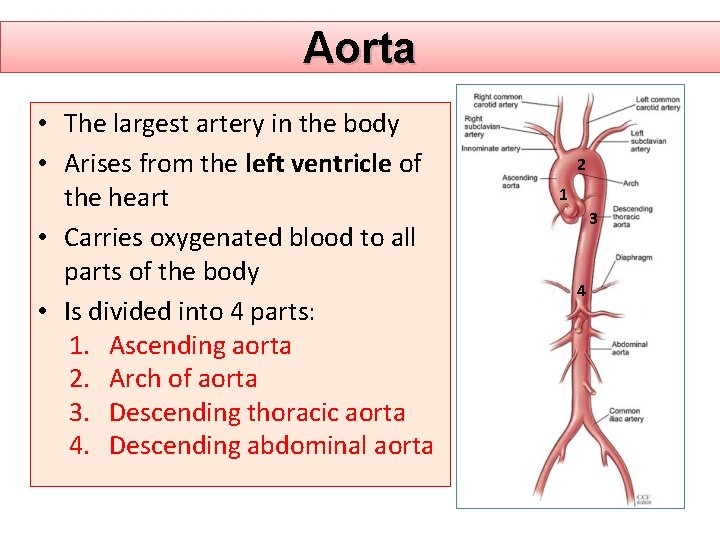
Aorta • The largest artery in the body • Arises from the left ventricle of the heart • Carries oxygenated blood to all parts of the body • Is divided into 4 parts: 1. Ascending aorta 2. Arch of aorta 3. Descending thoracic aorta 4. Descending abdominal aorta 2 1 3 4
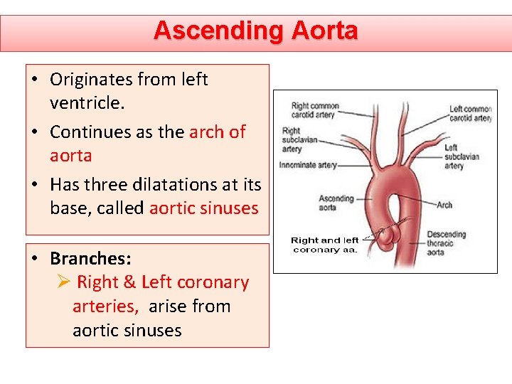
Ascending Aorta • Originates from left ventricle. • Continues as the arch of aorta • Has three dilatations at its base, called aortic sinuses • Branches: Ø Right & Left coronary arteries, arise from aortic sinuses
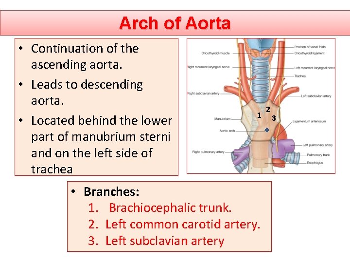
Arch of Aorta • Continuation of the ascending aorta. • Leads to descending aorta. • Located behind the lower part of manubrium sterni and on the left side of trachea 1 • Branches: 1. Brachiocephalic trunk. 2. Left common carotid artery. 3. Left subclavian artery 2 3
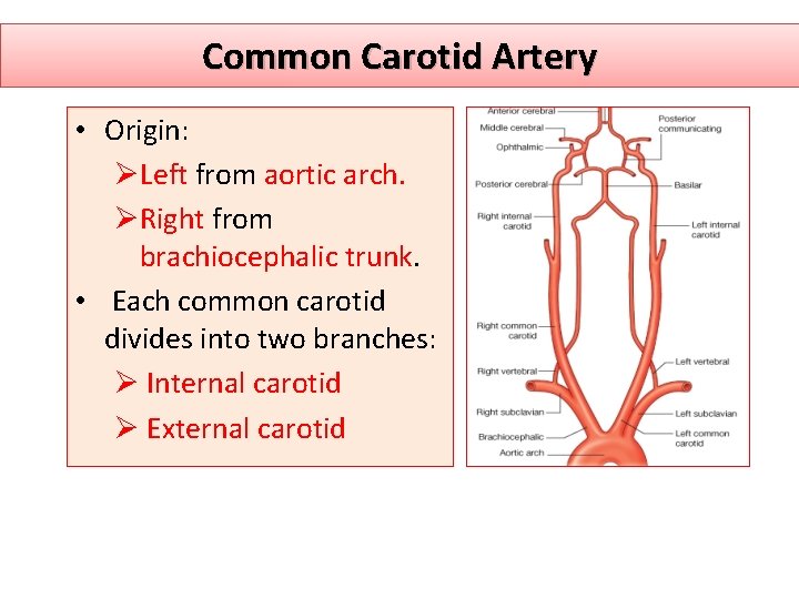
Common Carotid Artery • Origin: ØLeft from aortic arch. ØRight from brachiocephalic trunk. • Each common carotid divides into two branches: Ø Internal carotid Ø External carotid
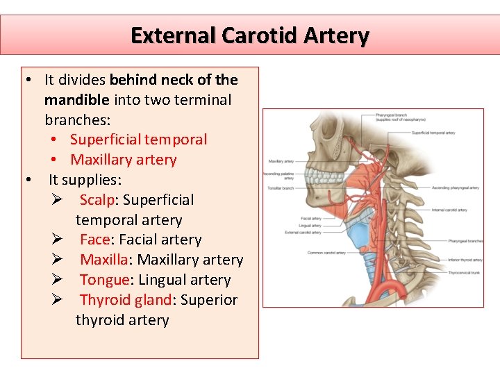
External Carotid Artery • It divides behind neck of the mandible into two terminal branches: • Superficial temporal • Maxillary artery • It supplies: Ø Scalp: Superficial temporal artery Ø Face: Facial artery Ø Maxilla: Maxillary artery Ø Tongue: Lingual artery Ø Thyroid gland: Superior thyroid artery
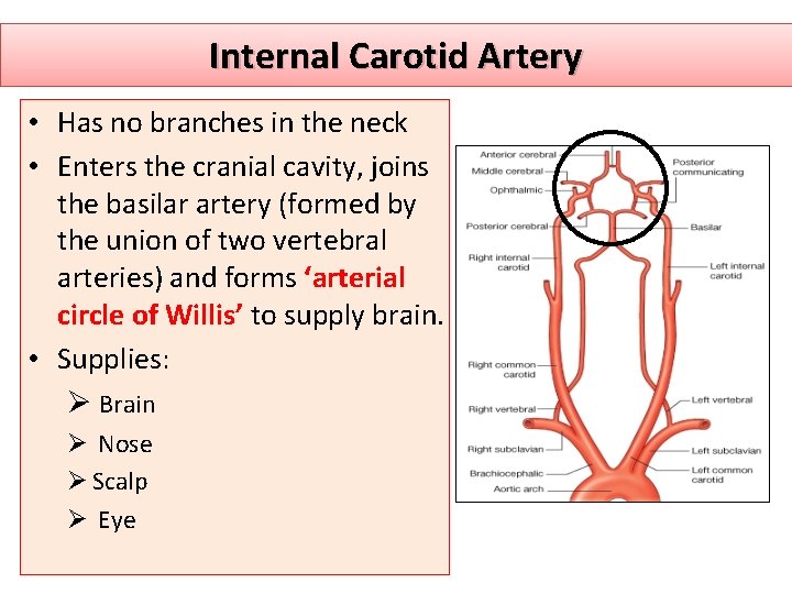
Internal Carotid Artery • Has no branches in the neck • Enters the cranial cavity, joins the basilar artery (formed by the union of two vertebral arteries) and forms ‘arterial circle of Willis’ to supply brain. • Supplies: Ø Brain Ø Nose Ø Scalp Ø Eye
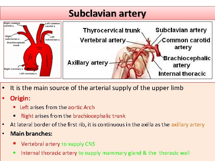
Subclavian artery • It is the main source of the arterial supply of the upper limb • Origin: § Left arises from the aortic Arch § Right arises from the brachiocephalic trunk • At lateral border of the first rib, it is continuous in the axilla as the axillary artery • Main branches: § Vertebral artery to supply CNS § Internal thoracic artery to supply mammary gland & the thoracic wall
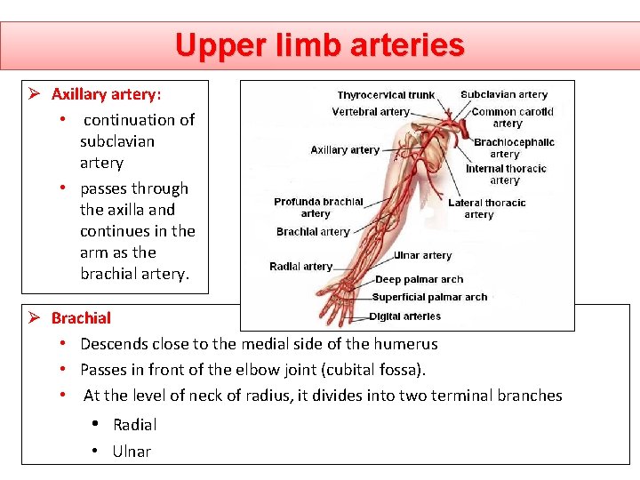
Upper limb arteries Ø Axillary artery: • continuation of subclavian artery • passes through the axilla and continues in the arm as the brachial artery. Ø Brachial • Descends close to the medial side of the humerus • Passes in front of the elbow joint (cubital fossa). • At the level of neck of radius, it divides into two terminal branches • Radial • Ulnar
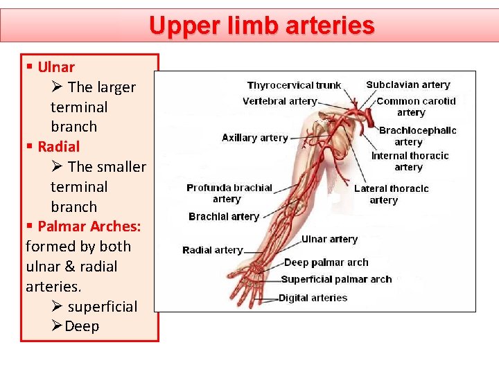
Upper limb arteries § Ulnar Ø The larger terminal branch § Radial Ø The smaller terminal branch § Palmar Arches: formed by both ulnar & radial arteries. Ø superficial ØDeep
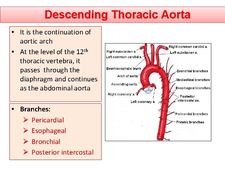
Descending Thoracic Aorta • It is the continuation of aortic arch • At the level of the 12 th thoracic vertebra, it passes through the diaphragm and continues as the abdominal aorta • Branches: Ø Pericardial Ø Esophageal Ø Bronchial Ø Posterior intercostal
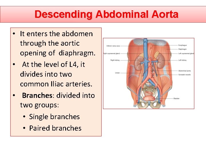
Descending Abdominal Aorta • It enters the abdomen through the aortic opening of diaphragm. • At the level of L 4, it divides into two common Iliac arteries. • Branches: divided into two groups: • Single branches • Paired branches
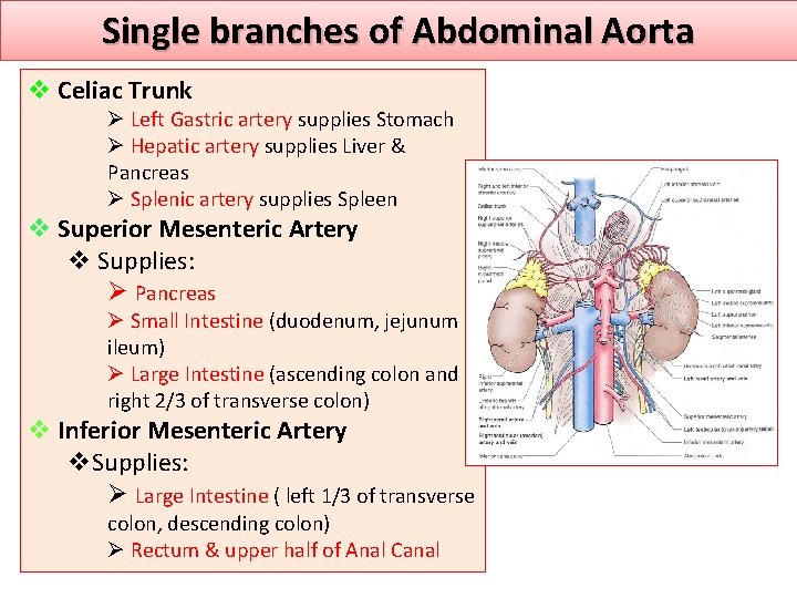
Single branches of Abdominal Aorta v Celiac Trunk Ø Left Gastric artery supplies Stomach Ø Hepatic artery supplies Liver & Pancreas Ø Splenic artery supplies Spleen v Superior Mesenteric Artery v Supplies: Ø Pancreas Ø Small Intestine (duodenum, jejunum & ileum) Ø Large Intestine (ascending colon and right 2/3 of transverse colon) v Inferior Mesenteric Artery v. Supplies: Ø Large Intestine ( left 1/3 of transverse colon, descending colon) Ø Rectum & upper half of Anal Canal
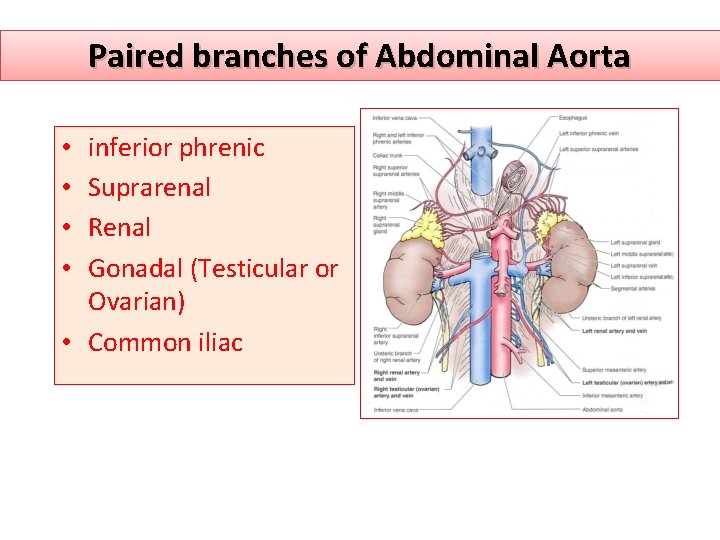
Paired branches of Abdominal Aorta inferior phrenic Suprarenal Renal Gonadal (Testicular or Ovarian) • Common iliac • •
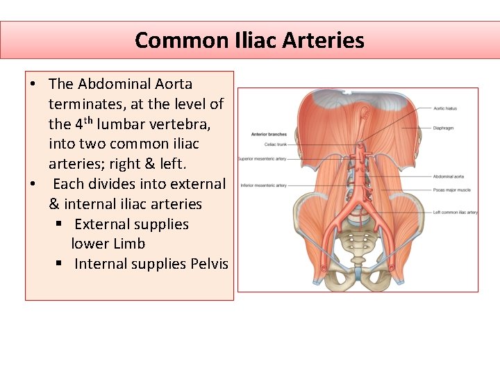
Common Iliac Arteries • The Abdominal Aorta terminates, at the level of the 4 th lumbar vertebra, into two common iliac arteries; right & left. • Each divides into external & internal iliac arteries § External supplies lower Limb § Internal supplies Pelvis
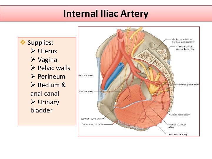
Internal Iliac Artery v Supplies: Ø Uterus Ø Vagina Ø Pelvic walls Ø Perineum Ø Rectum & anal canal Ø Urinary bladder
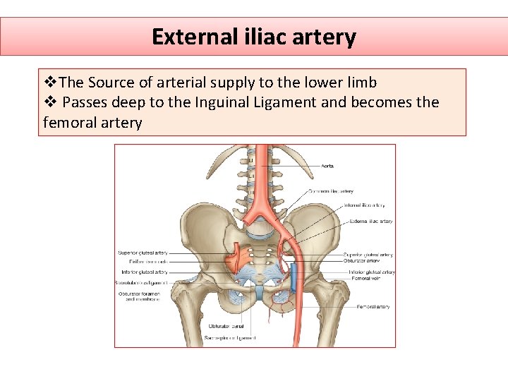
External iliac artery v. The Source of arterial supply to the lower limb v Passes deep to the Inguinal Ligament and becomes the femoral artery
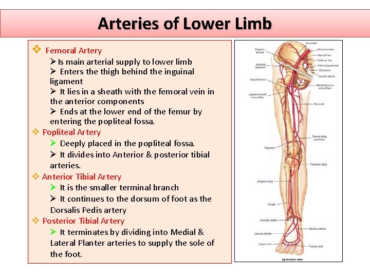
Arteries of Lower Limb v Femoral Artery ØIs main arterial supply to lower limb Ø Enters the thigh behind the inguinal ligament Ø It lies in a sheath with the femoral vein in the anterior components Ø Ends at the lower end of the femur by entering the popliteal fossa. v Popliteal Artery Ø Deeply placed in the popliteal fossa. Ø It divides into Anterior & posterior tibial arteries. v Anterior Tibial Artery Ø It is the smaller terminal branch Ø It continues to the dorsum of foot as the Dorsalis Pedis artery v Posterior Tibial Artery Ø It terminates by dividing into Medial & Lateral Planter arteries to supply the sole of the foot.
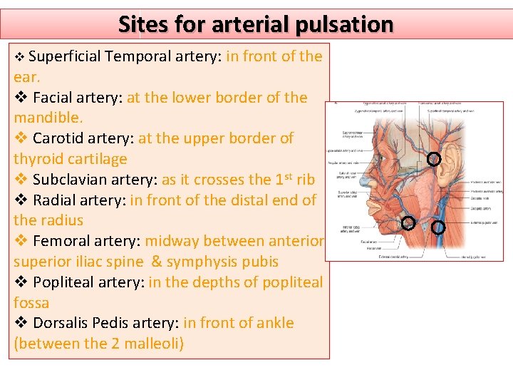
Sites for arterial pulsation v Superficial Temporal artery: in front of the ear. v Facial artery: at the lower border of the mandible. v Carotid artery: at the upper border of thyroid cartilage v Subclavian artery: as it crosses the 1 st rib v Radial artery: in front of the distal end of the radius v Femoral artery: midway between anterior superior iliac spine & symphysis pubis v Popliteal artery: in the depths of popliteal fossa v Dorsalis Pedis artery: in front of ankle (between the 2 malleoli)
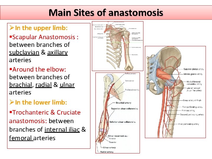
Main Sites of anastomosis ØIn the upper limb: §Scapular Anastomosis : between branches of subclavian & axillary arteries §Around the elbow: between branches of brachial, radial & ulnar arteries ØIn the lower limb: §Trochanteric & Cruciate anastomosis: between branches of internal iliac & femoral arteries

Thank you
- Slides: 23