Maj Muhammad Alamgir Khan Kidneys Two kidneys Bean


Maj Muhammad Alamgir Khan

Kidneys ► Two kidneys Bean shaped organs ► Weight 150 gm each ► Right kidney slightly lower than left ► Located ► Between T-12 and L-3 ► Retroperitoneal ►

4

Kidney

Cortex Medulla Renal papilla Minor calyx Pyramid Major calyx Renal pelvis Ureter

Nephron Functional unit of kidney

Parts of nephron ► Two main parts ► Renal corpuscle ► ► Formation of filtrate Renal tubule ► Convert the filtrate into urine

Renal tubule Renal corpuscle

Renal corpuscle

Renal corpuscle Bowman’s capsule Glomerulus Proximal tubule Distal tubule Cortex Afferent arteriole Cortical collecting tubule Arcuate artery Loop of henle • Descending limb • Thick ascending limb • Thin ascending limb Medullary collecting tubule

Parts of nephron ► Renal corpuscle ► Glomerulus ► ► Tuft of capillaries Bowman's capsule ► Blind end of renal tubule

Parts of nephron ► Renal tubule Proximal convoluted tubule ► Loop of henle ► Descending limb ► Thin ascending limb ► Thick ascending limb ► Distal convoluted tubule ► Collecting tubule ► Cortical collecting tubule ► Medullary collecting tubule ► ► Collecting duct

Efferent arteriole Glomerular capillaries Afferent arteriole Renal vein Peritubular capillaries

Renal blood flow ► About 22 % of cardiac output ~ 1100 ml/min ► Serves two purposes ► Metabolic needs (much greater than requirement) ► Urine formation (purification of blood) ►

22 % 78 %
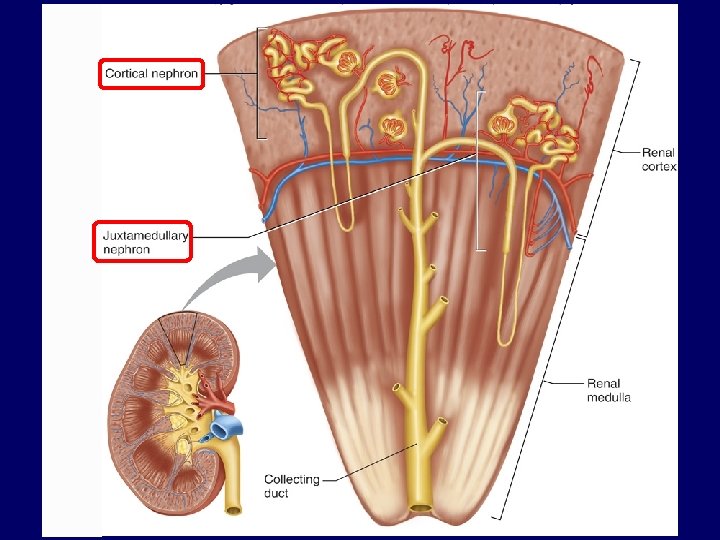
 Cortical nephron")
Types of nephron ► 2 types (based upon location of renal corpuscle) Cortical nephron ► Juxtamedullary nephron ►
 Renal corpuscle lies in")
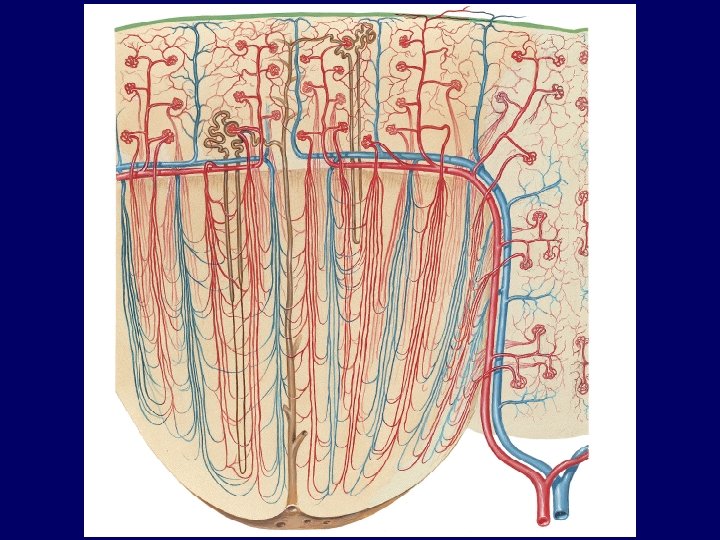
Types of nephron ► Cortical nephrons (70 - 80 %) Renal corpuscle lies in cortex ► More in number ► Short loop of henle ► Extensive blood supply ► ► Peritubular capillaries
 Renal corpuscle lies near")
Types of nephron ► Medullary nephrons (20 - 30 %) Renal corpuscle lies near corticomedullary junction ► less in number ► long loop of henle ► less blood supply ► ► ‘U’ shaped capillaries - vasa recta

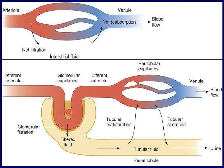
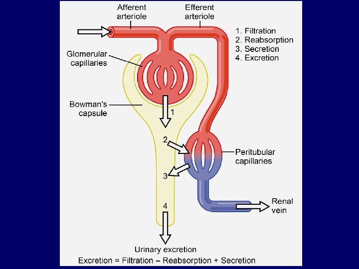
Urine formation ► Result of 3 processes Glomerular filtration ► Tubular reabsorption ► Tubular secretion ► Urine = Filtration - reabsorption + secretion

Urine formation Blood Filtrate Vein Urine

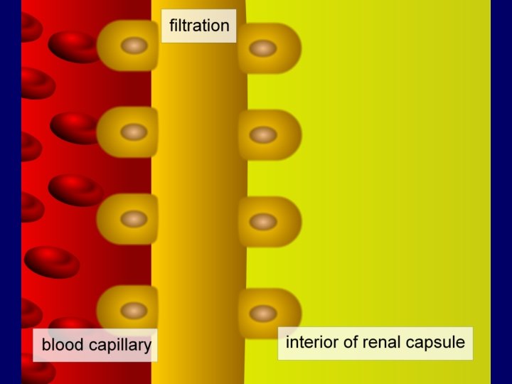
Glomerular filtration Filtration of protein free plasma from glomerular capillaries into bowman’s capsule ► 1 st step in urine formation ►

Glomerular filtration ► Glomerular filtrate ► Composition ► Same as plasma without proteins Osmolarity equal to plasma ► Fluid to be processed in renal tubule to form urine ►
 ► Volume of glomerular filtrate formed per minute ► 125")
Glomerular filtration rate (GFR) ► Volume of glomerular filtrate formed per minute ► 125 ml/min (180 l/day)
 ► Filtration fraction ► Fraction of renal plasma flow filtered")
Glomerular filtration rate (GFR) ► Filtration fraction ► Fraction of renal plasma flow filtered Filtration fraction = GFR Renal plasma flow 125 650 = ~ 0. 2

Glomerular filtration membrane ► 3 layers ► Capillary endothelial layer Fenestrated endothelium ► Negatively charged ► ► Basement membrane ► ► Negatively charged Podocyte layer (visceral layer of bowman’ capsule) Foot processes form ‘slit pores’ ► Negatively charged ►
 Basement membrane Capillary endothelium")
Visceral layer of bowman’s capsule (podocyte layer) Basement membrane Capillary endothelium

Glomerular filtration membrane Capillary endothelium Basement membrane Podocyte layer

Glomerular filtrate Slit pores Podocyte layer - --- - - Fenestrations - -

Glomerular filtration membrane
 ► Determinants of GFR Filtration coefficient (Kf) ► Net filtration")
Glomerular filtration rate (GFR) ► Determinants of GFR Filtration coefficient (Kf) ► Net filtration pressure ► GFR = Kf × Net filtration pressure

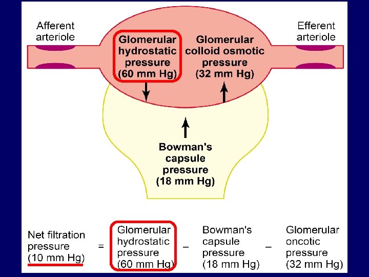
Determinants of GFR ► Net filtration pressure ► Pressures favoring filtration ► Glomerular capillary hydrostatic pressure (PG) ► ► 60 mm Hg Bowman’s capsule colloid osmotic pressure ( B) ► 0 mm Hg

Determinants of GFR ► Net filtration pressure ► Pressures opposing filtration ► Bowman’s capsule hydrostatic pressure(PB) ► ► 18 mm Hg Glomerular capillary colloid osmotic pressure ( G) ► 32 mm Hg

Determinants of GFR ► Net filtration pressure = PG - PB - G + B Net filtration pressure = 60 - 18 - 32 + 0 Net filtration pressure = 10 mm Hg
 10 mm Hg")
60 32 18 60 - (32+18) 10 mm Hg
 Physiologic regulator of GFR ►")
Determinants of GFR ► Glomerular capillary hydrostatic pressure (PG) Physiologic regulator of GFR ► Depends upon ► Arterial blood pressure ► Afferent arteriolar resistance ► Efferent arteriolar resistance ►
 ► arterial pressure ► ► Afferent")
Determinants of GFR ► Glomerular capillary pressure (PG) ► arterial pressure ► ► Afferent arteriolar constriction ► ► PG GFR Efferent arteriole ► Moderate constriction ► ► PG GFR Sever constriction ► G GFR

Determinants of GFR ► Effect of renal blood flow on GFR ► By affecting PG ► ► blood flow PG GFR By affecting filtration fraction & G ► blood flow filtration fraction G GFR

46
 ► GFR per mm Hg of net")
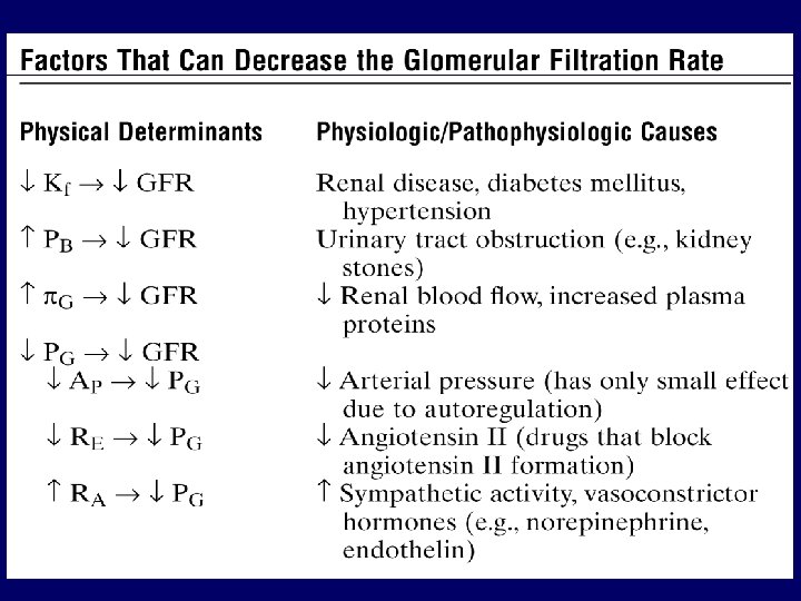
Determinants of GFR ► Filtration coefficient (Kf) ► GFR per mm Hg of net filtration pressure ► ► Kf = 125/10 = 12. 5 ml/min/mm Hg Product of Capillary permeability ► Capillary surface area ► ► Decreased in diseases Diabetes mellitus ► Hypertension ►

Regulation of GFR ► 3 primary regulators of GFR Autoregulation ► Neural control ► Hormonal & autacoid control ►
 ► Tubuloglomerular feedback ► ► Achieved by")
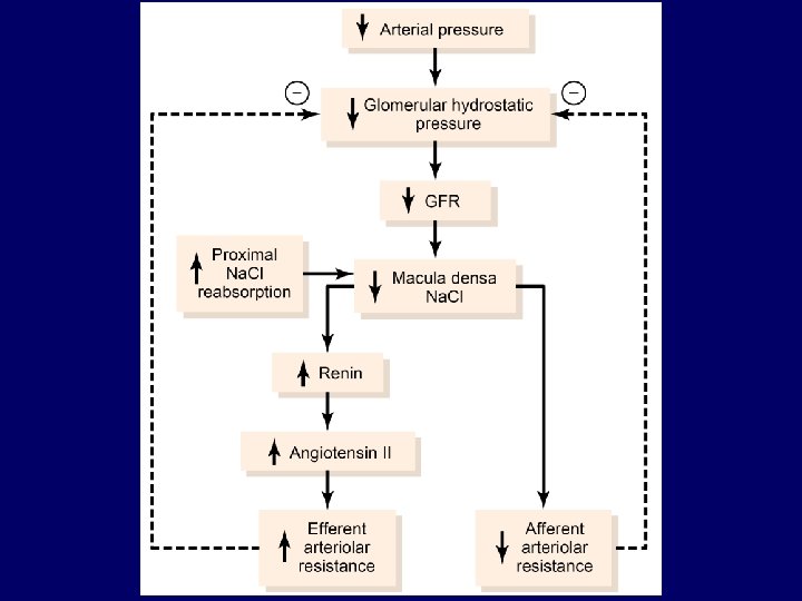
Regulation of GFR ► Autoregulation (intrinsic control) ► Tubuloglomerular feedback ► ► Achieved by juxtaglomerular apparatus Myogenic mechanism ► Intrinsic property of vascular smooth muscle
 ► Juxtaglomerular cells")
Juxtaglomerular apparatus ► Consists of Macula densa cells (renal tubular cells) ► Juxtaglomerular cells (afferent/efferent ► arteriolar cells) ► Lacis cells (Extraglomerular mesangial cells)

On next slide 53

Lacis cells 54

Afferent arteriole JG cells Lacis cells Macula densa cells JG apparatus Distal convoluted tubule

Juxtaglomerular apparatus ► Juxtaglomerular cells Present in afferent arterioles ► Some in efferent arterioles ► Contain granules of renin ► Also called granular cells ►

Juxtaglomerular apparatus ► Macula densa cells Present in initial part of distal convoluted tubule ► Sense change in Na. Cl delivery ►

Juxtaglomerular apparatus ► Function ► Maintains renal blood flow & GFR by tubuloglomerular feedback mechanism

► Tubuloglomerular feedback ► Alterations in GFR & renal blood flow induced by tubular flow rate

Juxtaglomerular apparatus ► Tubuloglomerular feedback Afferent arteriolar feedback ► Efferent arteriolar feedback ►

Lacis cells 61

Change in renal blood flow/GFR Change in Na. Cl delivery to DCT Macula densa cells sense the change Paracrine vasoactive agents Afferent arteriole Efferent arteriole Change in diameter Restoration of renal blood flow/GFR JG cells Renin release Angiotensin II
 64")
Direct effect of BP on GFR (without autoregulation) 64

Autoregulation in response to BP 65

Tubuloglomerular feedback ► Regulation of GFR/renal blood flow ► Over a wide range of arterial blood pressure ► 75 to 160 mm. Hg

75 160
")
Myogenic mechanism ► Contraction of vascular smooth muscle when stretched (due to arterial pressure) ► arterial pressure ( GFR & renal blood flow) stretch contraction resistance blood flow (restoration of GFR & renal blood flow)

Autoregulation Blunts BP induced changes in GFR/renal blood flow and maintains urine volume output

Regulation of GFR ► Neural control ► Sympathetic nervous system ► Mediated through baroreceptor reflex Alters afferent arteriolar resistance ► Contracts glomerular mesangial cells (stimulation) ► ► Kf ( glomerular capillary surface area) GFR

Regulation of GFR ► Hormonal & autacoid control ► Hormones & autacoids increasing GFR Endothelial derived nitric oxide ► Prostaglandins (PGE 2 & PGI 2) ► Bradykinin ► ► Hormones & autacoids decreasing GFR Norepinephrine ► Endothelin ► Angiotensin II ►

Regulation of urine volume output GFR = 125 ml/min ► T/reabsorption rate = 124 ml/min ► Urine flow rate = 1 ml/min ►

Regulation of urine volume output ► Tubuloglomerular feedback 1 line of defense ► st ► Glomerulotubular feedback 2 line of defense ► nd

Regulation of urine volume output Renal arteriolar pressure 2 nd line of defense Glomerulotubular feedback Glomerular capillary pressure GFR Solute reabsorption in proximal tubule Solute reabsorption in thick ascending limb Salt & fluid delivery to distal tubule (macula densa) 1 st line of defense Tubuloglomerular feedback

Regulation of urine volume output ► Tubuloglomerular & glomerulotubular feedbacks - not 100 % perfect Pressure diuresis ► Pressure natriuresis ►

- Slides: 76