
Maintenance of Electrolyte and Fluid Balance A A
Maintenance of Electrolyte and Fluid Balance A. A. J. Rajaratne
The loop of Henle: In the loop of Henle about 20% of filtered Na+, Cl-, and K+ are reabsorbed. Ca++, Mg++, and HCO 3 - are also reabsorbed. About 17% of filtered water is reabsorbed in Henle's loop. Water is reabsorbed from the descending limb; the ascending limb is impermeable to water.
The loop of Henle - contd: 1. Transport processes in thick ascending limb. Na+ enters the TAL epithelial cells across the luminal membrane via the Na/K/2 Cl transporter, which transports Cl- and K+ into the cell against their electrochemical potential gradients. The energy of the Na+ gradient is used for this process.
There is also a luminal Na/H exchanger, which exports H+ into the lumen and causes reabsorption of HCO 3 -. K+, Cl-, and HCO 3 - are transported across the basolateral membrane by other transport proteins. The luminal fluid in the TAL is electrically positive to the extracellular fluid of the basolateral membrane. This powers the reabsorption of Na+, K+, Ca++, and Mg++, partly via transcellular and partly via paracellular pathways.
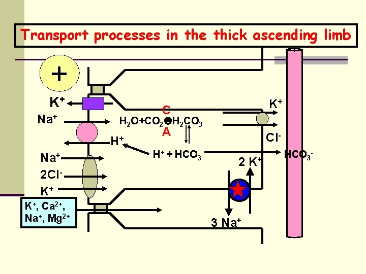
Transport processes in the thick ascending limb + K+ Na+ K+, Ca 2+, Na+, Mg 2+ A Cl- H 2 O+CO 2 H 2 CO 3 H+ Na+ 2 Cl. K+ C K+ H+ + HCO 3 2 3 Na+ K+ HCO 3 -
Significant absorption of solute occurs in the TAL, but water cannot follow due to the impermeability of the TAL to water. Thus the osmolarity of TAL fluid falls below isotonicity, reaching less than 150 milliosmolar.
The extrusion of Na+ and Cl- by the TAL contributes to an osmotic gradient in the medullary interstitium. The osmotic pressure is highest near the renal papillae and lowest near the corticomedullary junction. Since the descending limb is permeable to water (the ascending limb is not), water is reabsorbed from the descending limb as it descends through the osmotic gradient.
The distal tubule and the collecting tubule Reabsorb about 10% of filtered Na+ and Cl. Secrete K+ and H+. Reabsorb a variable amount of water.
The first part of the distal reabsorbs Na+, Cl-. and Ca 2+. tubule Since this segment of the distal tubule is impermeable to water, the luminal fluid becomes still more dilute, approaching 100 m. Osm. The luminal membrane has electrogenic Na+ channels (blocked by amiloride and triamterine diuretics) and a coupled Na+/Cl- transporter (blocked by thiazide diuretics).
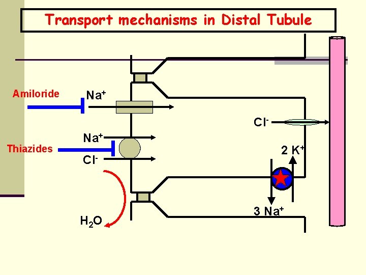
Transport mechanisms in Distal Tubule Amiloride Na+ Cl- Thiazides Na+ Cl- H 2 O 2 K+ 3 Na+
Last part of distal tubule and collecting tubule: Contain two types of epithelial cells: 1. Principal cells 2. Intercalated cells a. Principal cells in the last part of the distal tubule and in the cortical collecting tubule reabsorb Na+, Cl-, and water and usually secrete K+.
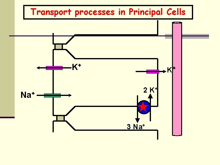
Transport processes in Principal Cells K+ Na+ K+ 2 K+ 3 Na+
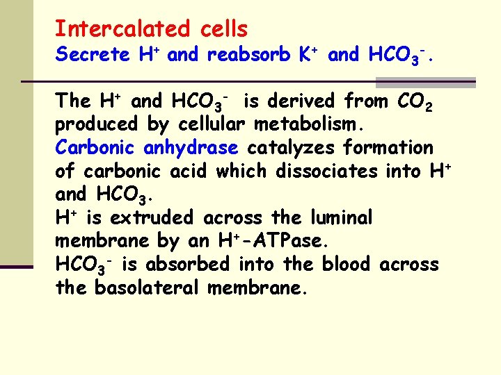
Intercalated cells Secrete H+ and reabsorb K+ and HCO 3 -. The H+ and HCO 3 - is derived from CO 2 produced by cellular metabolism. Carbonic anhydrase catalyzes formation of carbonic acid which dissociates into H+ and HCO 3. H+ is extruded across the luminal membrane by an H+-ATPase. HCO 3 - is absorbed into the blood across the basolateral membrane.
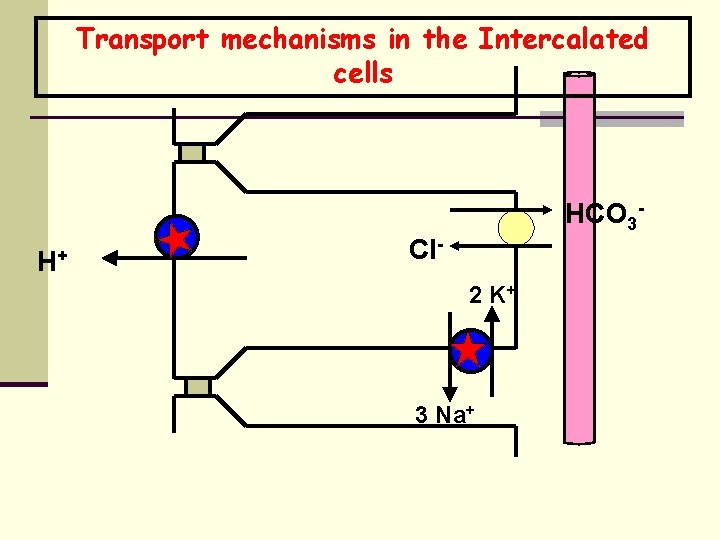
Transport mechanisms in the Intercalated cells H+ HCO 3 - Cl 2 K+ 3 Na+
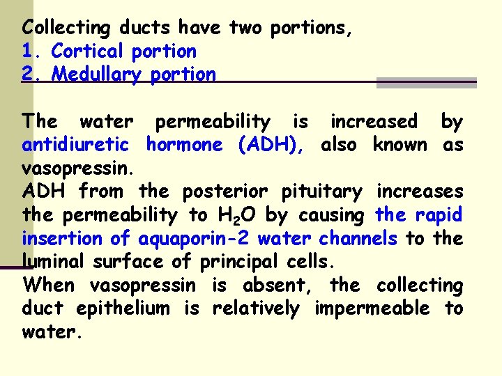
Collecting ducts have two portions, 1. Cortical portion 2. Medullary portion The water permeability is increased by antidiuretic hormone (ADH), also known as vasopressin. ADH from the posterior pituitary increases the permeability to H 2 O by causing the rapid insertion of aquaporin-2 water channels to the luminal surface of principal cells. When vasopressin is absent, the collecting duct epithelium is relatively impermeable to water.
Control of ECF osmolality and volume
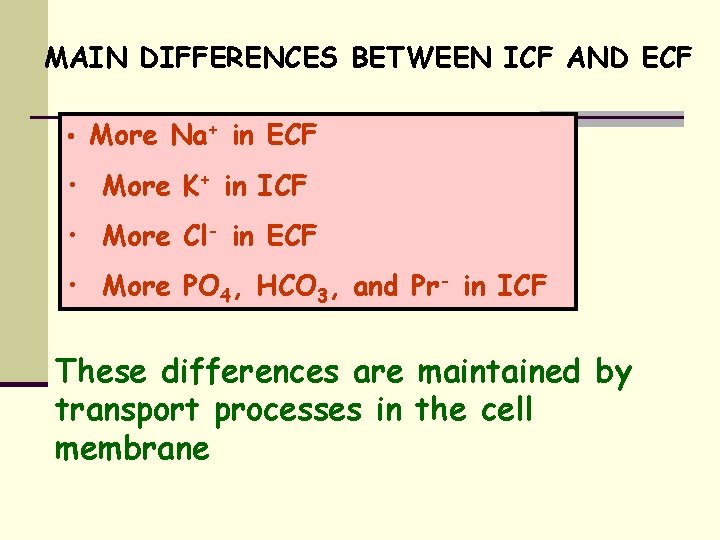
MAIN DIFFERENCES BETWEEN ICF AND ECF • More Na+ in ECF • More K+ in ICF • More Cl- in ECF • More PO 4, HCO 3, and Pr- in ICF These differences are maintained by transport processes in the cell membrane
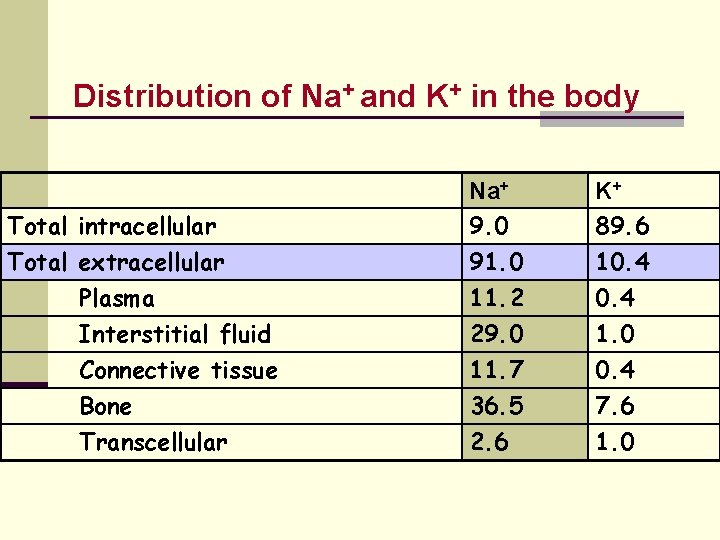
Distribution of Na+ and K+ in the body Total intracellular Total extracellular Plasma Interstitial fluid Connective tissue Bone Transcellular Na+ 9. 0 91. 0 11. 2 K+ 89. 6 10. 4 29. 0 11. 7 36. 5 2. 6 1. 0 0. 4 7. 6 1. 0
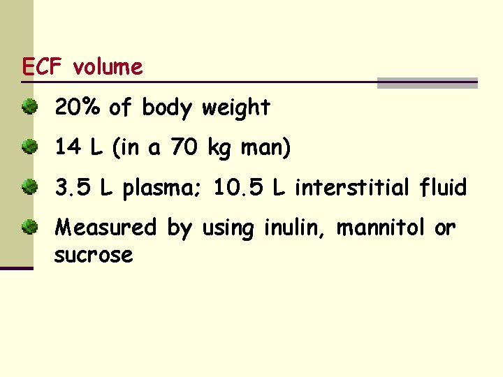
ECF volume 20% of body weight 14 L (in a 70 kg man) 3. 5 L plasma; 10. 5 L interstitial fluid Measured by using inulin, mannitol or sucrose
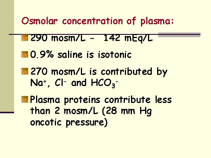
Osmolar concentration of plasma: 290 mosm/L - 142 m. Eq/L 0. 9% saline is isotonic 270 mosm/L is contributed by Na+, Cl- and HCO 3 Plasma proteins contribute less than 2 mosm/L (28 mm Hg oncotic pressure)
Ranges of salt and water intake and excretion: a. Salt intake from 50 mg to 25 g/day b. Water excretion from 400 ml to 25 l/day
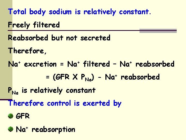
Total body sodium is relatively constant. Freely filtered Reabsorbed but not secreted Therefore, Na+ excretion = Na+ filtered – Na+ reabsorbed = (GFR X PNa) - Na+ reabsorbed PNa is relatively constant Therefore control is exerted by GFR Na+ reabsorption
Sensors: 1. Extrarenal baroreceptors Carotid sinuses Arteries Great veins Atria 2. Renal juxtaglomerular apparatus Efferents: 1. Renal sympathetic nerves 2. Macula densa renin angiotensin II aldosterone
Control of GFR: 1. Angiotensin II efferent arteriolar constriction PGC 2. Renal sympathetic nerves Na+ adrenergic receptors Constriction of afferent and efferent arterioles PGC
Osmoreceptor -ADH mechanisms
Renal handling of Na. Cl and water: Na. Cl & H 2 O are freely filterable at the glomerulus. There is extensive tubular reabsorption but no tubular secretion. Na+ reabsorption is driven by the basolateral Na+/K+-ATPase and is responsible for the major energy expenditure in kidney.
H 2 O permeability of the late DT: Water permeability of distal tubule and initial collecting tubule, is also extremely low. However under the influence of ADH it becomes highly water permeable. Further removal of solute in the EARLY DT presents the LATE DT with markedly hypotonic urine containing even less Na+ Removal of Na+ continues in the LDT and collecting system, so that the final urine may contain virtually no Na+.
Anti-diuretic hormone: ADH (antidiuretic hormone), vasopressin or arginine vasopressin (AVP) is the major regulator of urine osmolality and urine volume. ADH is a nonapeptide produced by neurons in the supraoptic and paraventricular nuclei of the hypothalamus. The axon terminals of these neurons reside in the posterior pituitary. ADH is stored in these axon terminals.
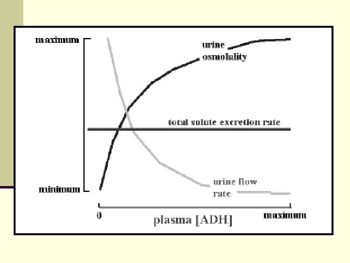
When ADH is released from the posterior pituitary it causes the kidney to produce urine that is high in osmolality and low in volume. In the absence of ADH the kidney tends to produce a large volume of urine with low osmolality. Total solute excretion is relatively constant over a wide range of urine flow rates and osmolalities.
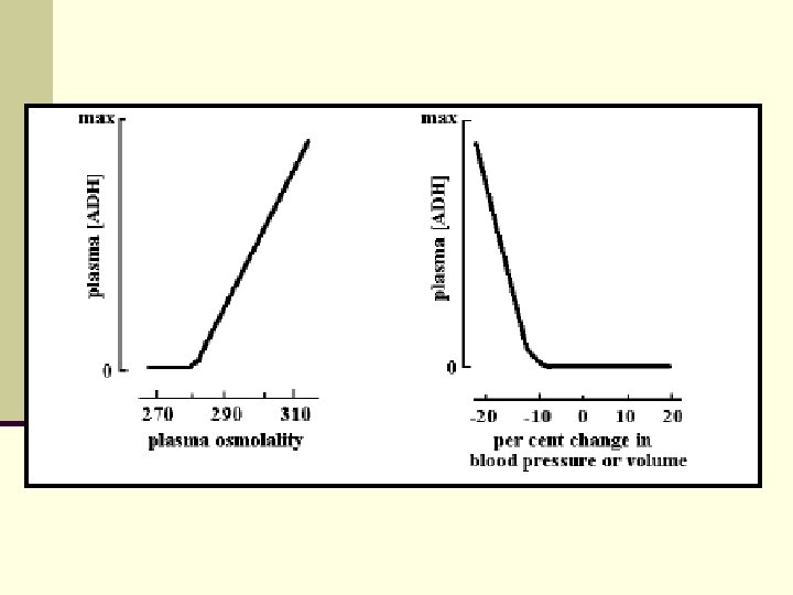
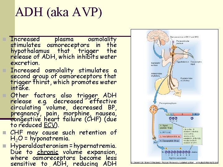
Control of ADH release: 1. Increased osmolality of ECF is a powerful stimulus for ADH release: a 1% change in osmolality induces significant increase in ADH release. Hypothalamic supra-optic and paraventricular nuclei respond to increased osmolality of ECF by producing ADH. As a result of this high sensitivity, responses to increased osmolality occur rapidly.
Control of ADH release: 2. Volume: In a volume-depleted individual, the release of ADH is more sensitive to increased osmolality. In a volume-expanded state, ADH release is less sensitive to increases in osmolality. 3. Decreased blood pressure or blood volume also enhance ADH release, but not with such high sensitivity: 5 to 10% changes are required to alter ADH secretion.
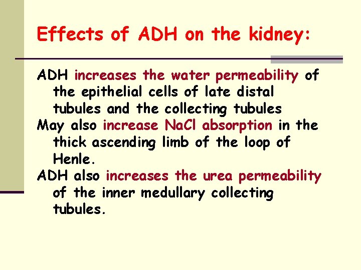
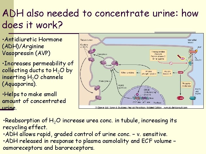
Effects of ADH on the kidney: ADH increases the water permeability of the epithelial cells of late distal tubules and the collecting tubules May also increase Na. Cl absorption in the thick ascending limb of the loop of Henle. ADH also increases the urea permeability of the inner medullary collecting tubules.
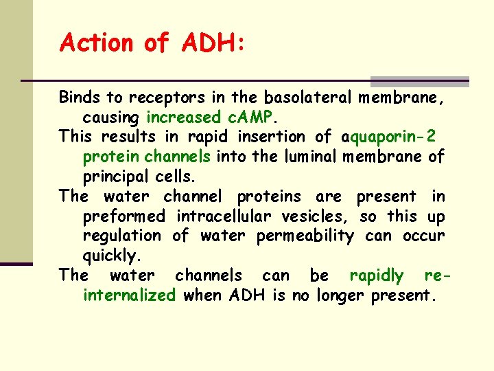
Action of ADH: Binds to receptors in the basolateral membrane, causing increased c. AMP. This results in rapid insertion of aquaporin-2 protein channels into the luminal membrane of principal cells. The water channel proteins are present in preformed intracellular vesicles, so this up regulation of water permeability can occur quickly. The water channels can be rapidly reinternalized when ADH is no longer present.
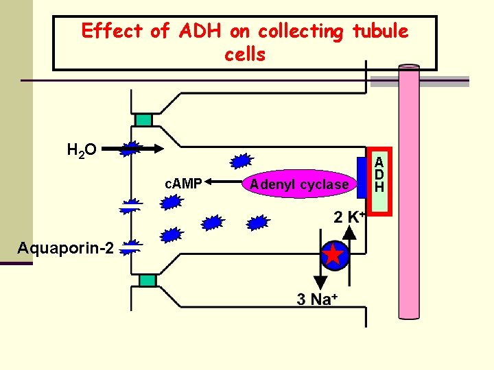
Effect of ADH on collecting tubule cells H 2 O c. AMP Aquaporin-2 Adenyl cyclase A D H
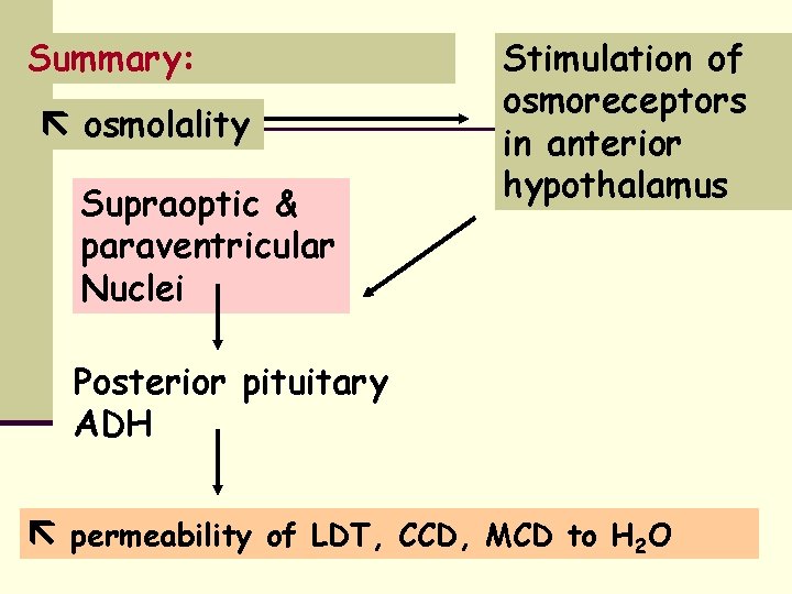
Summary: osmolality Supraoptic & paraventricular Nuclei Stimulation of osmoreceptors in anterior hypothalamus Posterior pituitary ADH permeability of LDT, CCD, MCD to H 2 O
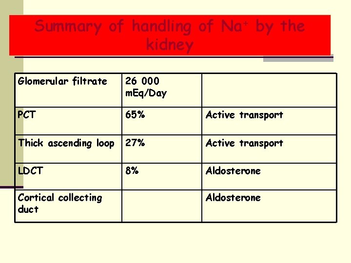
Summary of handling of Na+ by the kidney Glomerular filtrate 26 000 m. Eq/Day PCT 65% Active transport Thick ascending loop 27% Active transport LDCT 8% Aldosterone Cortical collecting duct Aldosterone
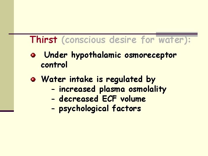
Thirst (conscious desire for water): Under hypothalamic osmoreceptor control Water intake is regulated by - increased plasma osmolality - decreased ECF volume - psychological factors
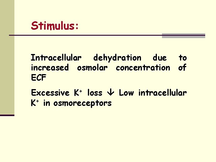
Stimulus: Intracellular dehydration due to increased osmolar concentration of ECF Excessive K+ loss Low intracellular K+ in osmoreceptors
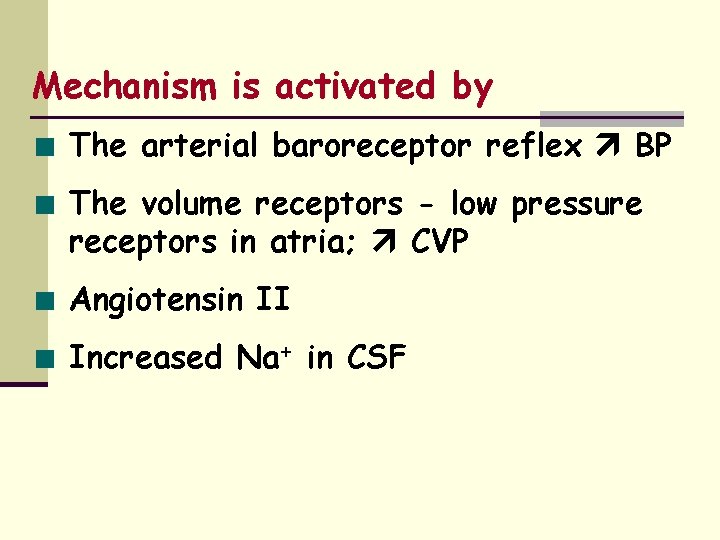
Mechanism is activated by The arterial baroreceptor reflex BP The volume receptors - low pressure receptors in atria; CVP Angiotensin II Increased Na+ in CSF
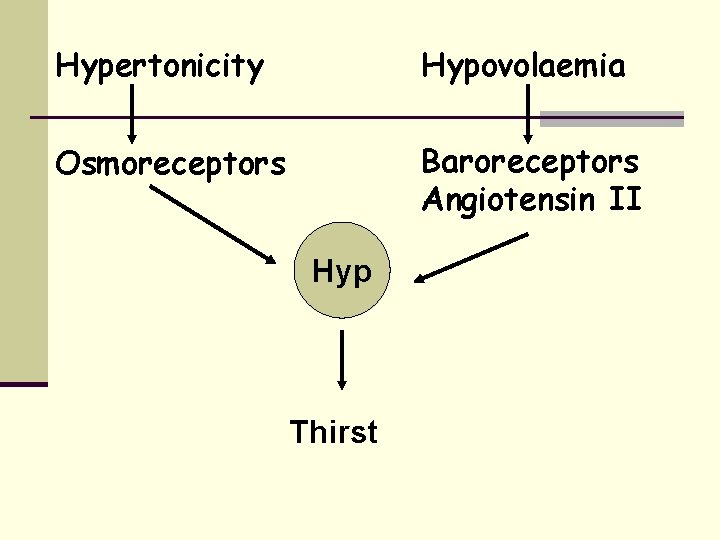
Hypertonicity Hypovolaemia Osmoreceptors Baroreceptors Angiotensin II Hyp Thirst
Other factors regulating water intake: Psycho-social Dryness of pharyngeal mucous membrane ? Gastrointestinal pharyngeal metering
Renin-angiotensin – aldosterone system
Renin: Produced by Juxtaglomerular cells – located in media of afferent arterioles Lacis cells – junction between afferent and efferent arterioles
Factors affecting renin secretion: Stimulatory Increased sympathetic activity via renal nerves Increased circulating catecholamines Prostaglandins Inhibitory Increased Na+ and Cl- reabsorption in macula densa Angiotensin II Vasopressin
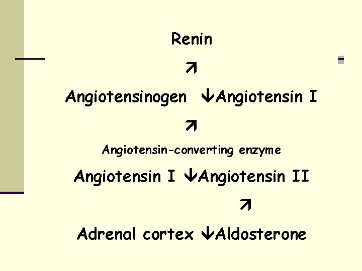
Renin Angiotensinogen Angiotensin I Angiotensin-converting enzyme Angiotensin II Adrenal cortex Aldosterone
Actions of angiotensin II Arteriolar vasoconstriction and rise in SBP and DBP On adrenal cortex to produce aldosterone Facilitates release of noradrenaline Contraction of mesangeal cells - GFR Brain - sensitivity of baroreflex
Actions of aldosterone: Increased reabsorption of Na+ from urine, sweat, saliva and GIT – ECF volume expansion Kidney Principal cells – increased amounts of Na+ are exchanged for K+ and H+
Salt appetite
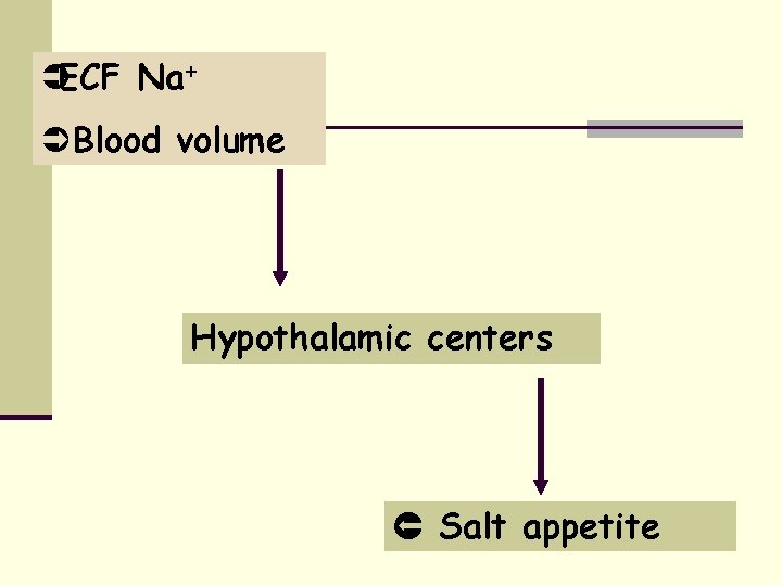
ÜECF Na+ Ü Blood volume Hypothalamic centers Salt appetite
Potassium excretion
Renal handling of K+: 800 m. Eq/day enter the filtrate 100 m. Eq/day is secreted PCT – reabsorption DCT and CD – both reabsorption and secretion
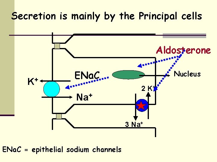
Secretion is mainly by the Principal cells Aldosterone K+ ENa. C Na+ Nucleus 2 K+ 3 Na+ ENa. C = epithelial sodium channels
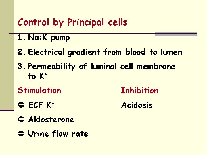
Control by Principal cells 1. Na: K pump 2. Electrical gradient from blood to lumen 3. Permeability of luminal cell membrane to K+ Stimulation Inhibition ECF K+ Acidosis Û Aldosterone Û Urine flow rate
How the kidney makes concentrated and dilute urine
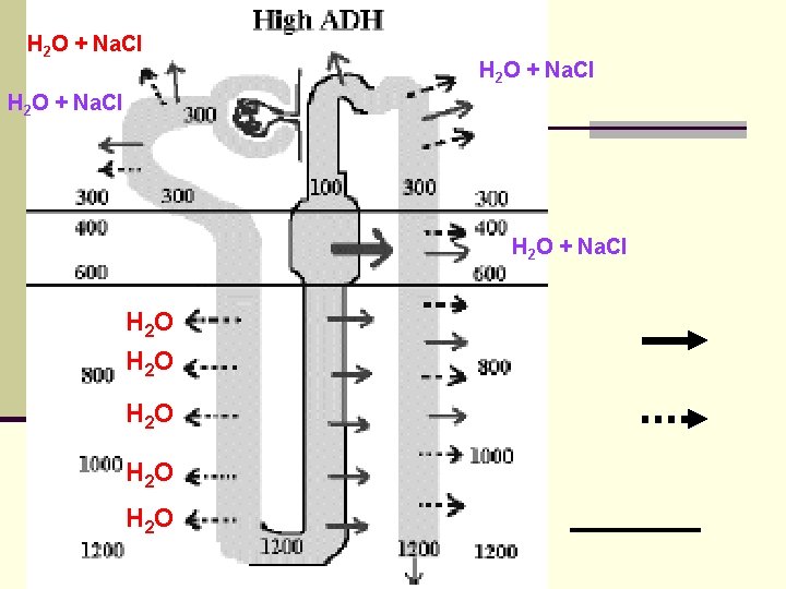
There is a gradient in osmolality in the medullary interstitium. Cortico-medullary junction 300 m. Os. M. Renal papilla 1200 m. Os. M in presence of ADH. About half of this is due to Na. Cl and the other half to urea. The vasa recta function as countercurrent exchangers, so that the blood flow does not wash out the gradients of Na. Cl and urea.
Effect of ADH: In the presence of ADH, the collecting tubule is highly permeable to water. As fluid in the collecting tubule descends through the medullary gradient, water is extracted, producing a low volume of urine that is high in osmolality (up to 1200 m. Os. M).
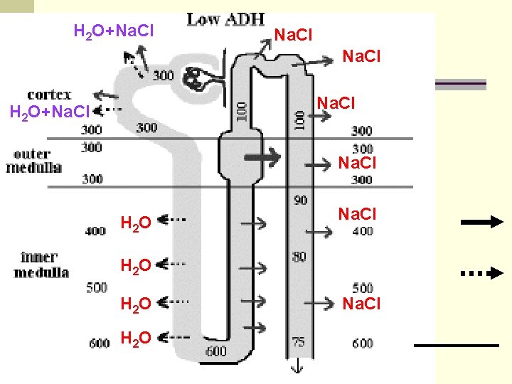
In the absence of ADH a large volume of dilute urine leaving the DCT (about 100 m. Os. M). In the absence of ADH, the collecting tubule is highly impermeable to water. The dilute fluid does not lose water as it descends through the medullary interstitium. Thus, volume does not decrease, nor does solute concentration increase, and a high volume of dilute urine is produced.
The Medullary Countercurrent System: The maximal concentration of urine produced ~ 1200 -1400 mosmol/l. Urea, sulphate, phosphate and other waste ~ 600 mosmol/day obligatory water loss ~ (600 mosmol/day)/(1300 mosmol/l) ~ 0. 46 l/day.
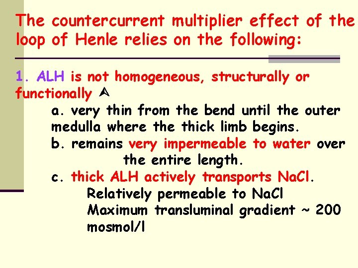
The countercurrent multiplier effect of the loop of Henle relies on the following: 1. ALH is not homogeneous, structurally or functionally a. very thin from the bend until the outer medulla where thick limb begins. b. remains very impermeable to water over the entire length. c. thick ALH actively transports Na. Cl. Relatively permeable to Na. Cl Maximum transluminal gradient ~ 200 mosmol/l
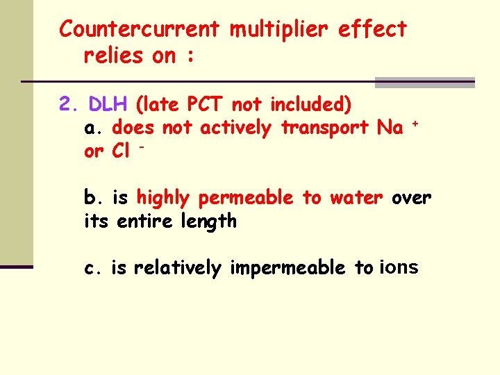
Countercurrent multiplier effect relies on : 2. DLH (late PCT not included) a. does not actively transport Na or Cl - + b. is highly permeable to water over its entire length c. is relatively impermeable to ions
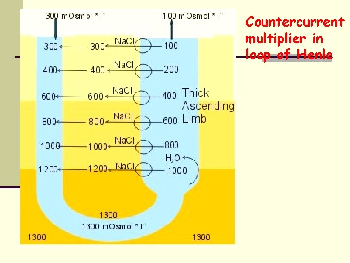
Countercurrent multiplier in loop of Henle
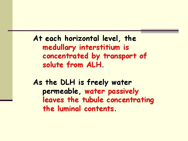
At each horizontal level, the medullary interstitium is concentrated by transport of solute from ALH. As the DLH is freely water permeable, water passively leaves the tubule concentrating the luminal contents.
These two processes proceed at each horizontal level so that the final concentration of solute deep in the medullary interstitium is ~ 1200 -1400 mosmol/l. The gradient at each horizontal level across the ALH remains at only 200 mosmol/l.
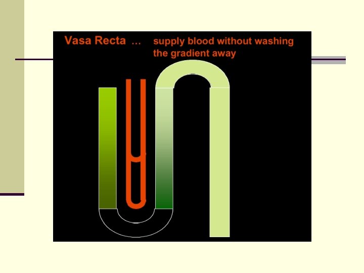
Role of the vasa recta: Form a countercurrent exchange system, having descending and ascending branches running in close proximity to the loop and each other. Countercurrent exchange in the vasa recta prevents washout of too much Na. Cl and urea.
Role of the vasa recta: As blood descends in the vasa recta, Na. Cl and urea enter the blood. As blood rises in the ascending limbs of the vasa recta, Na. Cl and urea diffuse out of the blood and back into the interstitium and the osmolality decreases to about 350 m. Os. M. Thus blood perfuses the medulla (flow is low), but relatively small amounts of Na. Cl and urea are removed from the medulla by the blood.
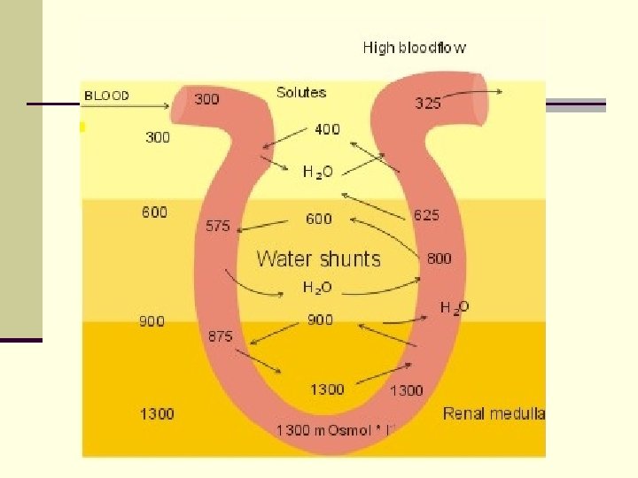
1. In the PT ~ 65% of filtered Na. Cl and water are reabsorbed but the urine remains iso-osmotic. 2. In the loop, water is reabsorbed from the DLH but a greater amount of Na. Cl is removed from the ALH, so that hypo -osmotic fluid enters the DT. 3. The early DT is always impermeable to water, therefore further dilutes the urine. This, with the ALH are referred to as "diluting segments”.
4. from the late DT, plasma ADH determines water permeability of the tubule. i. low [ADH] Little water reabsorbed, therefore these segments production of large volume of dilute urine. ii. high [ADH] By the end of the cortical CT's the urine is again iso-osmotic, and is further concentrated through the medulla to 1400 mosmol/l (containing ~ no Na. Cl)
5. Although Na. Cl in the medullary interstitium is essential for the concentrating ability of the kidney, the final urine may contain virtually none, the excreted solute being urea, creatinine, urate, K+, etc. 6. The excretion of large quantities of Na+ is always accompanied by the excretion of large amounts of water. 7. However, the excretion of large amounts of water does not necessitate the excretion of Na+, decreased [ADH]pl not significantly altering Na+ transport.
H 2 O + Na. Cl H 2 O H 2 O
H 2 O+Na. Cl H 2 O+Na. Cl H 2 O H 2 O Na. Cl
ADH also needed to concentrate urine: how does it work? • Antidiuretic Hormone (ADH)/Arginine Vasopressin (AVP) • Increases permeability of collecting ducts to H 2 O by inserting H 2 O channels (Aquaporins). • Helps to make small amount of concentrated urine. • Reabsorption of H 2 O increase urea conc. in tubule, increasing its recycling effect. • ADH allows rapid, graded control of urine conc. – v. sensitive. • ADH released in response to plasma osmolality and ECF volume – osmoreceptors and baroreceptors.
ADH (aka AVP) n Increased n n plasma osmolality stimulates osmoreceptors in the hypothalamus that trigger the release of ADH, which inhibits water excretion. Increased osmolality stimulates a second group of osmoreceptors that trigger thirst, which promotes water intake. Other factors also trigger ADH release e. g. decreased effective circulating volume, decreased BP, pregnancy, pain, morphine, nausea, congestive heart failure (CHF) (due to reduced ECV). CHF may cause such retention of H 2 O = hyponatremia. Hyperaldosteronism = hypernatremia. Due to chronic volume expansion, where osmoreceptors become less sensitive to ADH, reducing ADH
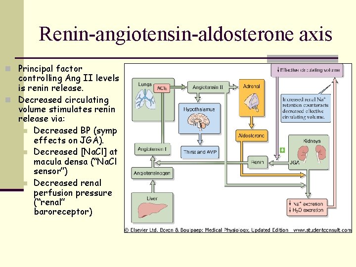
Renin-angiotensin-aldosterone axis n Principal factor controlling Ang II levels is renin release. n Decreased circulating volume stimulates renin release via: n Decreased BP (symp effects on JGA). n Decreased [Na. Cl] at macula densa (“Na. Cl sensor”) n Decreased renal perfusion pressure (“renal” baroreceptor)
Angiotensin II – important actions n Stimulation of aldosterone release from adrenal n n n cortex. Vasoconstriction of renal and other systemic vessels. Enhanced tubuloglomerular feedback – makes macula densa more sensitive. Enhance Na-H exchanger and Na channel function to promote Na reabsorption. Renal hypertrophy. Stimulates thirst and ADH release by acting upon hypothalamus.
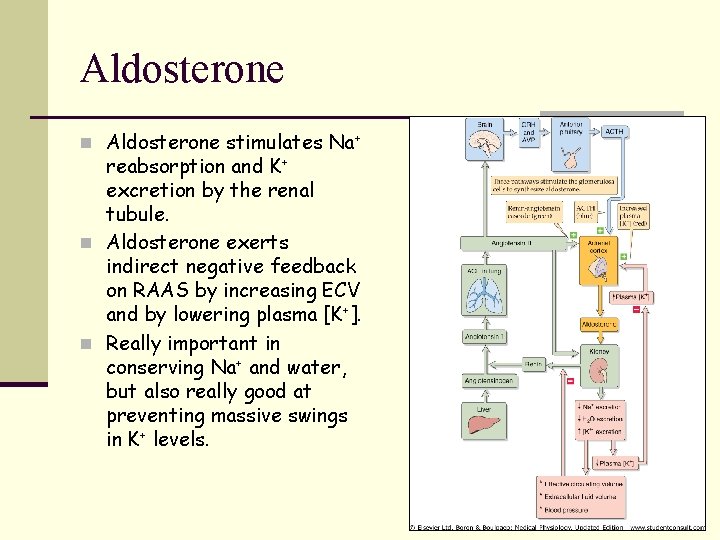
Aldosterone n Aldosterone stimulates Na+ reabsorption and K+ excretion by the renal tubule. n Aldosterone exerts indirect negative feedback on RAAS by increasing ECV and by lowering plasma [K+]. n Really important in conserving Na+ and water, but also really good at preventing massive swings in K+ levels.
Atrial Natriuretic Peptide (ANP) n ANP promotes natriuresis (loss of sodium). n Atrial myocytes synthesise, store and release n n ANP in response to stretch (low P volume sensor). Major effect is renal vasodilatation. Increased blood flow = increased GFR. Thus, more Na+ reaches macula densa. More Na+ excreted. May inhibit actions of renin, and generally opposes effects of angiotensin II.
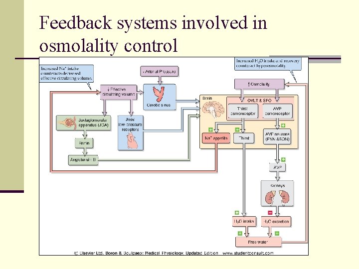
Feedback systems involved in osmolality control
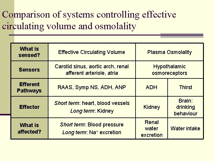
Comparison of systems controlling effective circulating volume and osmolality What is sensed? Effective Circulating Volume Plasma Osmolality Sensors Carotid sinus, aortic arch, renal afferent arteriole, atria Hypothalamic osmoreceptors Efferent Pathways RAAS, Symp NS, ADH, ANP ADH Thirst Effector Short term: heart, blood vessels Long term: Kidney Brain: drinking behaviour What is affected? Short term: Blood pressure Long term: Na+ excretion Renal water excretion Water intake
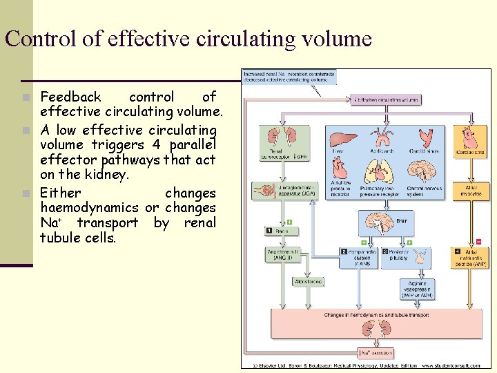
Control of effective circulating volume n Feedback control of effective circulating volume. n A low effective circulating volume triggers 4 parallel effector pathways that act on the kidney. n Either changes haemodynamics or changes Na+ transport by renal tubule cells.
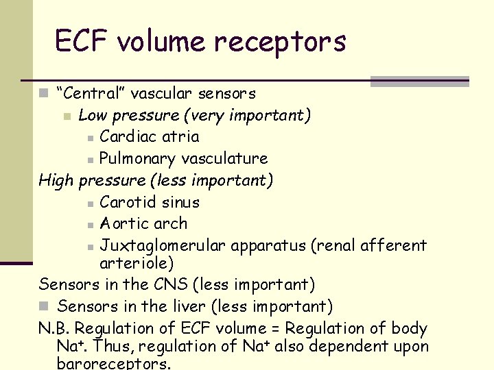
ECF volume receptors n “Central” vascular sensors Low pressure (very important) n Cardiac atria n Pulmonary vasculature High pressure (less important) n Carotid sinus n Aortic arch n Juxtaglomerular apparatus (renal afferent arteriole) Sensors in the CNS (less important) n Sensors in the liver (less important) N. B. Regulation of ECF volume = Regulation of body Na+. Thus, regulation of Na+ also dependent upon baroreceptors. n
- Slides: 84