Magnification Working Distance Resolution and Field of View


 The field of view is the circular")
 As you change from the low power")

 We need")


- Slides: 20
Magnification, Working Distance, Resolution and Field of View
Magnification 1 X Total Mag. 40 X Total Mag. 4 X Objective 2
Magnification 40 x Objective with Ocular provides 400 x Total Magnification 1 X Total Mag. 400 X Total Mag. 40 X Objective 3
Working Distance Working distance is the distance between the tip of the objective and the specimen. 4 ] Working Distance [
Resolution is the ability to distinguish between two points that are close together. Good resolution depends on both magnification and lens quality. Low Magnification Higher Resolution of individual legs 5
Lens Quality Improves Resolution 40 X Magnification Poor Quality Lens High Quality Lens Better Resolution 6
Field of View (F. O. V. ) The field of view is the circular area that is visible when you look through the microscope. Because the size of objects is different at each magnification, you have to calculate the diameters of the fields of view at each magnification this is also called “calibrating your microscope” 40 x 100 x 400 x
Field of View (F. O. V. ) As you change from the low power objective to the high power objective, the field of view changes. As the magnification increases, the area that you are viewing decreases. Magnification is inversely proportional to field of view In order to determine the size of the object that you are viewing, you need to understand the scale that corresponds to the magnification you are using.
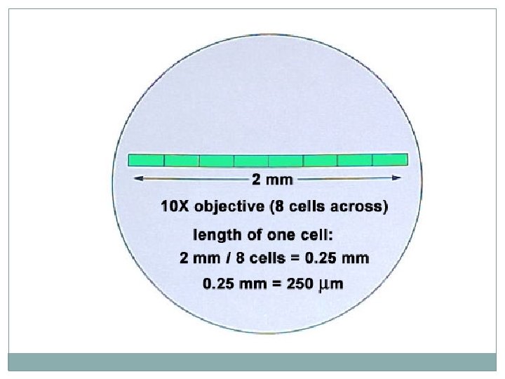
Day 4: Determining Field of View Copy the table in your science notebook. Eyepiece Magnification Objective Magnification Total Magnification Field of View width –diameter (mm) Field of View width - diameter (µm) 10 x 10 x You will need this table to estimate the size of the cells that you will be viewing in the next activities.
Measuring Field of View width for the low power objective (4 X) We need to find the field-of-view width: Using the low power objective (4 x), focus on a clear millimeter ruler Count the number of lines that you see in the field of view. (The distance from the center of one line to the center of the next line is 1 mm. ) This is the diameter of the field of view for the scanning objective.
Field of View
Low Magnification Large FOV High Magnification Small FOV As you increase magnification, you see less but in more detail.
Measure field of view with low magnification. Field of View is about 2. 5 mm diameter at 40 X total magnification. 14
Calculate Field of View at High Magnification = FOV low mag. X Mag. low 2. 5 mm X 4 X 0. 1 mm = = FOV high mag. X Mag. high FOV high mag. X 100 X FOV high mag. For example, if the FOV diameter is 2. 5 mm at 4 X, then at 100 X the FOV diameter is 0. 1 mm. 15
Calculating the Field of View Width – Low Power decreases
Calculating Field of View Width – High Power � decreases
Field of View Finally, fill in the table you created earlier today. Eyepiece Magnification Objective Magnification Total Magnification Field of View width –diameter (mm) Field of view width diameter (µm 10 x 4 x 40 x 10 x 100 x 1. 8 mm 0. 9 mm 10 x 400 x 0. 45 mm 0. 2225 mm 2. 2 mm In your notebook, answer the following: How can you use this table to estimate the size of cells that you view?
Example #1: ocular power = 10 x low power objective = 20 x high power objective = 50 x a) What is the highest magnification you could get using this microscope ? b) If the diameter of the low power field is 2 mm, what is the diameter of the high power field of view in mm? in micrometers ? c) If 10 cells can fit end to end in the low power field of view, how many of those cells would you see under high power ?
ANSWER to Example #1: ocular power = 10 x low power objective = 20 x high power objective = 50 x a) What is the highest magnification you could get using this microscope ? 500 x Ocular x high power = 10 x 50 = 500. (We can only use 2 lenses at a time, not all three. ) b) If the diameter of the low power field is 2 mm, what is the diameter of the high power field of view in mm ? . 8 mm The ratio of low to high power is 20/50. So at high power you will see 2/5 of the low power field of view (2 mm). 2/5 x 2 = 4/5 =. 8 mm in micrometers ? 800 micrometers To convert mm to micrometers, move the decimal 3 places to the right (multiply by 1000). . 8 mm x 1000 = 800 micrometers c) If 10 cells can fit end to end in the low power field of view, how many of those cells would you see under high power ? 4 cells. We can answer this question the same way we go about "b" above. At high power we would see 2/5 of the low field. 2/5 x 10 cells = 4 cells would be seen under high power.