Macroscopic microscopic views Gross appearance of an opened


- Slides: 13
Macroscopic & microscopic views
• Gross appearance of an opened cystic teratoma of the ovary – A DERMOID CYST. Note the presence of hair, sebaceous material, and tooth. You do not need a microscope to appreciate this tumor produces both connective tissue as well as epithelial derived elements.
A microscopic view of a similar tumor shows sebaceous glands, respiratory epithelium, bone, and bone marrow.
Necrotizing Enterocolitis
SEE PICTURES IN PREVIOUS SLIDE • Necrotizing enterocolitis. A, Postmortem examination in a severe case of NEC shows the entire small bowel is markedly distended with a perilously thin wall (usually this implies impending perforation). B, The congested portion of the ileum corresponds to areas of hemorrhagic infarction and transmural necrosis microscopically. Submucosal gas bubbles (pneumatosis intestinalis) can be seen in several areas (arrows), caused by gas forming bacteria.
• THIS is a brain from a child with jaundice The basal ganglia and thalamus are particularly susceptible. • Unconjugated bilirubin is water insoluble and lipophilic. It can cross the blood brain barrier and lead to kernicterus.
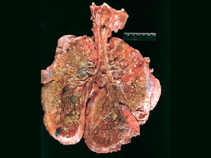
• Lungs of a patient dying of cystic fibrosis. There is extensive mucus plugging and dilation of the tracheobronchial tree. The pulmonary parenchyma is consolidated by a combination of both secretions and pneumonia—the green color associated with Pseudomonas infections.
• Unconjugated bilirubin is water insoluble and lipophilic. It can cross the blood brain barrier and lead to kernicterus. The basal ganglia and thalamus are particularly susceptible.
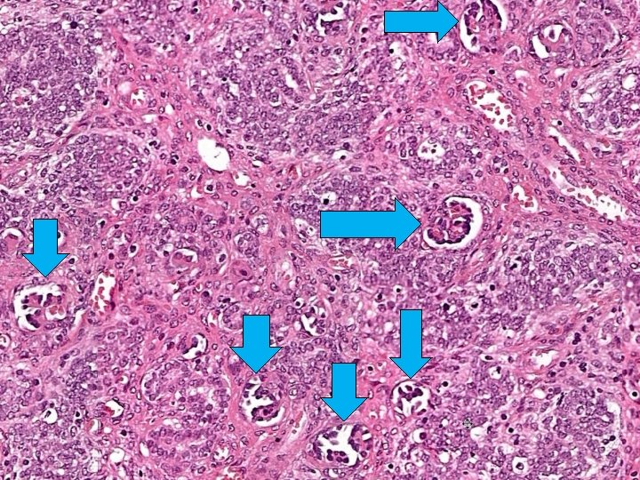
Pathology of Wilms Tumor • Gross – well circumscribed fleshy tan tumor – areas of hemorrhage and necrosis • Microscopic: triphasic appearance – Blastema: small blue cells – Epithelial elements: tubules & glomeruli – Stromal elements • Anaplasia – correlates with p 53 mutation and poor prognosis and resistance to chemotherapy
FATTY LIVER
FATTY LIVER