Macrophages T and B cells primary and secondary

Macrophages, T and B cells, primary and secondary immune organs, mucosal immune system

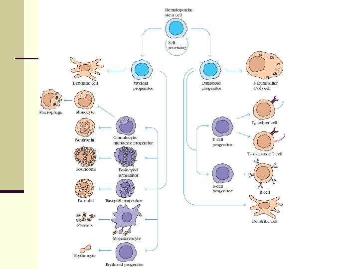
Macrophages n Terminal stage of monocyte-macrophage line differentiation n Monocyte-macrophage cells differentiate from myeloid precursor (developed from pluripotent stem cell bearing CD 34) in bone marrow n Matured monocytes are released to peripheral blood stream, then move in organs and develop into tissue macrophages
:")
Development of monocytes and macrophages is affected by various cytokines: n SCF(stem cell factor): produced by stromal cells → activation of stem cell n GM-CSF (granulocyte-monocyte colony stimulating factor): produced by bone marrow (BM) stromal cells, lymphocytes → stimulation of monocyte production n M-CSF (monocyte colony stimulating factor): produced by stromal cells, lymphocytes, endothelial and epithelial cells → production and maturation of monocytes n IL-3: produced by lymphocytes → production of monocytes (and other blood cells)
 and the rest in BM n")
Macrophages- development n Monocytes- in the blood (7%) and the rest in BM n Macrophages - in tissues

histiocytes

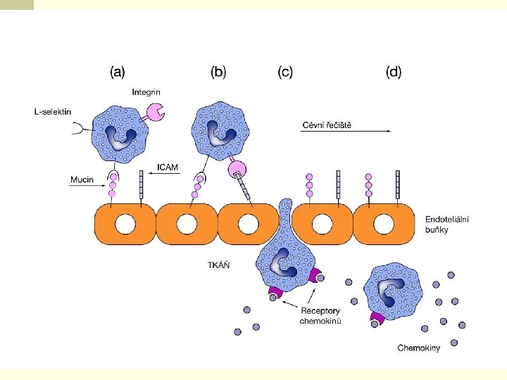
Macrophages n a monocyte enter damaged tissue through the endothelium of a blood vessel n a monocyte is attracted to damaged site by chemokines, triggered by stimuli including damaged cells, pathogens and cytokines released by macrophages n after migration of monocytes to the tissues, they differentiate into different forms of macrophages n macrophages survive several months

Macrophage surface molecules n MHC gp I, II assist in the presentation of antigen to T lymphocytes n CD 35 - complement receptor 1 (CR 1), binds complement C 3 b n Receptor for the Fc portion of Ig. G n CD 14 - receptor for bacterial lipopolysaccharides

Cytokines produced by macrophages n IL- 1 α, ß - stimulate both T and B cells, Ig synthesis, n n activation of other macrophages, sensitizing cells to IL-2 and IFN TNF- α - similar in function to IL-1 IL- 8 - secreted by activated macrophages - chemokine attracting neutrophils and T cells IL-12 - promotes induction of Th 1 cells, inhibits Th 2 cells IFN- α- activates host cells to induce enzymes inhibiting viral replication; increases expression of MHC gp I on host cells; activates NK cells, T cells, other macrophages

Functions of macrophages n Phagocytosis n Production of cytokines n Presentation of epitops with MHC gp II n Presentation of epitops with MHC gp I

Phagocytosis n a foreign substances are ingested n microbes are killed and digested n follows processing of antigenic epitopes and their presentation on the cell membrane

Macrophage - functions § Macrophages provide defense against tumor cells and human cells infected with fungi or parasites. § T cell becomes an activated effector cell after recognition of an antigen on the surface of the APC release chemical mediators → stimulation of macrophages
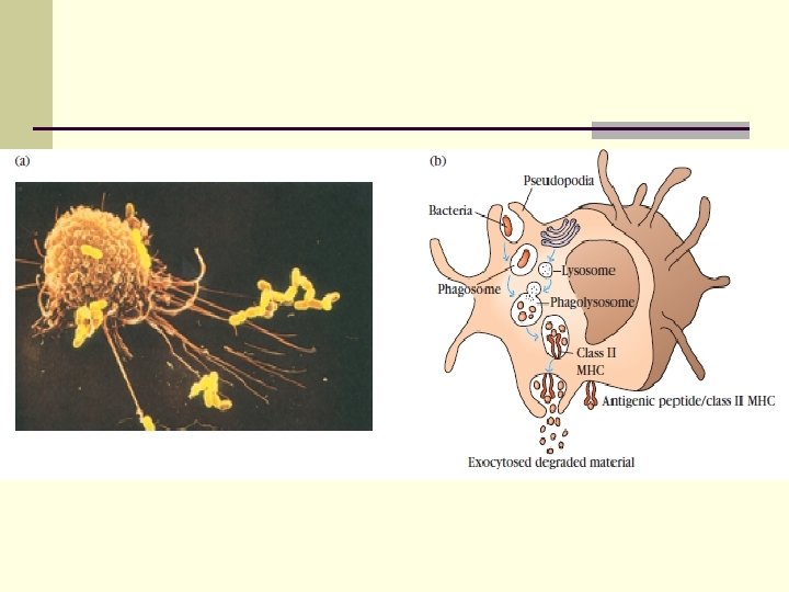

Presentation of epitopes with MHC gp II n After endocytosis and degradation of the antigen, presentation of its epitopes follows n epitope is connected to MHC gp II → cell surface → presentation to Th cells n MHC (Major Histocompatibility Complex) = complex of genes that governs the production of the major histocompatibility antigens - in humans termed HLAs (Human Leukocyte Antigens)

Presentation epitopes with MHC gp I n intracellular parasites are hydrolyzed in proteasomes of macrophages n their peptides are connected to TAP (Transporters Associated with antigen Processing molecules 1, 2), that carry the epitope and MHC gp I → presentation on the cell surface to Tc cells

Antigen presentation
 n DC mature after a contact with pathogen, then migrate to")
Dendritic Cells (DC) n DC mature after a contact with pathogen, then migrate to lymph nodes where antigen-specific immune response develops n DC are equipped with numerous cytoplasmic processes, allowing contact with up to 3000 T cells n In lymph nodes, the expression of MHC gp I and co- stimulatory molecules (CD 80, CD 86) on DC increases

Types of Dendritic Cells n Myeloid DC – similar to monocytes n Plasmocytoid DC – looks like plasma cells, but have certain characteristics like myeloid cells - production of huge amounts of interferons

Function of DCs n DCs are the most important APC n DCs can be easily infected by viruses → processing of viral proteins → their presentation in complex with MHC gp I → activation of Tc n DCs can ingest extracellular viral particles → their presentation in complex with MHC gp II → activation of Th 2 cells → help for B cells → production of antiviral antibodies n DCs can also be activated by apoptotic cells
 n Dendritic cells, macrophages, B cells n Antigen processing and")
Antigen Presenting Cells (APC) n Dendritic cells, macrophages, B cells n Antigen processing and its presentation to T cells in the complex with HLA class I or II n Providing additional signals to T cells which are necessary for their activation (CD 80, CD 86)

T cells: ontogenesis, surface markers. Subpopulations of T cells and their functions.

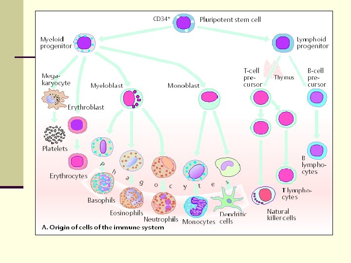
T lymphocytes - ontogenesis n Stem cell in BM gives rise to lymphoid precursor cell which matures into 3 types of lymphocytes: n T lymphocytes n B lymphocytes n Natural killer (NK) cells n Pro-thymocytes move to the thymus where continue the maturation into T lymphocytes n Maturation of B lymphocytes continues in BM
 proteins - molecules on")
Surface markers of T cells n CD (Cluster of Differentiation) proteins - molecules on the cells membrane, they allow the identification of cells n TCR - receptor for antigen n MHC gp I

CD proteins n allow an identification of T-cell subsets n CD 2 = adhesion molecule n CD 3 = important in intracellular signaling (initiation of immune response); closely associated with TCR n CD 5, 7 n CD 4, 8 = are expresed on subclasses of mature T cells; CD 4 reacts with MHC gp II, CD 8 reacts with MHC gp I on macrophages n CD 28 – molecule that provides co-stimulatory signals, binds CD 80 and 86

Maturation of T lymphocytes Consist of three types of processes: n Proliferation of immature cells n Expression of antigen receptors genes n Selection of lymphocytes

TCR n Antigen receptors are encoded by several gene n n segments that recombine during lymphocyte maturation Heterodimer consisting of 2 nonidentical polypeptide chains linked together by disulfide bonds > 95% T cells express the αß heterodimer, 5% γδ TCR heterodimer is noncovalently associated with the γ, δ, ε chains of the CD 3 molecule complex TCR-CD 3 makes contact with both the Ag and MHC gp
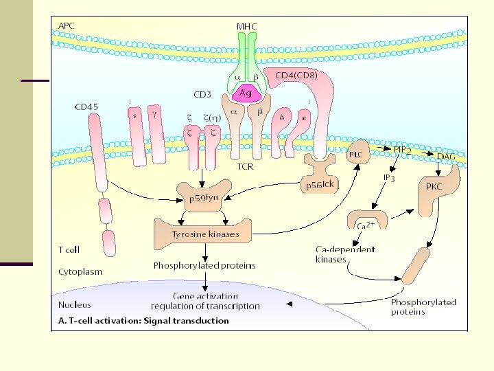

Subpopulation of T cells n Subpopulation of T cells are defined according to their particular function and their CD membrane markers n T cytotoxic cells (Tc) CD 8+ - recognize the foreign epitope in association with class I MHC molecules n T helper cells (Th) CD 4+ - recognize the epitopes in association with class II MHC molecules
 n cause lysis of target cell; active against")
T cytotoxic lymphocytes (Tc; CD 8+) n cause lysis of target cell; active against tumors, virus- infected cells, transplanted allogeneic tissue n release TNF → decrease of proteosynthesis n recognize the foreign epitope in association with MHC gp I molecules n Destroy target cells by perforins (create pores in the cell membrane → cell lysis) and granzymes (degradation of essential macromolecules)
 n recognize the epitopes in association with MHC")
T helper lymphocytes (Th; CD 4+) n recognize the epitopes in association with MHC gp II n help for B cells to produce antibodies and help for phagocytes to destroy ingested microbes n subsets of Th cells: Th 1, Th 2 cells

Regulatory T cells n Express CD 4, CD 25, Fox. P 3 n Regulate the activation or effector function of other T cells n Are necessary to maintain tolerance to self antigens n Production of IL-10, TGF-b

The role of thymus. Positive and negative selection of T lymphocytes.

The role of thymus n In thymus, lymphocyte precursors from the bone marrow become thymocytes, and subsequently mature into T cells n Once matured, T cells migrate from the thymus and constitute the peripheral T cell repertoire responsible for specific cell response

Phases of thymocyte maturation n A rare population of hematopoietic progenitors enters the thymus from the blood, and expands to a large population of immature thymocytes n Immature thymocytes each produce distinct T cell receptors by a process of gene rearrangement. n This process is error-prone, and some thymocytes fail to make functional T cell receptors, whereas other thymocytes make T cell receptors that are autoreactive

Positive and negative selection n Immature thymocytes undergo a process of selection, based on the specificity of their T cell receptors. n This involves selection of T cells that are functional (positive selection), and elimination of T cells that are autoreactive (negative selection)

Positive selection of T cells Entrance of precursor T cells into thymus from the blood 2. Presentation of self-antigens in complexes with MHC molecules on the surface of cortical epithelial cells to thymocytes 3. Only those thymocytes which bind the MHC/antigen complex with adequate affinity will receive a vital "survival signal" 4. The other thymocytes die (>95%) 1.

Negative selection of T cells 1. Thymocytes that survive negative selection migrate towards the thymic cortex and medulla 2. Presentation of self-antigen in complex with MHC molecules on antigen-presenting cells 3. Thymocytes that react inappropriately strongly with the antigen receive an signal of apoptosis

B-lymphocytes - ontogenesis, surface markers, function.

B-lymphocytes are an essential component of the adaptive immune system n Maturation of B cells takes place in BM n B cell originates from stem cell and need to be in touch with the stromal cells in the bone marrow n Stromal cells produce SCF (stem cell factor) necessary for development at early period, IL-7 necessary at later period of maturation n Ig gene rearrangements and the appearance of surface markers identify the stage of B-cell development

Development of B lymphocytes n Lymphoid progenitor → pro-B cells n During maturation from pro-B cells into pre-B cells: Ig genes of the heavy chain recombine; pre-B cells express pre-BCR n During maturation from pre-B cells into B cells: Ig genes of the light chain recombine n Immature B cells express membrane Ig. M n Mature B cells express membrane Ig. M and Ig. D = BCR and are able to respond to antigen in peripheral lymphoid tissues

Negative selection n If an immature B cell binds an antigen in the bone marrow with high affinity → further maturation is stopped and B cell dies by apoptosis n Negative selection eliminates potentially dangerous cells that can recognize and react against self antigens n B cells that survive this selection process leave the bone marrow through efferent blood vessels

B-lymphocytes – surface markers n CD 10 - immature B cells, malignant cells n CD 35 - receptor for the C 3 b of the complement n CD 19 - characteristic marker of B cells n CD 20 - typical surface antigen of Ig-positive B lymphocytes n Ig. M, Ig. D - antigen receptors = BCR n MHC gp II - antigen-presenting molecules

B-lymphocytes – functions n After stimulation B lymfocytes convert into the plasma cells and produce antibodies against soluble antigens n Other functions are : antigen presentation cooperation with complement system

Primary immune organs and their role in the immune system.

Primary immune organs n Bone marrow n Thymus n are organs of development, differenciation and maturation of immune cells and elimination of autoreactive cells n T and B lymphocytes mature and become competent to respond to antigens in PIOs

Bone marrow is the central cavity of bone and the site of generation of all circulating blood cells in adults, including immature lymphocytes, and the site of B-cell maturation. n The pluripotent stem cell gives rise to the progenitors of all immune cells n Production of the cells takes place in the spaces divided by vascular sinuses n Endothelial cells of the sinuses produce cytokines n Sinuses are bordered by reticular cells
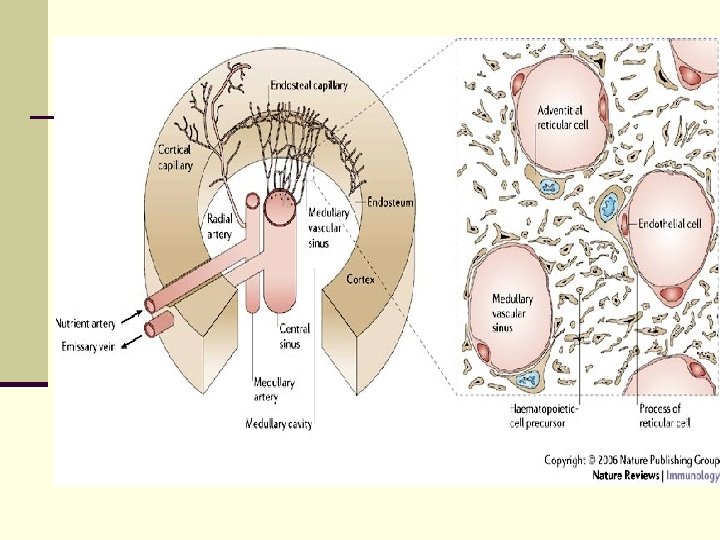

Differentiation in the BM n Differentiation from the stem cell is influenced by: n n membrane interaction between the stem cells and the stromal cells cytokines (CSF, IL-3, thrombopoetin, erythropoetin)

Thymus n is located between the sternum and the major vessel trunks n It consist of two lobes n Each lobe is surrounded by a capsule and is divided into lobules, which are separated from each other by strands of connective tissue = trabeculae

Structure of the thymus Each lobule is organized into two compartments: - the cortex (outer compartment) – contains lymphocytes that proliferate - the medulla (inner compartment)- mature lymphocytes, Hassall´s corpuscles
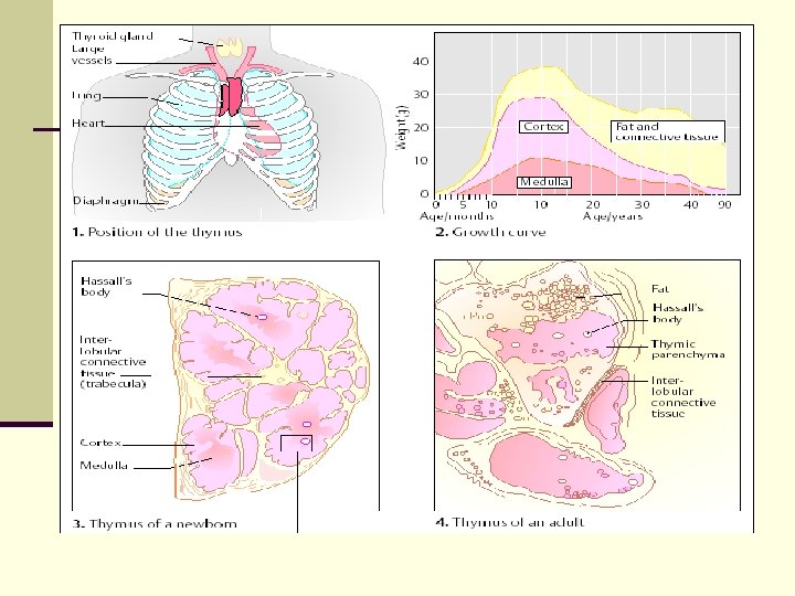

Thymus - morphology Various kinds of stromal cells: n thymic epithelial cells – production of thymulin, thymopoetin, thymosin that influence the maturation of T cells n dendritic cells n macrophages n The thymus contain a large number of blood vessels and efferent lymphoid vessels that drain into the mediastinal lymph nodes

Secondary immune organs - structure and function of lymphatic node and spleen.

Secondary immune organs • consist of the spleen, the lymph nodes, the mucosal and cutaneous immune system • are organized to optimize interactions of antigens, APCs and lymphocytes • are places of the development of adaptive immune responses spleen lymphatic nodes tonsils MALT appendix Peyer´s patches

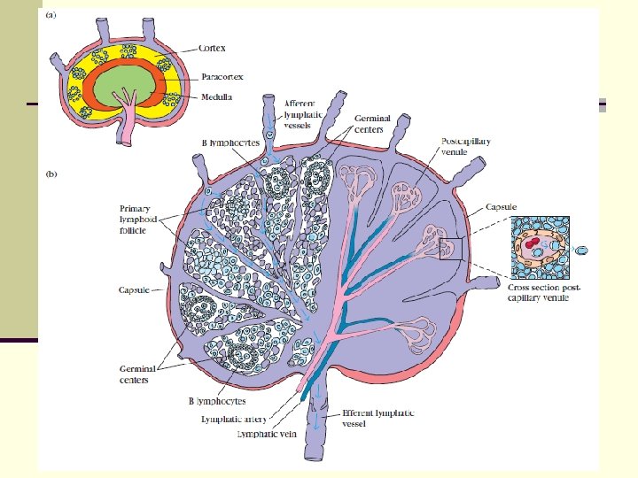
Lymphatic node n are nodular aggregates of lymphoid tissues located along lymphatic channels throughout the body n Lymph comes from tissues and most parenchymal organs to the lymph nodes n Lymph contains a mixture of substances absorbed from epithelia and tissues n As the lymph passes through lymph nodes, APCs in the LN are able to sample the antigens of microbes that may enter through epithelia into tissues

Lymphatic node • lymph circulates to the lymph node via afferent lymphatic vessels and drains into the node just beneath the capsule in a space called the subcapsular sinus • the subcapsular sinus drains into trabecular sinuses and finally into medullary sinuses • the sinus space is criss-crossed by the pseudopods of macrophages which act to trap foreign particles and filter the lymph • the medullary sinuses converge at the hilum and lymph then leaves the lymph node via the efferent lymphatic vessel

Lymphatic node- medulla The medullary cords are cords of lymphatic tissue, and include plasma cells and T cells • The medullary sinuses are vessel-like spaces separating the medullary cords; contain histiocytes (= immobile macrophages) and reticular cells. • Lymph flows to the medullary sinuses from cortical sinuses, and into efferent lymphatic vessels

Lymphatic node - cortex Contains lymphoid follicles = accumulation of Blymphocytes and follicular dendritic cells When a lymphocyte recognizes an antigen, B cells become activated and migrate to germinal centers = to the secondary nodule

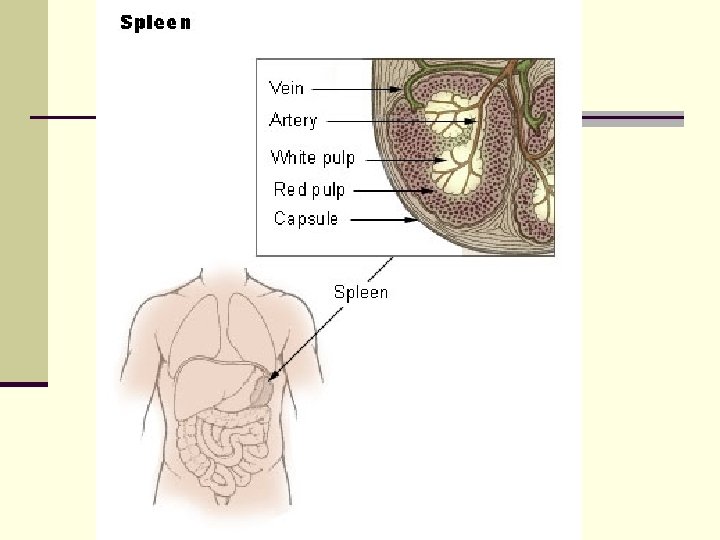
Spleen is a secondary lymphoid organ located high in the left abdominal cavity n is surrounded by a capsule, which sends trabeculae into the interior to form a compartmentalized structure n there are two types of compartments -red pulp and white pulp with a marginal zone in between n is NOT supplied by afferent lymphatics

Spleen n Red pulp : place of mechanical filtration and elimination of senescent red and white blood cells and microbes n White pulp : T lymphocytes CD 4+, CD 8+ are around arterioles (periarteriolar lymphoid sheaths), B lymphocytes are in the folicles; final maturation of B lymphocytes course in germinal center of secondary folicles

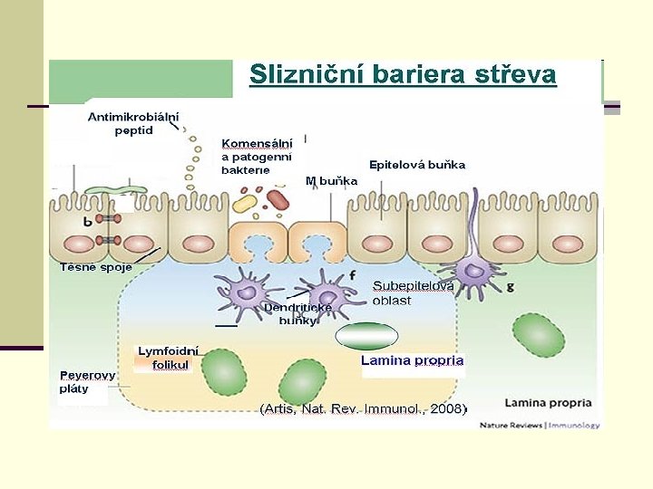
Mucosal immune system n MALT = mucosa-associated lymphoid tissue n GALT = gut-associated lymphoid tissue n BALT = bronchus-associated lymphoid tissue n GIT, respiratory, and urogenital systems are lined by mucous membranes n Includes clusters of lymphoid cells in lamina propria of intestinal villi n contains a very large population of plasma cells that synthesize Ig. A antibodies

M cells n are epithelial cells that are specialized for the transport antigen from the lumen of the respiratory, GIT, and urogenital tracts to the underlying MALT n contain a characteristic pocket filled with B cells, T cells, and macrophages n are found at inductive sites that overlie organized lymphoid follicles in the lamina propria n antigens are endocytosed and transported within vesicles from the luminal membrane to the pocket membrane, where the vesicles fuse and deliver their contents to antigen-presenting cells

Secretory Ig. A n daily production of secretory Ig. A into mucus secretions exceeds that of any other class of immunoglobulin (5 -15 g each day) n is an important line of defense for mucosal surfaces against bacteria n binding of secretory Ig. A to bacteria and viruses also prevents attachment to mucosal epithelial cells, thereby inhibiting infection and colonization

Cutaneous immune system n Epidermis contains keratin cells that produce IL-1, 6 and TNF during inflamation; and IL-10, TGF-β during healing n Dermis contains fibroblasts that produce collagen, remove apoptotic cells ------------------------------
- Slides: 69