Macromolecules Chapter 5 Macromolecules n Large complex molecules


 Pentoses (C")
 Pentoses (C")

 Dehydration reaction in the")
, glycogen (animal), cellulose (plant),")
 and glucose ring structures Glucose (b) Starch: 1– 4 linkage")
 The structure of the chitin monomer. (b) Chitin forms the")


 Glycerol (a) Dehydration reaction in the synthesis of a fat")
 Fat molecule (triacylglycerol)")


 Side chains Peptide bond Backbone (b) Amino end")
 Valine (Val or V) Alanine (Ala")


 Thymine (T, in DNA) Uracil")
 Ribose (in RNA) (c) Nucleoside components:")


- Slides: 49
Macromolecules Chapter 5
Macromolecules n Large complex molecules n Carbohydrates, proteins, lipids & nucleic acids
Macromolecules n Polymer n Large molecule n Carboydrates, proteins, nucleic acids n Monomers n Smaller repeating units n Build polymers
Macromolecules n Monomers are connected by a dehydration reaction n Condensation reaction
Fig. 5 -2 a HO 1 2 3 H Short polymer HO Unlinked monomer Dehydration removes a water molecule, forming a new bond HO 1 2 H 3 H 2 O 4 H Longer polymer (a) Dehydration reaction in the synthesis of a polymer
Macromolecules n Polymers are broken apart into monomers by hydrolysis n Bonds are broken by adding water
Fig. 5 -2 b HO 1 2 3 4 Hydrolysis adds a water molecule, breaking a bond HO 1 2 3 (b) Hydrolysis of a polymer H H H 2 O HO H
Carbohydrates n Simple sugars to complex polymers n Stored energy n Structure
Carbohydrates Monosaccharides n Single sugar n – Glucose, fructose, galactose n n n Molecules contain carbon, hydrogen & oxygen in a 1: 2: 1 ratio CH 2 O End in –ose Aldose: aldehyde sugar Ketose: ketone sugar
Fig. 5 -3 a Aldoses Trioses (C 3 H 6 O 3) Pentoses (C 5 H 10 O 5) Hexoses (C 6 H 12 O 6) Glyceraldehyde Ribose Glucose Galactose
Fig. 5 -3 b Ketoses Trioses (C 3 H 6 O 3) Pentoses (C 5 H 10 O 5) Hexoses (C 6 H 12 O 6) Dihydroxyacetone Ribulose Fructose
Carbohydrates n Dissaccharides n Two monosaccharides combined n Glycoside linkage: n Covalent bond between two sugars n Sucrose (glucose & fructose) n Maltose (glucose & glucose) n Lactose (glucose & galactose)
Fig. 5 -5 1– 4 glycosidic linkage Glucose Maltose (a) Dehydration reaction in the synthesis of maltose 1– 2 glycosidic linkage Glucose Fructose (b) Dehydration reaction in the synthesis of sucrose Sucrose
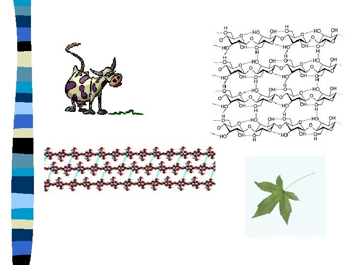
Carbohydrates n Polysaccharides n Many monosaccharides combined n Starch (plant), glycogen (animal), cellulose (plant), chitin
Fig. 5 -7 (a) and glucose ring structures Glucose (b) Starch: 1– 4 linkage of glucose monomers Glucose (b) Cellulose: 1– 4 linkage of glucose monomers
Fig. 5 -10 (a) The structure of the chitin monomer. (b) Chitin forms the exoskeleton of arthropods. (c) Chitin is used to make a strong and flexible surgical thread.
Lipids n Insoluble in water – Hydrophobic n Store energy n Make membranes
Lipids n Fats – Unsaturated – Saturated n Phospholipids – Glycerol, phosphate, fatty acid n Steroids
Fats n Glycerol and fatty acids n Triglyceride n 3 fatty acids attached to a glycerol
Fatty acid (palmitic acid) Glycerol (a) Dehydration reaction in the synthesis of a fat
Fig. 5 -11 b Ester linkage (b) Fat molecule (triacylglycerol)
Fig. 5 -12 Structural formula of a saturated fat molecule Stearic acid, a saturated fatty acid (a) Saturated fat Structural formula of an unsaturated fat molecule Oleic acid, an unsaturated fatty acid (b) Unsaturated fat cis double bond causes bending
Phospholipids n Cell membrane n Tails are hydrophobic n Hydrophilic head
Fig. 5 -14 Hydrophilic head Hydrophobic tail WATER
Steroids
Steroids
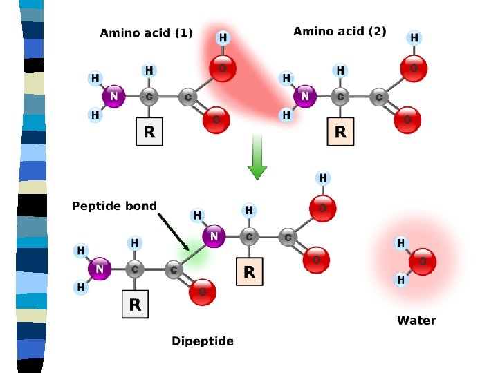
Proteins n Amino acids n Building blocks of proteins n 20 different amino acids n Peptide bond n Polypeptide
Fig. 5 -18 Peptide bond (a) Side chains Peptide bond Backbone (b) Amino end (N-terminus) Carboxyl end (C-terminus)
Fig. 5 -17 Nonpolar Glycine (Gly or G) Valine (Val or V) Alanine (Ala or A) Methionine (Met or M) Leucine (Leu or L) Trypotphan (Trp or W) Phenylalanine (Phe or F) Isoleucine (Ile or I) Proline (Pro or P) Polar Serine (Ser or S) Threonine (Thr or T) Cysteine (Cys or C) Tyrosine (Tyr or Y) Asparagine Glutamine (Asn or N) (Gln or Q) Electrically charged Acidic Aspartic acid Glutamic acid (Glu or E) (Asp or D) Basic Lysine (Lys or K) Arginine (Arg or R) Histidine (His or H)
Protein Function n Enzyme catalysis n Defense n Transport n Support n Motion n Regulation n Storage
Protein structure n Primary n Secondary n Tertiary n Quaternary
Primary Structure n Sequence of amino acids
Secondary Structure n Hydrogen bonds between amino acids n Pleats or helix
Tertiary structure n Attraction between side chains n Hydrophobic interaction n Disulfide bridges n Ionic bonds n Hydrogen bonds
Quaternary structure n Two or more polypeptide chains aggregate
Sickle cell anemia
Fig. 5 -22 Normal hemoglobin Primary structure 1 2 3 4 5 6 7 Secondary and tertiary structures subunit Function Normal hemoglobin (top view) Secondary and tertiary structures Val His Leu Thr Pro Val Glu 1 2 3 Normal red blood cells are full of individual hemoglobin moledules, each carrying oxygen. 6 7 subunit Sickle-cell hemoglobin Function Molecules interact with one another and crystallize into a fiber; capacity to carry oxygen is greatly reduced. 10 µm Red blood cell shape 5 Exposed hydrophobic region Molecules do not associate with one another; each carries oxygen. 4 Quaternary structure Sickle-cell hemoglobin Quaternary structure Primary structure Val His Leu Thr Pro Glu 10 µm Red blood cell shape Fibers of abnormal hemoglobin deform red blood cell into sickle shape.
Structure n Denaturation: n Alter, unravel shape of protein n Temperature, p. H, salt n Chaperonins: n Proteins that help with structure
Nucleic Acids n DNA and RNA n Transfer & store genetic information n Nucleotides are the subunits n Nitrogenous base n 5 carbon sugar n Phosphate group
Nucleic Acids n Pyrimidines n Cytosine, thymine and uracil n Single carbon ring n Purines n Adenine, guanine n Double ring structure
Nitrogenous Bases
Fig. 5 -27 c-1 Nitrogenous bases Pyrimidines Cytosine (C) Thymine (T, in DNA) Uracil (U, in RNA) Purines Adenine (A) Guanine (G) (c) Nucleoside components: nitrogenous bases
Fig. 5 -27 c-2 Sugars Deoxyribose (in DNA) Ribose (in RNA) (c) Nucleoside components: sugars
DNA n n n n Double helix Sugar-phosphate backbone is on the outside of helix Run in opposite direction Antiparallel Base pairs held together by hydrogen bonds Adenine-thymine (uracil) Cytosine-guanine
DNA
DNA
Fig. 5 -28 5' end 3' end Sugar-phosphate backbones Base pair (joined by hydrogen bonding) Old strands Nucleotide about to be added to a new strand 3' end 5' end New strands 5' end 3' end