m RNA t RNA y r RNA CA


- Slides: 77

m. RNA, t. RNA y r. RNA CA García Sepúlveda MD Ph. D Laboratorio de Genómica Viral y Humana Facultad de Medicina, Universidad Autónoma de San Luis Potosí 1

Contents • Central Dogma • m. RNA – Prokaryote – Eukaryote – Extrachromosomal (Mitochondrial) • • • t. RNA Translation m. RNA Life Cycle Polycistronic and monocistronic m. RNAs Prokaryotic and eukaryotic m. RNAs 2

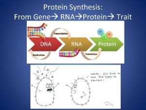
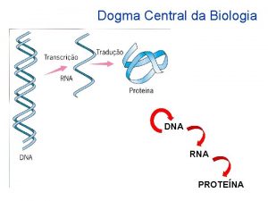
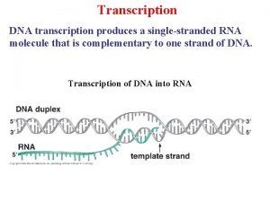
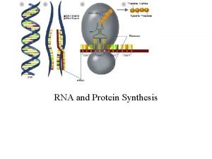
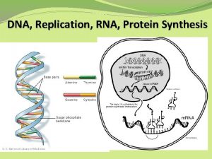
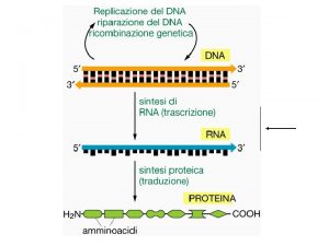
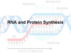
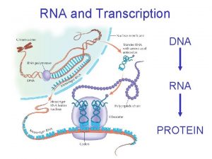
Central Dogma • Genetic information is expressed in two stages: 1. - Transcription 2. - Translation • Generation of ss. RNA from DNA template • Catalysed by RNA Polymerases • Generates: • m. RNA • t. RNA • r. RNA • Occurs in prokaryotes and eukaryotes by essentially identical processes 3

Central Dogma • Genetic information is expressed in two stages: 1. - Transcription 2. - Translation • Synthesis of a protein • m. RNA as a template • t. RNAs convert codon information into amino acid sequence • Catalysed by r. RNA 4

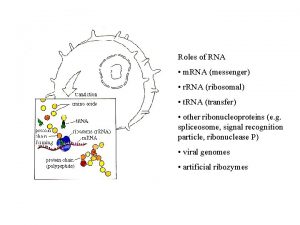
RNAs Coding RNAs Noncoding RNAs (Large) Small noncoding RNAs (sn. RNAs) sn. RNAs come in many varieties & form ribonucleoproteins (RNP). 1. - small nucleolar RNPs (sno. RNPs) process r. RNA in nucleolus. 2. - sn. RNAs splice pre-m. RNAs in the spliceosome. 3. - t. RNAs. 4. - pre-micro. RNAs (regulate gene expression by degrading m. RNA or blocking translation initiation site. 5

m. RNA • m. RNAs are single stranded RNA molecules • They are copied from the TEMPLATE strand of the gene, to give the SENSE strand in RNA • They are transcribed from the 5’ to the 3’ end • They are translated from the 5’ to the 3’ end 6

m. RNA • m. RNAs are single stranded RNA molecules • They are copied from the TEMPLATE strand of the gene, to give the SENSE strand in RNA • They are transcribed from the 5’ to the 3’ end • They are translated from the 5’ to the 3’ end 7

m. RNA • m. RNAs are single stranded RNA molecules • They are copied from the TEMPLATE strand of the gene, to give the SENSE strand in RNA • They are transcribed from the 5’ to the 3’ end • They are translated from the 5’ to the 3’ end 8

m. RNA • m. RNAs are single stranded RNA molecules • They are copied from the TEMPLATE strand of the gene, to give the SENSE strand in RNA • They are transcribed from the 5’ to the 3’ end • They are translated from the 5’ to the 3’ end 9

m. RNA • m. RNAs are single stranded RNA molecules • They are copied from the TEMPLATE strand of the gene, to give the SENSE strand in RNA • They are transcribed from the 5’ to the 3’ end • They are translated from the 5’ to the 3’ end 10

m. RNA • m. RNAs are single stranded RNA molecules • They are copied from the TEMPLATE strand of the gene, to give the SENSE strand in RNA • They are transcribed from the 5’ to the 3’ end • They are translated from the 5’ to the 3’ end 11

m. RNA Prokaryotes • They can code for one or many proteins (translation of products) in prokaryotes (polycistronic) 12

m. RNA Eukaryotes • They encode only one peptide(each) in eukaryotes (monocistronic). • Polyproteins are observed in eukaryotic viruses, but these are a single translation product, cleaved into separate proteins after translation. 13

m. RNA Synthesis (transcription) • Catalysed by RNA Polymerase • Stages: initiation, elongation and termination • Initiation is at the Promoter sequence • Regulation of gene expression is at the initiation stage • Transcription factors binding to the promoter regulate the rate of initiation of RNA Polymerase 14

m. RNA Synthesis (Prokaryotes) • 5’ and 3’ ends are unmodified • Ribosomes bind at ribosome binding site (do not require a free 5’ end) • Can contain many open reading frames (ORFs) • Translated 5’ 3 • Transcribed and translated together 15

m. RNA Life Cycle • m. RNA is synthesised by RNA Polymerase • Translated (once or many times) • Degraded by RNAses • Steady state level depends on the rates of both synthesis and degradation Production Degradation 16

m. RNA Life Cycle • m. RNA is synthesised by RNA Polymerase • Translated (once or many times) • Degraded by RNAses • Steady state level depends on the rates of both synthesis and degradation Production Degradation 17

m. RNA Life Cycle • m. RNA is synthesised by RNA Polymerase • Translated (once or many times) • Degraded by RNAses • Steady state level depends on the rates of both synthesis and degradation Production Degradation 18

m. RNA Life Cycle • m. RNA is synthesised by RNA Polymerase • Translated (once or many times) • Degraded by RNAses • Steady state level depends on the rates of both synthesis and degradation Production Degradation 19

m. RNA, Eukaryotes • Linear RNA structure • 5’ and 3’ ends are modified • 5’ Gppp. G cap • 3’ poly A tail • Transcribed, spliced, capped, poly Adenylated in the nucleus, exported to the cytoplasm 20

Translation in Eukaryotes • Translated from 5’ 3’ end in cytoplasm • Ribosomes bind at 5’ cap, and do require a free 5’ end • Can contain only one translated open reading frames (ORF). • Only first open reading frame is translated 21

Translation in Eukaryotes • Translated from 5’ 3’ end in cytoplasm • Ribosomes bind at 5’ cap, and do require a free 5’ end • Can contain only one translated open reading frames (ORF). • Only first open reading frame is translated 22

Translation in Eukaryotes • Translated from 5’ 3’ end in cytoplasm • Ribosomes bind at 5’ cap, and do require a free 5’ end • Can contain only one translated open reading frames (ORF). • Only first open reading frame is translated 23

m. RNA, 5' Capping • The 5' cap is a specially altered nucleotide end to the 5' end of precursor messenger RNA and, as a special exception, caliciviruses (Norwalk virus). • Actue epidemic gastroenteritis (nursery, hospital and cruises). } 30 nm 24

m. RNA, 5' Capping • Consists of a guanine nucleotide connected to the m. RNA via an unusual 5' to 5' triphosphate linkage. • This guanosine is methylated on C 7 position directly after capping by a methyl transferase. • It is referred to as a 7 -methylguanosine cap, abbreviated m 7 G. 25

m. RNA, 5' Capping • Further modifications include the methylation of the 2' hydroxy-groups of the first 3 ribose sugars of the 5' end of the m. RNA. • Functionally the 5' cap looks like the 3' end of an RNA molecule (the 5' carbon of the cap ribose is bonded, and the 3' unbonded). • Offers resistance to 5' exonucleases. 26

m. RNA, 5' Capping process 1) One of the terminal phosphate groups is removed (phosphatase), leaving two terminal phosphates. 2) GTP is added to the terminal phosphates (guanylyl transferase), losing two phosphate groups (from the GTP) in the process. This results in the 5' to 5' triphosphate linkage. 3) The guanine is methylated (by a methyl transferase). 4) Other methyltransferases are optionally used to carry out methylation of 5' proximal nucleotides. 27

m. RNA, 5' Capping process 1) One of the terminal phosphate groups is removed (phosphatase), leaving two terminal phosphates. 2) GTP is added to the terminal phosphates (guanylyl transferase), losing two phosphate groups (from the GTP) in the process. This results in the 5' to 5' triphosphate linkage. 3) The guanine is methylated (by a methyl transferase). 4) Other methyltransferases are optionally used to carry out methylation of 5' proximal nucleotides. 28

m. RNA, 5' Capping process 1) One of the terminal phosphate groups is removed (phosphatase), leaving two terminal phosphates. 2) GTP is added to the terminal phosphates (guanylyl transferase), losing two phosphate groups (from the GTP) in the process. This results in the 5' to 5' triphosphate linkage. 3) The guanine is methylated (by a methyl transferase). 4) Other methyltransferases are optionally used to carry out methylation of 5' proximal nucleotides. 29

m. RNA, 5' Capping process 1) One of the terminal phosphate groups is removed (phosphatase), leaving two terminal phosphates. 2) GTP is added to the terminal phosphates (guanylyl transferase), losing two phosphate groups (from the GTP) in the process. This results in the 5' to 5' triphosphate linkage. 3) The guanine is methylated (by a methyl transferase). 4) Other methyltransferases are optionally used to carry out methylation of 5' proximal nucleotides. 30

m. RNA, 5' Capping process 1) The Capping Enzyme Complex (CEC) is bound to the RNA polymerase II before transcription starts. 2) As soon as the 5' end of the new transcript emerges the enzymes transfer to it and caps it. 3) The enzymes for capping can only bind to RNA polymerase II. 31

m. RNA, 3' Polyadenylation Covalent linkage of a poly(Adenine) tail to most m. RNA (eukaryotic). The poly-A tail protects the m. RNA from exonucleases and is important for transcription termination, for export of the m. RNA from the nucleus, and for translation. Some prokaryotic m. RNAs also are polyadenylated. Polyadenylation occurs after transcription of DNA into RNA in the nucleus. Poly-A signal is transcribed, m. RNA is cleaved (endonuclease) in a special site (AAUAAA) then 50 to 250 adenine residues are added. This reaction is catalyzed by polyadenylate polymerase. 32

m. RNA, 3' Polyadenylation Cleavage and Polyadenylation Specificity Factor (CPSF) & Cleavage Stimulation Factor (Cst. F), both of which are multiprotein complexes, start bound to the rear of the advancing RNA polymerase II. 33

m. RNA, 3' Polyadenylation As RNA polymerase II advances over the adenylation signal sequence, CPSF & Cst. F transfer to the pre-m. RNA. CPSF binds to the AAUAAA sequence. Cst. F binds to the GU or U rich sequence following it. 34

m. RNA, 3' Polyadenylation CPSF & Cst. F promote cleavage 35 nt after AAUAAA sequence. Polyadenylate Polymerase (PAP) immediately starts adding Adenines. Cleavege does not take place before PAP has bound. Nuclear Polyadenylate Binding Protein (PABPN 1) immediately binds to the new poly -A tag. 35

m. RNA, 3' Polyadenylation PAB displaces CPSF. PAP continues to add 100 – 250 adenines (depending on the organism). PABPN 1 acts as a molecular ruler, specifying when polyadenylation should stop. 36

Mitochondrial m. RNA = Prokaryote • • Single stranded Polycistronic (many ORFs) Unmodified 5’ and 3’ ends Transcribed and translated together • Show similarity to prokaryote genes and transcripts 37

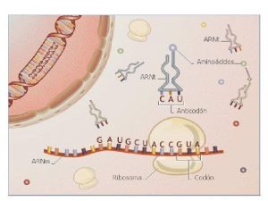
t. RNA • t. RNA first hypothesized by Francis Crick. • Small RNA chain that transfers a specific amino acid to a growing polypeptide chain in the ribosome • Has a 3' terminal site for amino acids (whose linkage depends on aminoacyl t. RNA synthetase). • Contains a three base region called the anticodon that complements the codon on the m. RNA. • Each type of t. RNA molecule can be attached to only one type of amino acid. • t. RNA molecules bearing different anticodons may also carry the same amino acid (degenerecy). 38

t. RNA Structure • t. RNA has primary structure (sequence), secondary structure (cloverleaf), and tertiary structure (L-shape). • Small RNAs 75 - 85 bases in length • Highly conserved secondary and tertiary structures • Each class of t. RNA charged with a single amino acid • Each t. RNA has a specific trinucleotide anti-codon for m. RNA recognition • Conservation of structure and function in prokaryotes and eukaryotes 39

t. RNA Structure 1) The 5'-terminal phosphate group. 2) The acceptor stem (7 bp) composed by the 5'-terminus base paired to the 3'-terminus (contains non-Watson. Crick base pairs). 3) The CCA tail is at the 3' end of the t. RNA molecule (important for the recognition of t. RNA by enzymes critical in translation). NOTE: In prokaryotes, the CCA sequence is transcribed. In eukaryotes, the CCA sequence is added. 4) The D arm (18 bp) ends in a loop which contains dihydrouridine. 5) The anticodon arm (ca 17 bp) contains the anticodon. 6) The T arm (17 bp) contains T C sequence ( = pseudouridine). 7) Modified (methylated) bases occur in several positions outside the anticodon. First anticodon base sometimes modified to inosine or ). 40

t. RNA Structure 41

t. RNA Codons & Anti-codons An anticodon is a unit made up of three nucleotides that complement the three bases of the codon on the m. RNA. 42

t. RNA Codons & Anti-codons Different base triplets (codons) in m. RNA code for different amino acids. 43

t. RNA Codons & Anti-codons The Genetic Code is degenerate 44

t. RNA Codons & Anti-codons The Genetic Code is degenerate Serine can be: UCU, UCC, UCA, UCG, AGU & AGC Each t. RNA contains a specific anticodon triplet sequence that can base-pair to one or more codons for an amino acid. For example, one codon for Serine is AGU; the anticodon being UCA. Some anticodons can pair with more than one codon due to wobble base pairing. When the first nucleotide of the anticodon is either inosine or pseudouridine (can hydrogen bond to different bases). 45

t. RNA Aminoacylation Aminoacylation is the process of covalently adding an aminoacyl group to a compound t. RNA). Each t. RNA is aminoacylated (charged) with a specific amino acid by an aminoacyl t. RNA synthetase. There is normally a single aminoacyl t. RNA synthetase for each amino acid. There can be more than one t. RNA, and more than one anticodon, for an amino acid. Recognition of the t. RNA by the synthetases depends on the anticodon, and the acceptor stem. 46

t. RNA genes Organisms have varying amounts of t. RNA genes. C. elegans has 29, 647 genes of which 620 code for t. RNA. Saccharomyces cerevisiae has 275 t. RNA genes in its genome. In the human genome there are: 4, 421 non-coding RNA genes (which include t. RNA genes). 22 mitochondrial t. RNA genes 497 nuclear genes encoding cytoplasmic t. RNA molecules and 324 t. RNA-derived putative pseudogenes. 47

t. RNA genes Cytoplasmic t. RNA genes are grouped into 49 families according to their anticodon features. t. RNA genes are found on all chromosomes, except 22 and Y. High clustering on 6 p and 1 is observed (140 t. RNA genes). t. RNA molecules are transcribed (in eukaryotic cells) by RNA polymerase III, unlike messenger RNA which is transcribed by RNA polymerase II. pre-t. RNAs contain introns; in bacteria these self-splice, whereas in eukaryotes and archaea they are removed by t. RNA splicing endonuclease. 48

Ribosomes Prokaryotic ribosomes are smaller than most of the eukaryotes. The ribosomes in eukaryote mitochondria resemble those in bacteria. The function of ribosomes is the assembly of proteins (translation). Ribosomes catalyze the assembly of individual amino acids into polypeptide chains. They use m. RNA as a template to join a correct sequence of amino acids. This reaction uses adapters called t. RNA. 49

Ribosomes First observed in the mid-1950 s by Romanian cell biologist George Palade using an electron microscope as dense particles or granules Nobel Physiology, 1974. The term "ribosome" was proposed by scientist Richard B. Roberts in 1958 Ribonucleic body (soma) 50

Ribosomes All ribosomes are composed of two subunits that separate when translation terminates and reunite when an new initiation complex is formed. Small subunit m. RNA t. RNA Large subunit 51

Session #20 -22 Translation Introduction • The basic form of the ribosome is conserved, but there appreciable variations in the overall size and proportions of RNA and protein in the ribosomes of bacteria, eukaryotic cytoplasm, and organelles. Al ribosomes consists of two subunits, each of which contains a major r. RNA and a number of small proteins. The large subunit may also contain smaller RNAs. 52

Svedberg Units Briefly Theodor Svedberg (1884 -1971). Nobel laureate (Chemistry, 1926) for his work on colloids and ultracentrifugation. Colloid = Mechanical mix (milk) with a dispersed and continuous phase. Svedberg, non-SI unit used to characterize the behaviour of a particle in ultracentrifugation. S = unit of time amounting to 10 -13 s or 100 femtoseconds. S not additive, since the sedimentation rate is associated with the shape & size of the particle. When two particles bind togethere is a loss of surface area, when measured separately they will have Svedberg values that do not add up. 53

Session #20 -22 Translation Introduction • The ribosomal proteins are known as r-proteins. • With the exception of one protein present at four copies per ribosome, there is one copy of each protein. 54

Session #20 -22 Translation Phylogenetics • The ribosomes of higher eukaryotes are larger than those of bacteria. • Total content of both RNA & protein is greater • Major RNA molecules are longer (18 S & 28 S r. RNAs) • Possess more proteins. • RNA is the predominant component. 55

Session #20 -22 Translation Phylogenetics • The ribosomes of higher eukaryotic cytoplasm are larger than those of bacteria. • Total content of both RNA & protein is greater • Major RNA molecules are longer (18 S & 28 S r. RNAs) • Possess more proteins. • RNA is the predominant component. 56

Session #20 -22 Translation Phylogenetics • The ribosomes of higher eukaryotic cytoplasm are larger than those of bacteria. • Total content of both RNA & protein is greater • Major RNA molecules are longer (18 S & 28 S r. RNAs) • Possess more proteins. • RNA is the predominant component. 57

Session #20 -22 Translation Phylogenetics • In prokaryotes: – Ribosomes = 2. 5 MDa – r. RNA = 66% of the ribosomal mass • In eukaryotes (mammals): – Ribosomes = 4. 2 MDa – r. RNA = 60% of the ribosomal mass • Organelle ribosomes are distinct from eukaryotic cytosol ribosomes and take varied forms. • In some cases, they are almost the size of bacterial ribosomes and have 70% RNA; in other cases, they are only 60 S and have <30% RNA. 58

Ribosomes Prokaryotic Ribosome is 70 S and composed of Large (50 S) and Small (30 S) subunits. Large subunit composed of: 23 S r. RNA (2900 nt) 5 S r. RNA (120 nt) 31 -34 proteins. Small subunit composed of: 16 S r. RNA (1540 nt) 21 proteins. 59

Ribosomes Eukaryotic Ribosome is 80 S and composed of Large (60 S) and Small (40 S) subunits. Large subunit composed of: 5. 8 S r. RNA (160 nt) 28 S r. RNA (4700 nt) 5 S r. RNA (120 nt) ~49 proteins Small subunit composed of: 18 S r. RNA (1900 nt) ~33 proteins 60

Ribosomes The ribosomes of chloroplasts & mitochondria also consist of large and small subunits bound together with proteins into one 70 S particle (as prokaryotes do). 61

Ribosomes are about 20 nm in diameter. Can be studied with EM. Curiously, r. RNA genes in a cluster ! 62

Session #20 -22 Translation Transmission Electron Microscopy • The complete 70 S ribosome has an asymmetric construction. • The partition between the head and body of the small subunit is aligned with the notch of the large subunit, so that the platform of the small subunit fits into the large subunit. • There is a cavity between the subunits which contains important sites. 63

Ribosome location Classified as "free" or "membrane-bound". Free ribosomes move about the cytoplasm (within the cell membrane). Proteins formed from free ribosomes are used within the cell. Membrane-bound ribosomes place newly produced polypeptides directly into the endoplasmic reticulum. Usually produce proteins that are used within the cell membrane or are expelled from the cell via exocytosis. Proteins containing disulfide bonds using cysteine cannot be produced outside of the lumen of the endoplasmic reticulum. 64

Ribosomes Composed of 60% ribosomal RNA and 30% ribosomal proteins (known as a Ribonucleoprotein or RNP). Py. Mol 65

Ribosomes Crystallographic work has shown that there are no ribosomal proteins close to the reaction site for polypeptide synthesis. This suggests that the protein components of ribosomes act as a scaffold that may enhance the ability of r. RNA to synthesize protein rather than directly participating in catalysis. 66

Ribosomes Their active sites (A, P & E) are made of RNA, so ribosomes are now classified as "ribozymes". 67

t. RNA binding to ribosome The ribosome has three binding sites for t. RNA molecules: the A, P and E sites. A= aminoacyl-t. RNA site P= Peptidyl-t. RNA site E= Egress site 68

t. RNA binding to ribosome The ribosome has three binding sites for t. RNA molecules: the A, P and E sites. A= aminoacyl-t. RNA site P= Peptidyl-t. RNA site E= Egress site During translation the A site binds an incoming aminoacyl-t. RNA as directed by the codon currently occupying this site. This codon specifies the next amino acid to be added to the growing peptide chain. The A site is only working after the first aminoacyl-t. RNA has attached to the P site. 69

t. RNA binding to ribosome The ribosome has three binding sites for t. RNA molecules: the A, P and E sites. A= aminoacyl-t. RNA site P= Peptidyl-t. RNA site E= Egress site The P-site codon is occupied by peptdyl-t. RNA (a t. RNA with multiple amino acids attached). The P site is actually the first to bind to aminoacyl t. RNA. This t. RNA in the P site carries the chain of amino acids that has already been synthesized. 70

t. RNA binding to ribosome The ribosome has three binding sites for t. RNA molecules: the A, P and E sites. A= aminoacyl-t. RNA site P= Peptidyl-t. RNA site E= Egress site The E site is occupied by the empty t. RNA as it is about to exit the ribosome. 71

t. RNA binding to ribosome The ribosome has three binding sites for t. RNA molecules: the A, P and E sites. A= aminoacyl-t. RNA site P= Peptidyl-t. RNA site E= Egress site The E site is occupied by the empty t. RNA as it is about to exit the ribosome. Agrawal animation Sannuga animation 72

Ribosomes Their active sites (A, P & E) are made of RNA, so ribosomes are now classified as "ribozymes". Py. Mol 73

Ribosomes Prokaryote and eukaryote ribsome differences lie outside of functional parts (A, P & E sites) and therefore “redundant insertions”. The differences are exploited by pharmaceuticals to create antibiotics that destroy a bacterial without harming the cells of the infected person. Even though mitochondria possess similar ribosomes they are not affected by these antibiotics (double membrane). Antibiotics such as macrolides, aminoglycosides and others: anisomycin cycloheximide chloramphenicol tetracycline streptomycin erythromycin puromycin, etc. 74

Ribosomes & Antibiotics Structure of the antibiotic gentamicin C 1 a bound to its r. RNA target. Gentamicin, an aminoglycoside antibiotic, binds within the major groove of the RNA, which is located in the decoding site of the bacterial ribosome. Aminoglycosides cause misreading of the genetic code. Binding of the drug causes a conformational change in ribosomal RNA that disrupts high-fidelity reading of the genetic code. 75

Ribosomes & Antibiotics Structure of the aminoglycoside paromomycin bound to the bacterial r. RNA decoding site. The sites that lead to resistance are highlighted with purple spheres. The N 7 methylation at G 1405 only causes resistance to aminoglycosides like gentamicin that contact this position directly. 76

Ribosomes& Translation Ribosomes are the workhorse for protein biosythesis= TRANSLATION 77
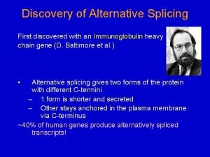
 Alternative rna splicing
 Adar1 rna
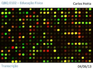
 Microarray analysis
 Rna
Rna Dna and rna
 Rna polymerase 1 2 3
 Dna rna protein trait
 Rantai dna yang berperan sebagai beton untuk rna adalah
 Rna ülesanne
 Arn mensajero caracteristicas
 Dna number of strands
 Biologia molecular
 Dehydration synthesis dna
 Northern blot rna
 Where are ribosomal rna produced
 Dna to rna rules
 Esquema dna
 Rna
Rna Rna transfer
 Atp rna
 Extraccion de rna
 Difference between dna and rna extraction
 Order of bases in dna
 Rna
Rna Rna splicing
 On and off
 Color 040613
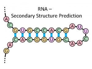
 Rna secondary structure
 Rna or dna
 Spectrophotometer blank
 Dna transcription
 Rna
Rna Rna secondary structure prediction
 Chargaff
 Rna polimerasi
 Overview of transcription and translation
 Codon chart
 Proteinas histonas
 Dna rna
 Rna
Rna Noncoding rna
 Rna quality control
 Gene interactions
 Rna
Rna Atp ester bond
 Dna mrna trna amino acid chart
 Rna
Rna Jalview
 Synthesis of rna
 Dna and rna similarities
 Illumina sequencing chemistry
 Site:slidetodoc.com
 Rna secondary structure prediction
 Rna transfer
 Colacca
 Arn de transferencia
 Dna to protein steps
 Nitrogenous bases in dna and rna
 Rna transfer
 Rna transcription
 Replication vs transcription venn diagram
 Rna codon chart
 Corona virus dna or rna
 Somatic cells vs germ cells
 Dogma msc
 What provides instructions for protein synthesis
 Che cos'è rna
 Dna and rna
 Rasi rna
 Nucleic acid made up of
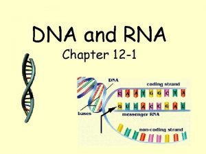
 Chapter 12 dna and rna
 Alternative rna splicing
 How does the mrna molecule leave the nucleus?
 Pseudoknot structure
 How does rna differ from dna? *
 Messenger rna sequence
 Tiosianato
 Small rna
