M Phase Cell Division Divided into two parts


 to become")

 centromere one chromatid b One chromosome (duplicated) two sister")


 in cell same chromosome (duplicated) prior to mitosis &")


- Slides: 29
M Phase – Cell Division • Divided into two parts – Mitosis • division of the cell’s nucleus – Cytokinesis • division of the cell’s cytoplasm
M Phase – Cell Division Chromatin • all of the chromosomes in the nucleus Chromosome • a single piece of DNA in the nucleus Chromatids • two identical “sister” replicated chromosomes, held together by a centromere
Chromosomes Chromatin Individual Chromosomes
Chromosome Visibility Chromosomes become visible during cell division as they condense (coil) to become more manageable during separation.
Please flip your notes to the opposite side of the page.
Chromosomes a One chromosome (unduplicated) centromere one chromatid b One chromosome (duplicated) two sister chromatids
Flip your page again. Attempt to label each blank on your notes. 1. ___________ 5. _________ 4. ___________ 2. _________ 3. ___________
Now correct each label on your notes. 1. chromosome 5. centromere 4. chromatid 2. sister chromatids 3. chromatid
Maintaining Chromosome Number • When cells divide, each cell duplicates an entire copy of its chromatin. • When the cell divides, each “daughter cell” receives one complete copy of the chromatin. • This allows each cell to have the same number of total chromosomes.
Maintaining Chromosome Number chromosome (unduplicated) in cell same chromosome (duplicated) prior to mitosis & cytokinesis chromosome (unduplicated) in daughter cell at interphase
Maintaining Chromosome Number • The two new daughter cells will be genetic twins, and they will genetically be identical to the cell they came from.
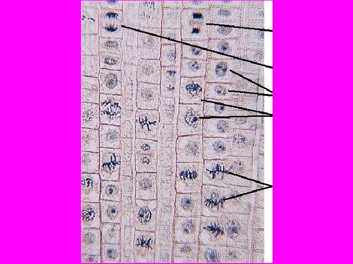
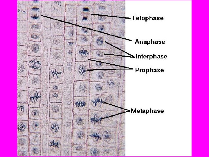
Mitosis • Mitosis occurs in 4 phases: 1. 2. 3. 4. Prophase Metaphase Anaphase Telophase
Mitosis • Prophase
Mitosis • Prophase – Chromatin condenses into individual chromosomes – Nuclear envelope breaks down – Centrioles separate • Centriole – specialized structure that aids in cell division
Mitosis • Metaphase
Mitosis • Metaphase – Chromosomes line up across the center of the cell – Each chromosome is connected to a spindle fiber at its centromere • Spindle fibers – centrioles and microtubules fibers that move chromosomes
Mitosis • Anaphase
Mitosis • Anaphase – Sister chromatids separate into individual chromosomes and are moved apart – As the chromatids are no longer connected, the centromeres that connected them no longer exist.
Mitosis • Telophase
Mitosis • Telophase – Chromosomes gather at opposite ends of the cell and lose their distinct shapes – Two new nuclear envelopes form – Spindle dissolves – Cells begin to separate in preparation for cytokinesis
What is visible in mitosis that is unable to be seen in interphase? I I P M A Mitosis T I
Cytokinesis • the cytoplasm of the cell is divided in half • the cell membrane grows to surround each new daughter cell
Mitosis & Cytokinesis 1. Interphase 2. Prophase 3. Metaphase 5. Late Anaphase 6. Telophase 7. Cytokinesis 4. Early Anaphase 8. Interphase
Mitosis & Cytokinesis Animation
Cytokinesis in Plants • the cell is divided in half by a cell plate – a newly formed cell wall
Mitosis in Animals vs. Plant Cells • Differently shaped cells – Plants • Square-ish – Animals • Round-ish • Differences in cytokinesis – No cell wall/cell plate in animal cells
Summary • Why is mitosis important? In other words, why is it important that a cell can copy its DNA (during Synthesis) and divide its DNA (during Cell Division)?