
LYMPHOSCINTIGRAPHY Sentinel Node Ramin Sadeghi M D Nuclear

- Slides: 99
LYMPHOSCINTIGRAPHY Sentinel Node Ramin Sadeghi, M. D. Nuclear Medicine Specialist Associate Professor DSNMC Nuclear Medicine Research Center (NMRC; MUMS) www. DSNMC. ir
Lymphoscintigraphy l Indications: • Evaluation of lymphedema • Sentinel node detection • Melanoma • Breast cancer • Evaluation of lymphedema • Evaluation of chyle stasis www. DSNMC. ir
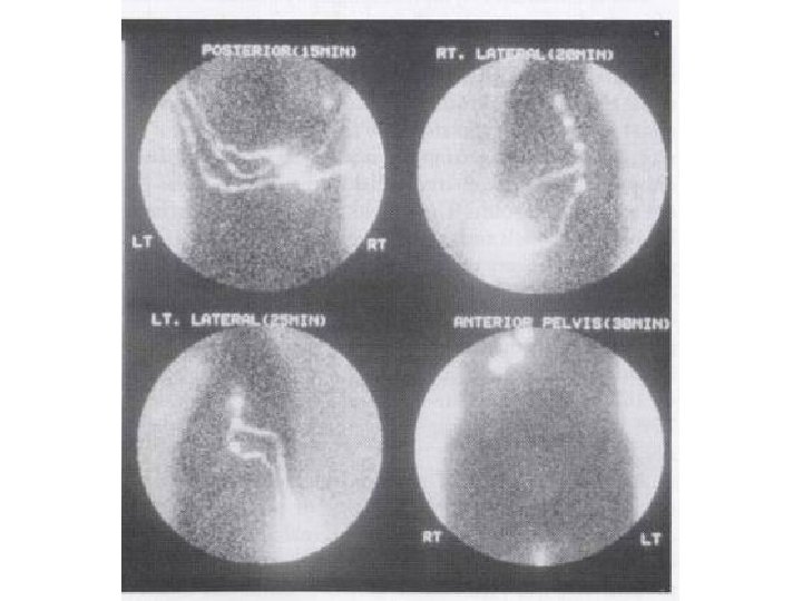
Lymphoscintigraphy l l l Normal • Prompt cephalic migration of radiocolloid to the illioinguinal lymph node groups in 10 -30 min. Primary lymphedema • Marked decreased visualization of the ilioinguinal nodes of the affected side as well as an absence of any diffuse interstitial activity. Secondary lymphedema • Marked interstitial accumulation of radiocolloid (diffuse activity throughout the involved extremity and poor visualization of primary channels) www. DSNMC. ir
SENTINEL NODE DETECTION www. DSNMC. ir
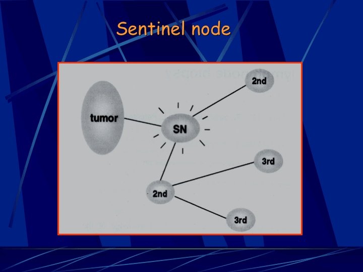
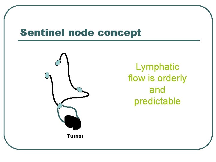
Sentinel node concept Lymphatic flow is orderly and predictable Tumor
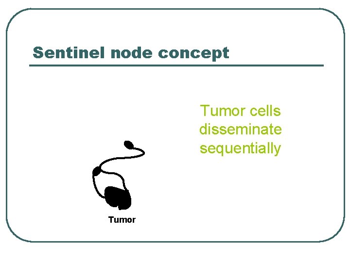
Sentinel node concept Tumor cells disseminate sequentially Tumor
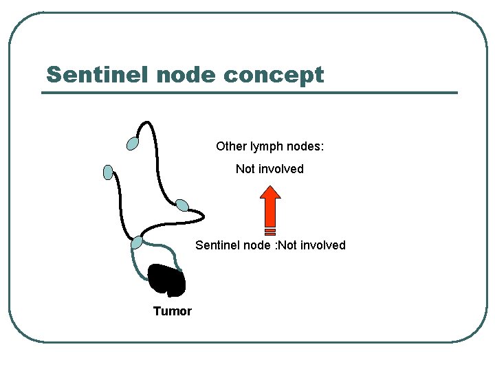
Sentinel node concept Other lymph nodes: Not involved Sentinel node : Not involved Tumor
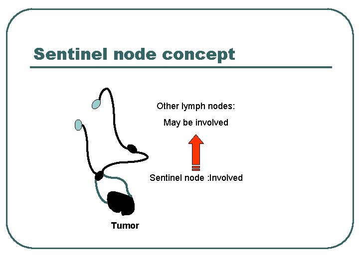
Sentinel node concept Other lymph nodes: May be involved Sentinel node : Involved Tumor
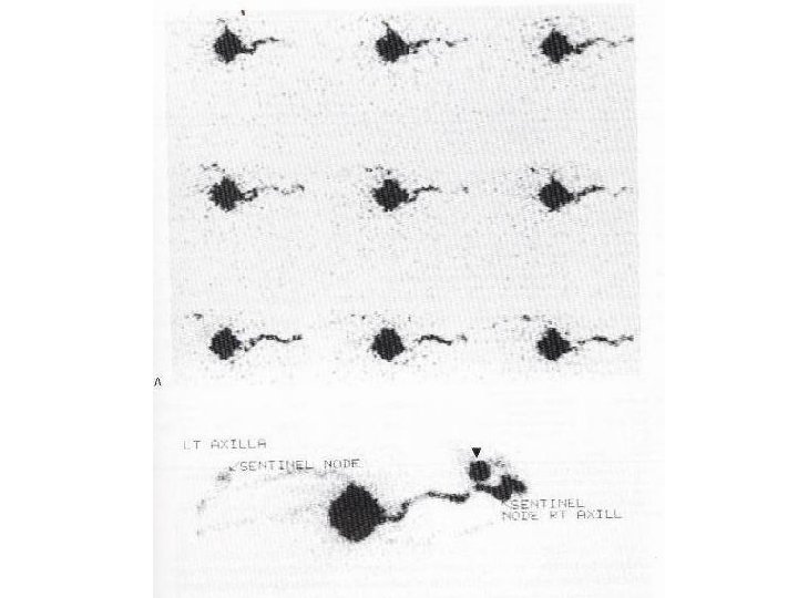
Sentinel node detection l Probe detection l Blue dye detection • Alone • With imaging • Alone • With probe detection
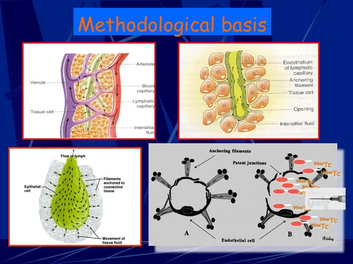
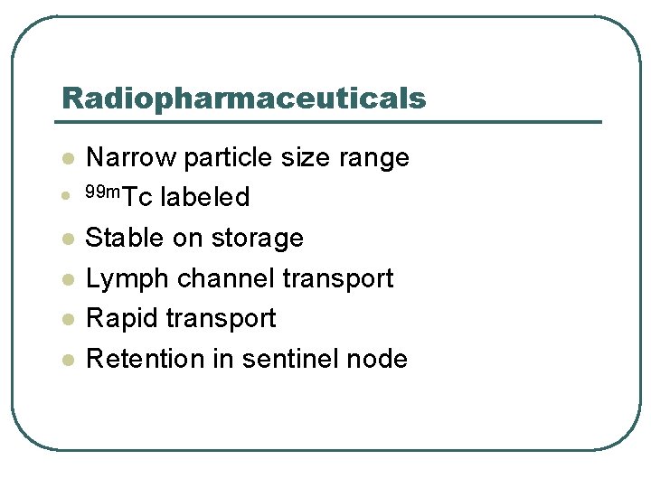
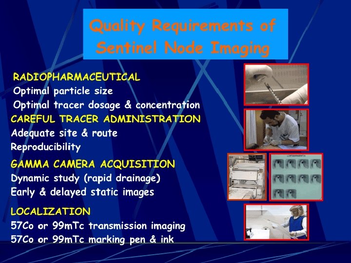
Radiopharmaceuticals l l l Narrow particle size range 99 m. Tc labeled Stable on storage Lymph channel transport Rapid transport Retention in sentinel node
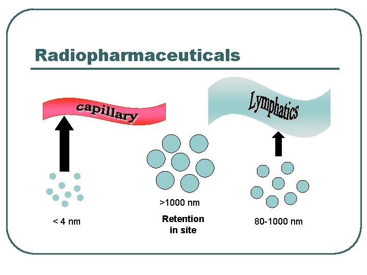
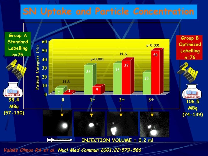
Radiopharmaceuticals >1000 nm < 4 nm Retention in site 80 -1000 nm
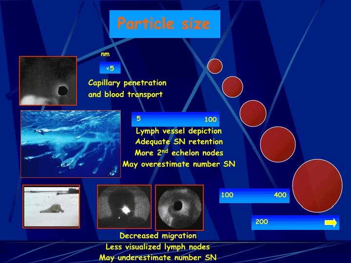
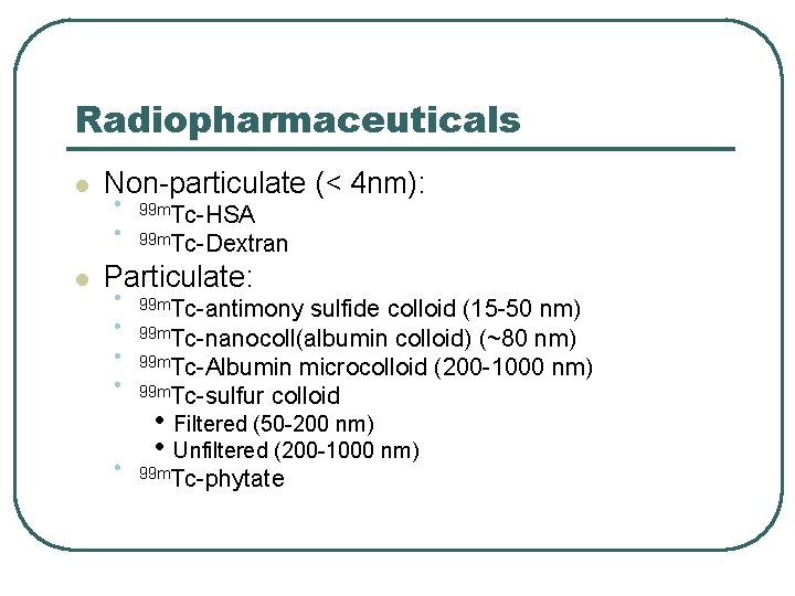
Radiopharmaceuticals l l Non-particulate (< 4 nm): • • 99 m. Tc-HSA 99 m. Tc-Dextran Particulate: • • • 99 m. Tc-antimony sulfide colloid (15 -50 nm) 99 m. Tc-nanocoll(albumin colloid) (~80 nm) 99 m. Tc-Albumin microcolloid (200 -1000 nm) 99 m. Tc-sulfur colloid • Filtered (50 -200 nm) • Unfiltered (200 -1000 nm) 99 m. Tc-phytate
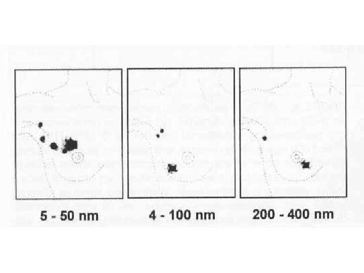
Radiopharmaceuticals The smaller the size of tracer, the more lymph nodes are detected
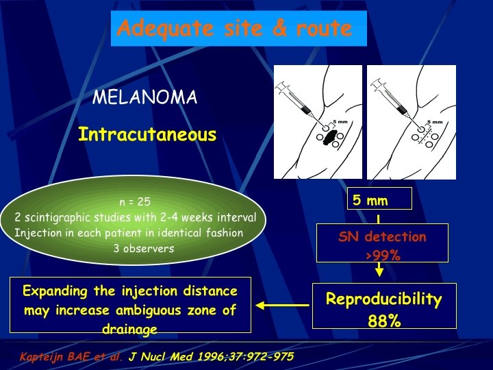
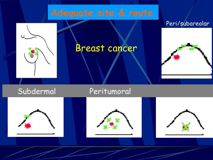
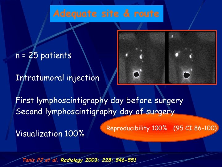
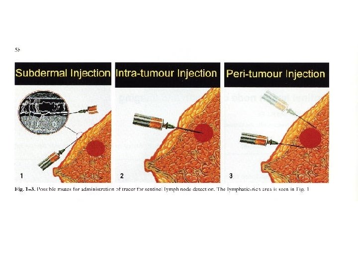
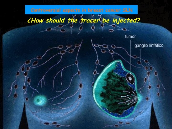
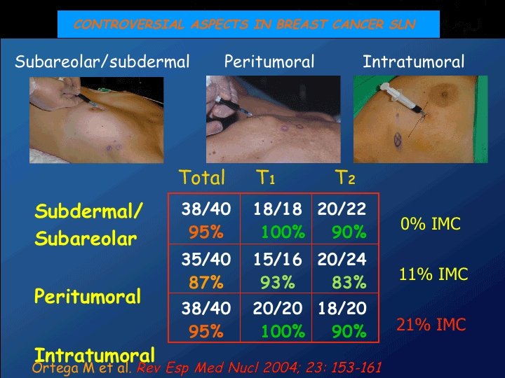
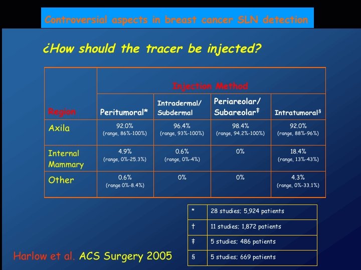
Injection site • Melanoma – Intradermal • Breast cancer – Subdermal – Intra-tumoral – Peri-areolar – Subdermal + peri-tumoral
Injection site (cont. ) • Subdermal injection is sub-optimal for internal mammary nodes • Subdermal + peri-tumoral is the preferred method of injection
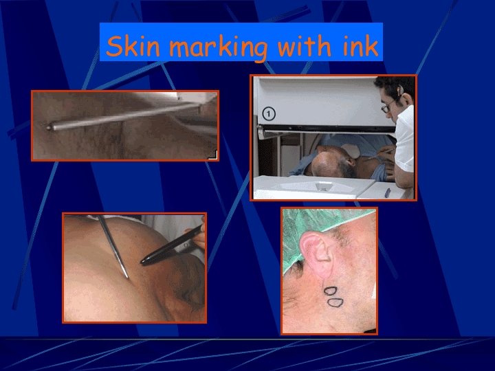
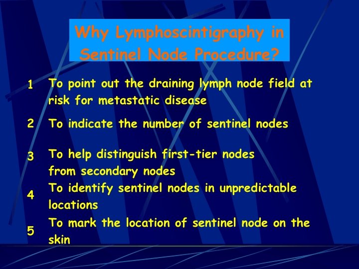
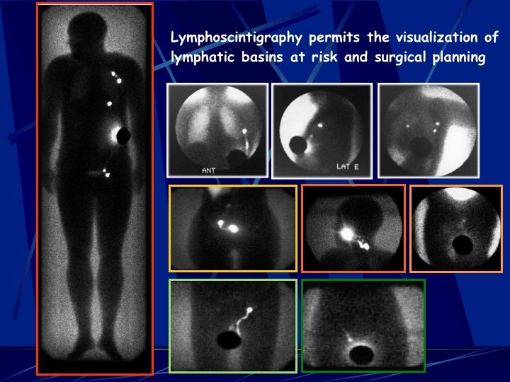
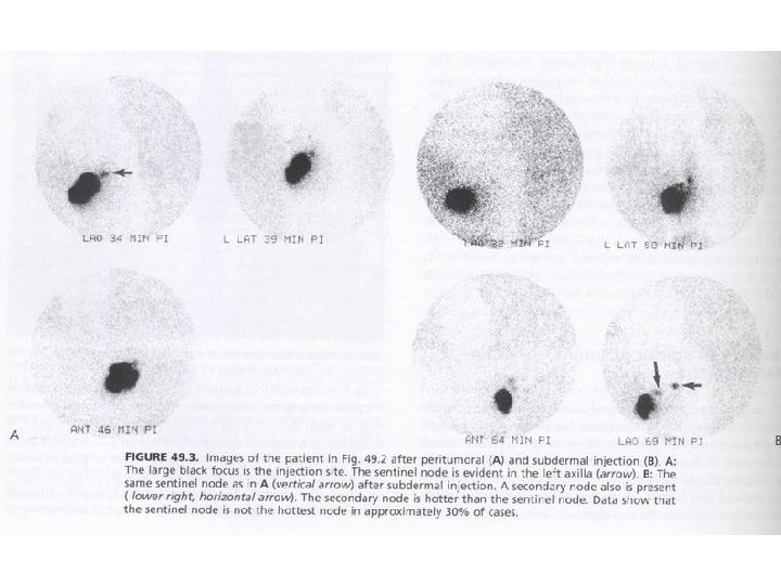
Imaging • Why imaging is necessary? – Helpful in incision planning – Smaller incision is performed – Surgical time is shortened – Not all hot lymph nodes should be excised
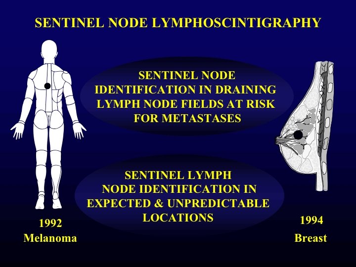
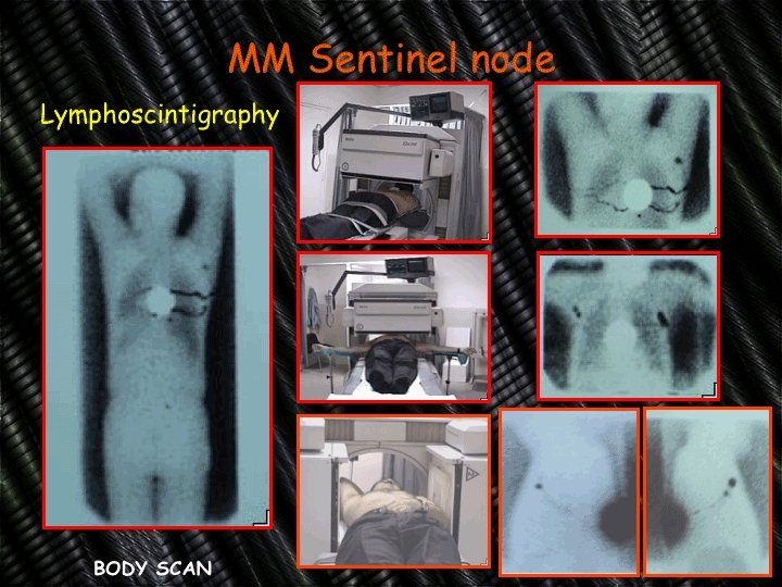
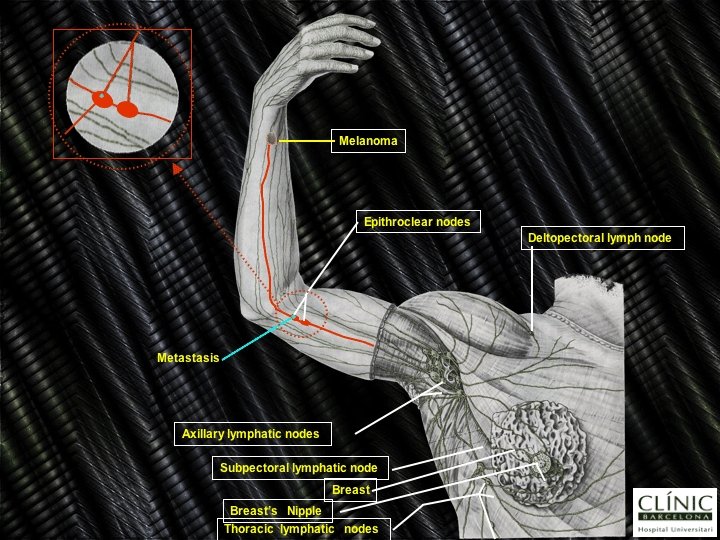
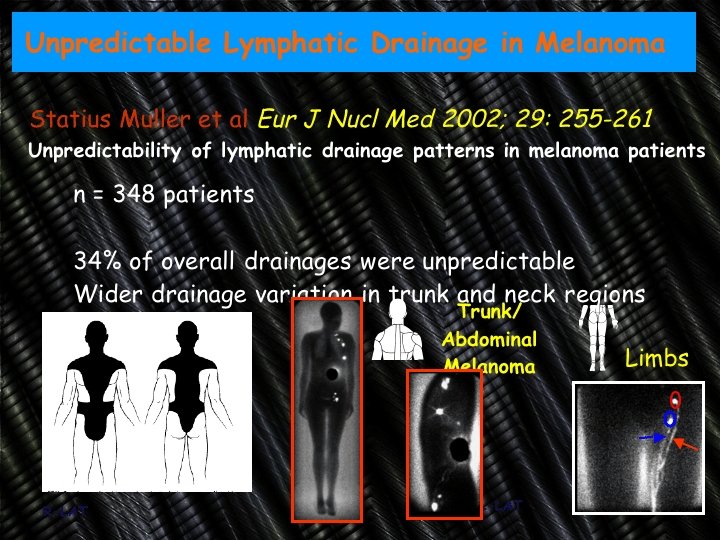
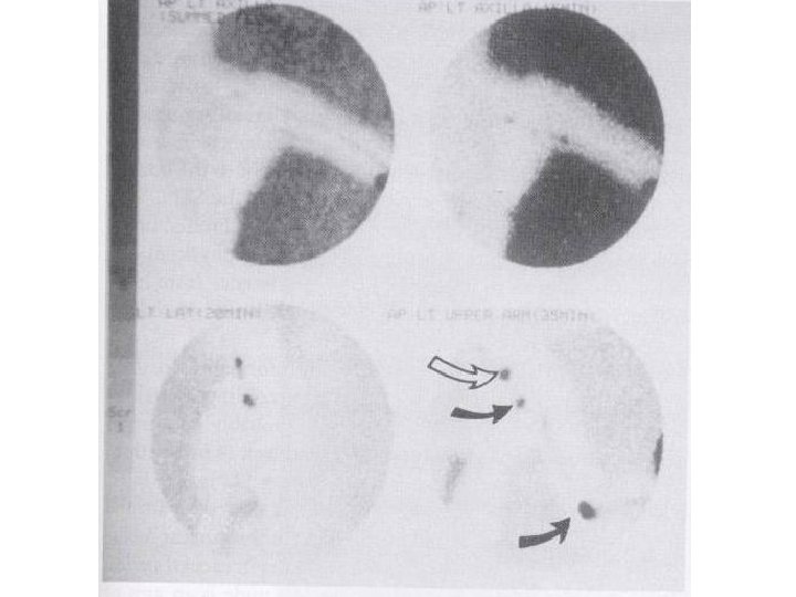
Melanoma l l In more than 10% of melanoma, lymphatic drainage goes to 2 or 3 node groups Sometimes lymphatic drainage is unpredictable: • Around Sappey’s line
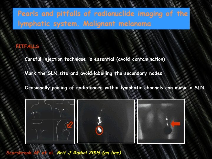
Melanoma: Technique l Radiotracer injections: l Avoid radioactive contamination Gentle finger massage 20% of the activity is absorbed systemically l l • 0. 1 m. L - 3. 7 MBq (100 μCi) Tc-99 m SC • 4– 8 peritumoral intradermal injections • Within 1 cm from the melanoma
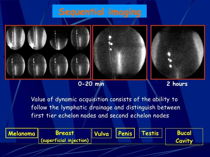
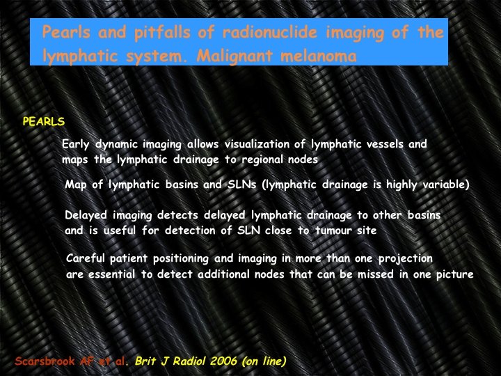
Melanoma: Technique l l Dynamic imaging • 30 sec/f for 2– 30 min and/or sequential static images every 5 min for up to 1 h or until the sentinel lymph node is identified. In-transit nodes • For extremity lesions, the knee or elbow regions in the field of view.
Melanoma l l In Head & Neck injection should not be inferior to the tumor Sentinel node is not the hottest node in 30% of cases
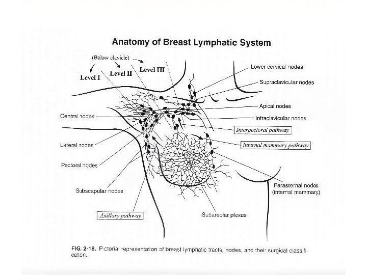
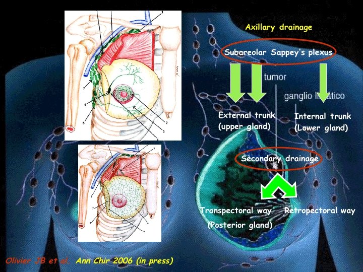
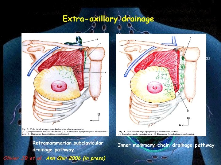
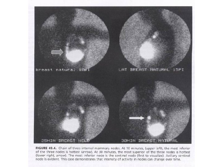
Breast carcinoma: Lymphatic drainage l There are 3 pathways: l 9% have regions with exclusive drainage to internal mammary nodes Never to the opposite axilla or contralateral internal mammary l • Axillary • Internal mammary • Supra or infra-clavicular
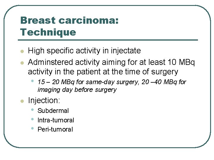
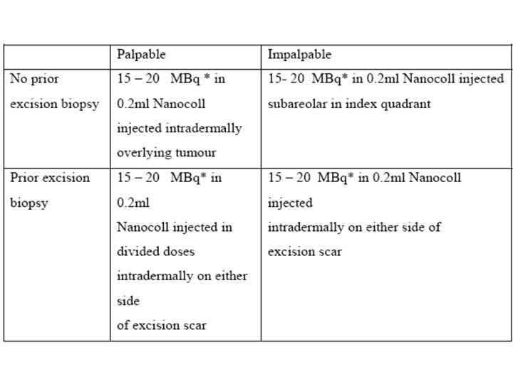
Breast carcinoma: Technique l l High specific activity in injectate Adminstered activity aiming for at least 10 MBq activity in the patient at the time of surgery • l 15 – 20 MBq for same-day surgery, 20 – 40 MBq for imaging day before surgery Injection: • • • Subdermal Intra-tumoral Peri-tumoral
Breast carcinoma: Technique (cont. ) l Injection: • The only difference is internal mammary node • l visualization Subdermal + peritumoral is the preferred method After injection: • Hot towel placement on the breast • Massaging the injection site
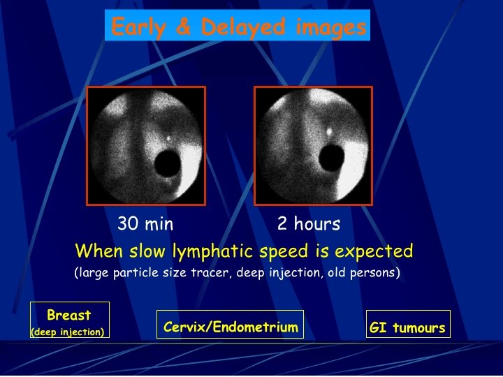
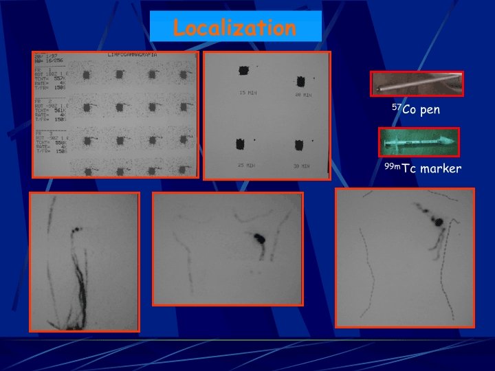
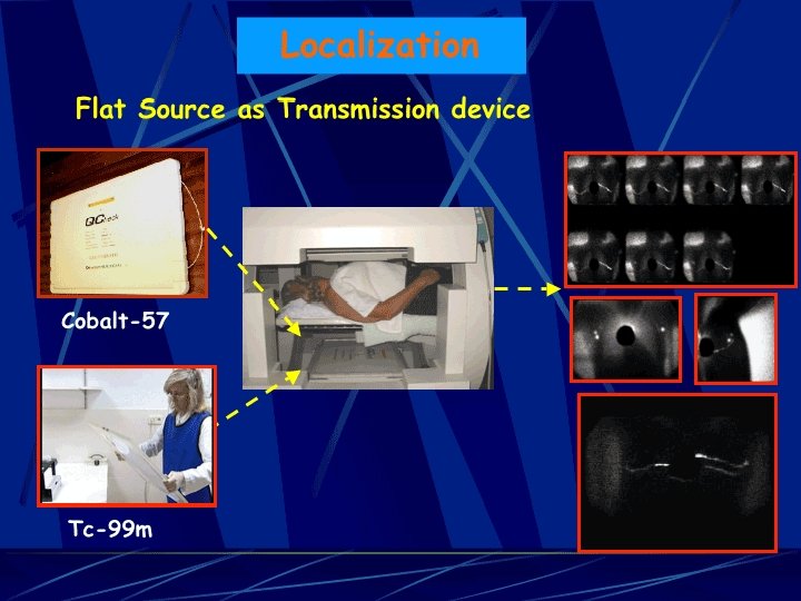
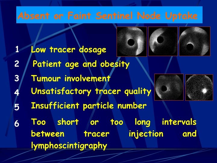
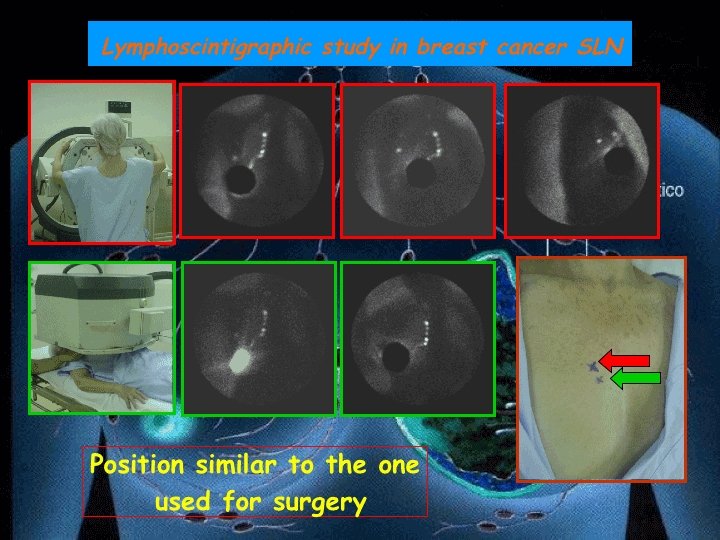
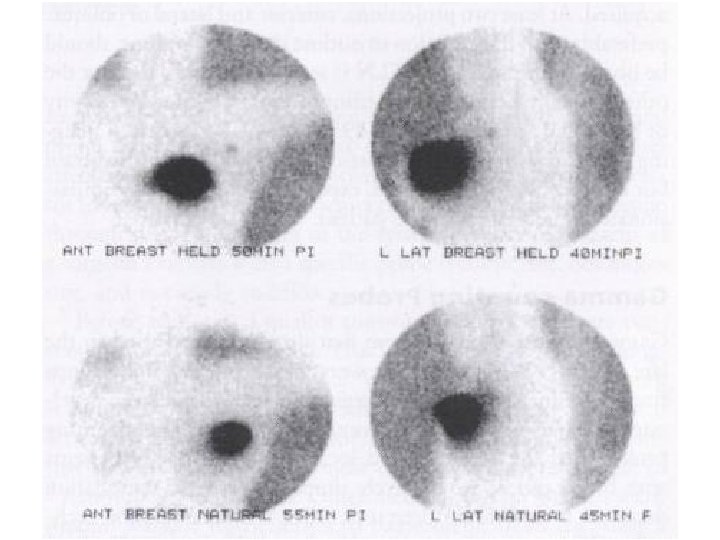
Breast Technique l Emission: • • • l l Anterior oblique 5 min (400, 000 – 500, 000 counts), 30 degree. Lateral 5 min - with arm abducted Anterior (if internal mammary nodes visualised) Transmission (Co-57 flood source) • Anterior oblique, lateral and anterior (if anterior emission image acquired) after their emission image If all images negative further imaging either 6 hrs post injection or next morning
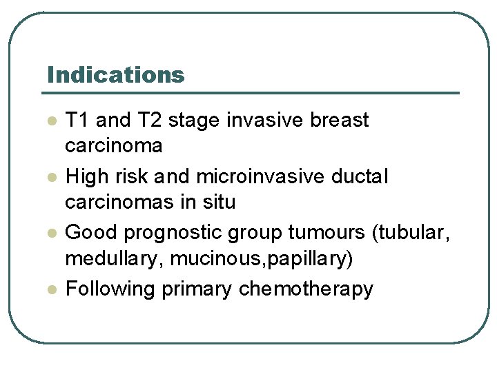
Indications l l T 1 and T 2 stage invasive breast carcinoma High risk and microinvasive ductal carcinomas in situ Good prognostic group tumours (tubular, medullary, mucinous, papillary) Following primary chemotherapy
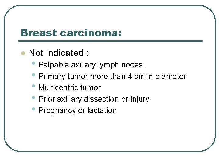
Breast carcinoma: l Not indicated : • Palpable axillary lymph nodes. • Primary tumor more than 4 cm in diameter • Multicentric tumor • Prior axillary dissection or injury • Pregnancy or lactation
Breast carcinoma: Technique Technical aspects should be followed carefully to have a successful lymphoscintigraphy
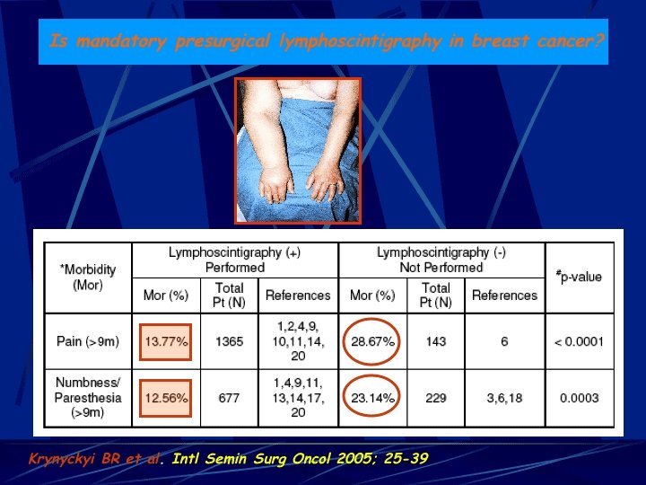
Conclusion l Lymphoscintigraphy • Becoming a standard procedure for several • • malignancies Is >95% successful in experienced hands Can decrease morbidity of lymph node dissection significantly www. DSNMC. ir