Lymphoid organs Primary lymphoid organs Bone marrow Thymus


Lymphoid organs Primary lymphoid organs • Bone marrow • Thymus Secondary lymphoid organs • Lymph node • Spleen • Tonsils
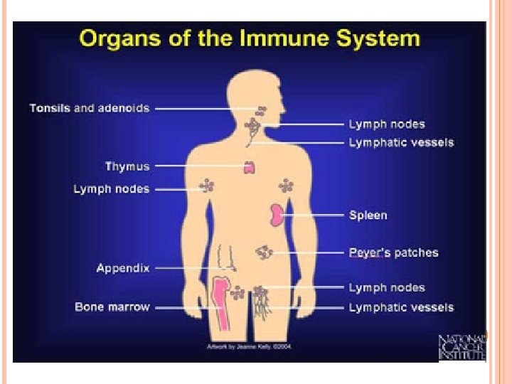

LYMPHATIC ORGAN Tonsils lymph node Spleen

Lymph nodes 1. Lymph nodes are bean-shaped, encapsulated structures 2. generally 2– 10 mm in diameter 3. distributed throughout the body along the course of the lymphatic vessels, in the chest, neck, pelvis, axilla (armpit), groin region, and in association with the blood vessels of the intestines

Lymph nodes are divided into two region: A. Cortix regions B. Medulla regions

Structure of lymph node q The lymph node consists of lymphoid follicles in an outer portion called the "cortex. “, and the inner portion called the "medulla, " which is surrounded by the cortex on all sides except for a portion known as the "hilum. “ q The hilum presents as a depression on the surface of the lymph node, which makes the spherical lymph node, bean-shaped or ovoid. q The efferent lymph vessel , arteries and veins supplying the lymph node with blood enter and exit through the hilum.
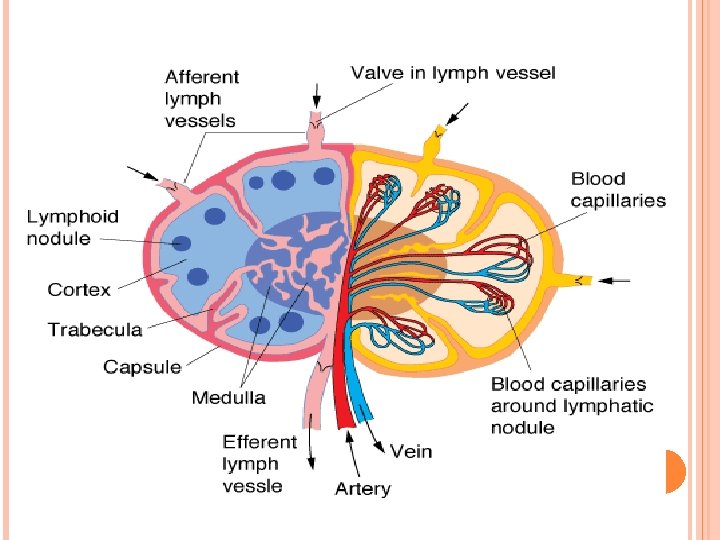
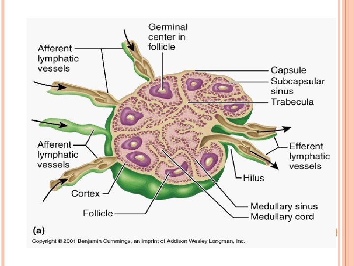

A. Cortex 1. Lies deep to the capsule from which it is separated by a subcapsular sinus 2. Is incompletely subdivided into compartments by connective tissue septa derived from the capsule

The cortex consists of the following components: 1. Lymph node germinal center 2. Lymph nodule 3. Sinusoids 4. Trabecula and lymph sinus 5. Paracortex
 Lymphoid nodules are composed mainly of : 1. B cells. 2. T cells.")
(a) Lymphoid nodules are composed mainly of : 1. B cells. 2. T cells. 3. Follicular dendritic cells. 4. Macrophages. 5. Reticular cells.
 Sinusoids : Are Endothelium-lined spaces that extend along the capsule and trabeculae and")
(b) Sinusoids : Are Endothelium-lined spaces that extend along the capsule and trabeculae and are known as subcapsular and cortical sinusoids, respectively.
 The paracortex: 1. located between the cortex and the medulla. 2. the region")
(c) The paracortex: 1. located between the cortex and the medulla. 2. the region where circulating Lymphocytes reach to lymph nodes via post capillary.

B. The medulla 1. lies deep to the paracortex and cortex it is composed of : a. Medullary sinusoids b. Medullary cords.

a. Medullary sinusoids: • are endothelium-lined spaces supported by reticular fibers and reticular cells and frequently contain macrophages. • Medullary sinusoids receive lymph from the cortical sinuses. b. Medullary cords: • are composed of lymphocytes and plasma cells.

Function of lymph nodes 1. Lymph nodes have the primary function of producing lymphocytes which help protect the body against microorganisms and harmful foreign particles. 2. Lymph nodes normally swell when they are actively responding to infection as they fill with the

Tonsils: are aggregation of lymphoid tissue. Lymphoid tissue: is connective tissue characterized by a rich population of lymphocytes Location : The site of the tonsils at the opening of both the respiratory and digestive tracts provides a unique and strategic position from which to initiate immunological protection against airborne and ingested pathogens.

Tonsils These lymphoid masses are named according to their location as : 1. Palatine tonsils 2. Pharyngeal tonsil 3. Lingual tonsil
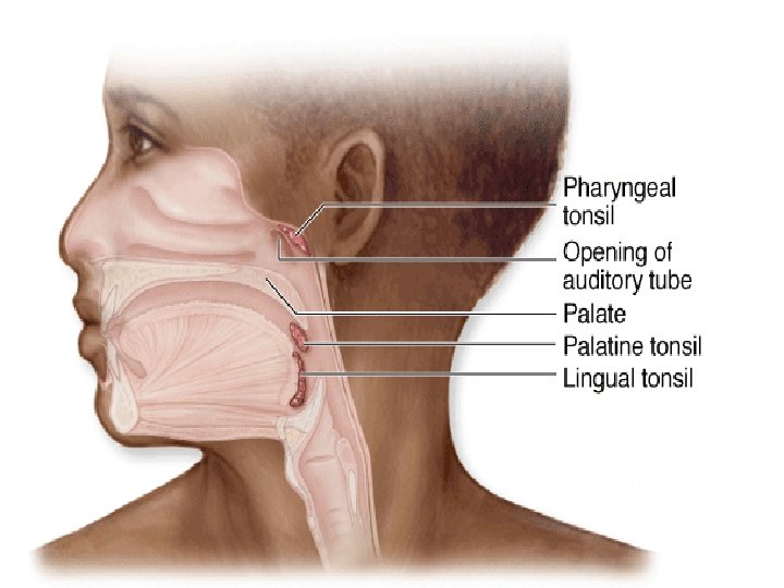
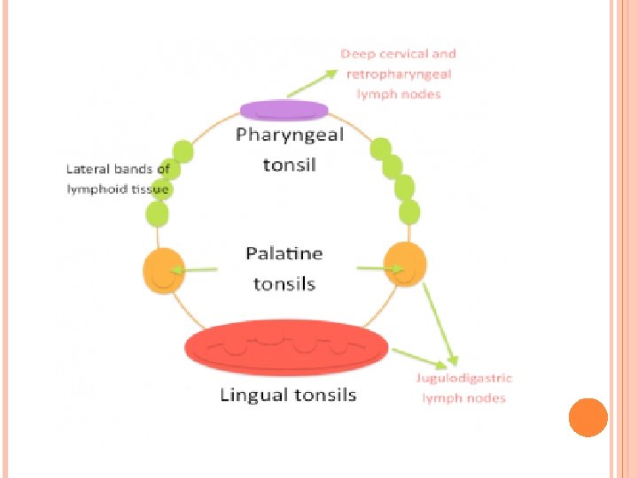

1. Palatine tonsils These are paired ovoid masses of lymphoid tissue Location: Located Paired at oropharynx

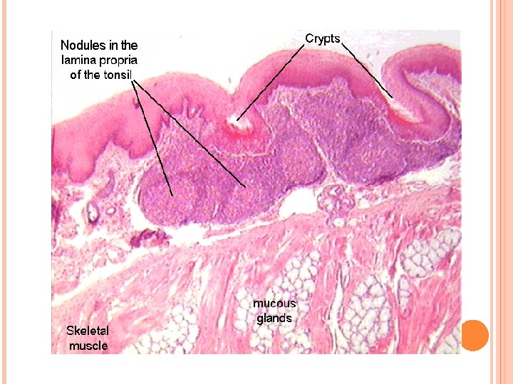
Structure: • covered by stratified squamous epithelium • The tonsils have many invaginations which form blind crypts • Tonsillar crypts (lined by a continuous surface of epithelium) • Each crypt surrounded by a zone of varying thickness consisting of mass of lymphoid tissue • Nodule • Capsule : fibrous connective tissue

2. Pharyngeal tonsil: This is an accumulation of lymphoid tissue Location: In the midline on the roof of the nasopharynx

Structure : • Covered by pseudostratified columnar ciliated epithelium • Patches of stratified squamous epithelium may be present • No crypts • Lymphoid tissue • Thin capsule

3. Lingual tonsil: Location : Posterior third of tongue Structure: • One or more wide crypts lined by a stratified squamous epithelium • Lymphatic nodules

Tonsil Location In the midline on Pharyng the roof of the eal nasopharynx Lingual Posterior third of tongue Paired at Epithelium Crypts Ciliated pseudostratified columnar epithelium typical Numerous folds of the respiratory of pharyngeal tract, with some epithelium, not patches of stratified true crypts squamous epithelium observed Multiple monocryptic Stratified squamous non'units' with a single shallow keratinising crypt each 10 -30 deep and sometimes Stratified

Spleen • Spleen is the largest lymphoid organ in the body • It is the only organ specialized for filtering blood

Spleen Structure: • connective tissue capsule • Trabeculae • sinuses (filled with blood instead of lymph) • Splenic pulp
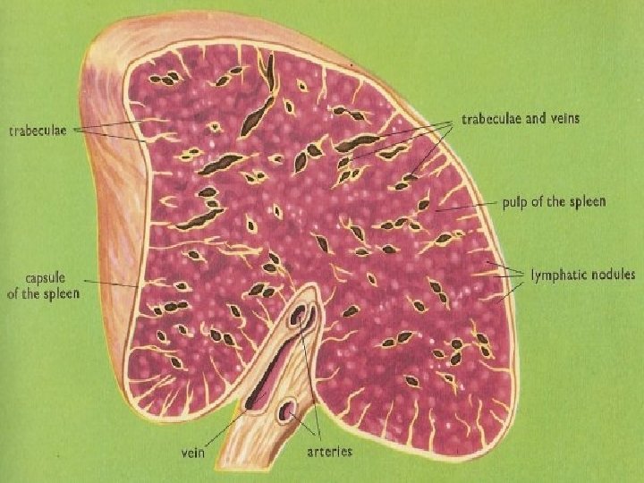

Spleen Splenic pulp: • The spaces between the trabeculae are occupied by a soft sponge like tissue known as the splenic pulp. • If a freshly cut spleen is examined by naked eye , the splenic pulp have different colors(red or white) in various region.
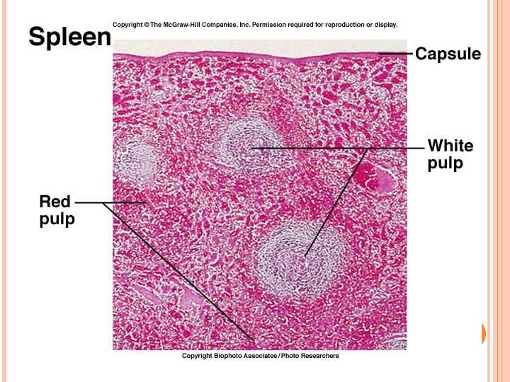

Spleen Splenic pulp is divided into the following two kinds: 1. Red pulp 2. White pulp

Spleen 1. Red pulp red pulp consists of: • Blood-filled sinusoids • Splenic cords.

Spleen 2. White pulp The small masses of white pulp consist of: • lymphoid nodules • Periarteriolar lymphoid sheathes

Spleen Function of spleen: 1. Filtration of blood 2. Production of lymphocytes 3. Destruction of old erythrocytes 4. Storage of blood

Thank you
- Slides: 38