Lymphatic system The lymphatic system is a part


 Tunica media (")


")

: Central arteriole Germinal")


 The surrounding medullary tissue in thymus")


- Slides: 36
Lymphatic system The lymphatic system is a part of the circulatory system, comprising a network of lymphatic vessels that carry a clear fluid called lymph (from Latin lympha "water goddess"
Lymphatic system It composed of lymphatic vessels & organs Lymphatic capillaries, which are the smallest vessels collects the tissue fluid which is called the lymph as soon as it inters these capillaries. lymph is relatively acellular & watery fluid, so that the lymph of those finest lymphatic radicals is almost devoid of cells. Lymphatic capillaries unite to form larger & then largest vessels which empty into the veins, thus considered not closed vascular ring as in case of blood vascular system. Lymphatic organs are located along the course of the lymphatic vessels & give various-sized lymphocytes to the lymph passing through them.
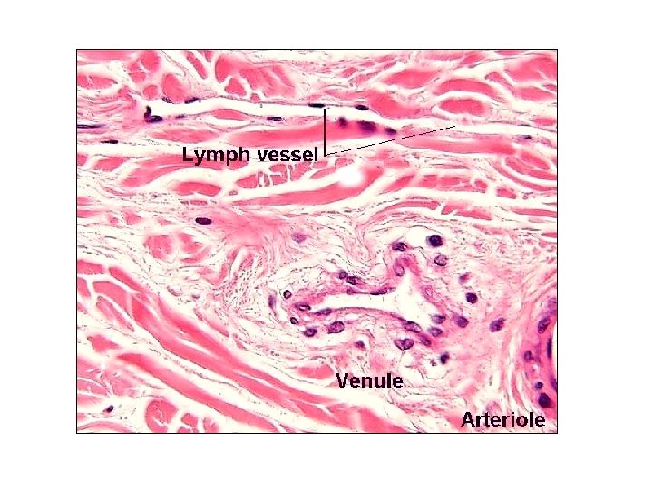
Lymphatic capillaries: These are thin-walled tubular structures of irregular calibers with blind, rounded or swollen ends. Best example for them found in the intestine forming the central lacteal. It forms expanded networks of considerable size around the solitary lymphatic nodules of the intestine, thyroid & mammary glands. *They differ from blood capillaries by 1. their irregular calibers, 2. slightly larger & thinner endothelial cells (their nuclei cannot distinguished from the nuclei of the fibroblasts) and 3. without layer of pericytes.
Lymphatic capillary blood venules endothelium Lymphatic capillary blood capillary
Lymphatic capillary Striated border Goblet cell Simple columnar epithelium Intestinal villus Lacteal lumen in Lymphatic capillary lacteals communicate with a fine network of lymphatic vessels which ramify on the surface of the mesentery. Have valves, the absorbed oil globules, of food, slowly pass to thoracic duct
Lymphatic capillary Simple columnar epithelium Goblet cell Lacteal lumen in Lymphatic capillary Intestinal villus
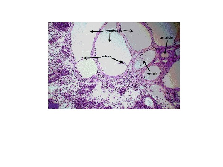
Larger lymphatic vessels: The lymph passes from the capillary networks into these vessels which have slightly thicker walls & are provided with valves. They are covered with thin collagenous bundles, elastic fibers & a few smooth muscle fibers arranged tangentially or transversely to the vessel. Vessels of greater than 0. 2 mm have thicker walls, characterized with 3 indistinct layers: • Tunica intima which consists of endothelium & a thin layer of longitudinal interlacing elastic fibers. • Tunica media which have several layers of mainly circular & a few longitudinal smooth muscle fibers & several thin elastic fibers. • Tunica adventitia which is the thickest layer consists of interlacing collagenous & elastic fibers & smooth muscle fiber bundles.
Valves They occur in pairs, placed on opposite sides of the vessel with their free edges pointing in the direction of the lymph flow. They are folds of tunica intima, consists of thin basal connective tissue surrounded by 2 layers of endothelium.
Large lymphatic vessels Larger lymphatic vessels come together & form two main trunks: Right lymphatic duct: it is the smallest & carries the lymph from the upper right portion of the body. Thoracic duct: it carries the lymph from the other parts of the body. The walls of the large ducts are different from the large veins by their greater developed tunica media & less distinct division into 3 layers.
Tunica intima consist of endothelial lining, several thin layers of cells & elastic fibers which are condensed near the junction site with media forming a layer similar to internal elastic lamina. Tunica media composed of transverse smooth muscle bundles which are penetrated by elastic fibers comes from the internal lamina. Tunica adventitia composed of longitudinal collagenous fibers, interlacing elastic fibers & a few longitudinal smooth muscle bundles. It consist of many blood vessels which may extends to the outer part of tunica media.
aorta Thoracic duct This duct passes upwards beside the vertebral column, and pours its contents into the left jugular vein in the neck
Thoracic duct aorta
Thoracic duct
Thoracic duct Endothelial lining (Thin layers of cells & elastic fibers) Tunica media ( smooth muscle bundles +elastic fibers) Tunica adventitia ( longitudinal collagen fibers + elastic fibers + longitudinal smooth muscle fibers) + blood vessels
Passage of lymph from the tissues into the lymphatics Injection of certain dyes (Chinese ink) revealed that the lumen of the lymphatics does not communicate directly with the tissue spaces. The tissue fluid must pass through the endothelial cytoplasm or through narrow intercellular clefts between the endothelial cells to reach the lymphatic lumen. Inflammation can increase the permeability of the local lymphatics to certain dyes.
Lymphatic organs Lymph node, spleen, thymus, tonsils & solitary lymphatic nodules.
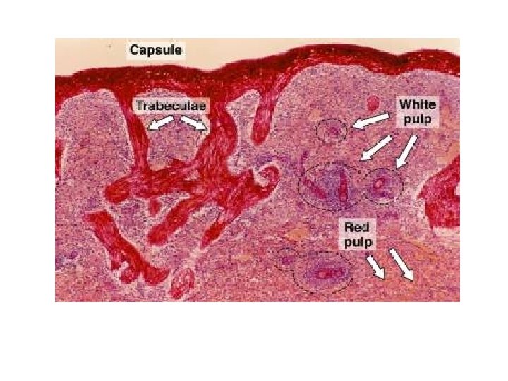
Red pulp Spleen Splenic nodules (white pulp)
Spleen Capsule
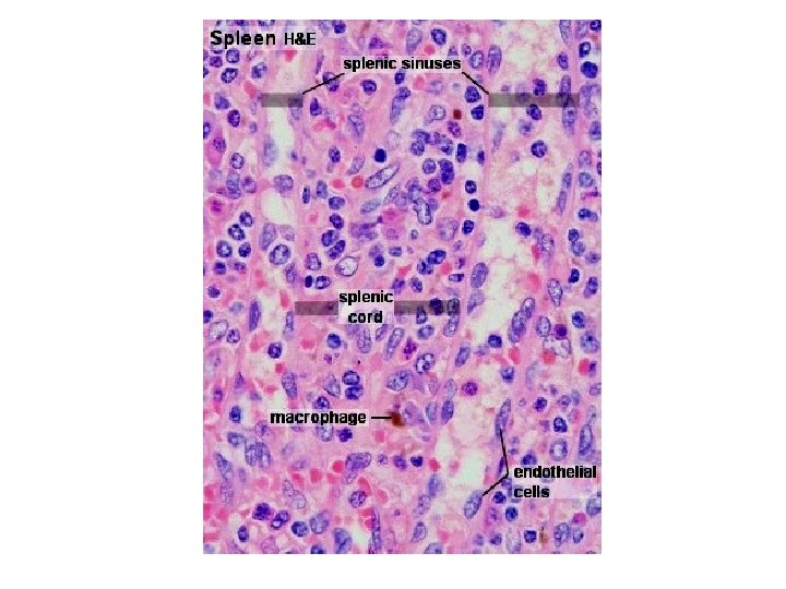
Spleen Red pulp: Splenic sinuses Splenic cords Splenic nodules (white pulp): Central arteriole Germinal center of the lymphatic nodule
Thymus capsule thymus lobule: cortex + medulla Blood vessels thymus lobules Interlobular C. T. trabeculae
Thymus Hassall corpuscles
Thymus Interlobular C. T. trabeculae medulla cortex Blood vessels Hassall corpuscles
Thymus Arrows showing Epithelial cells Hassall corpuscles
Thymus Hassall corpuscles Eosinophilic epithelial cells (concentrically arranged) The surrounding medullary tissue in thymus lobule
Lymph node Cortex Capsule Medulla Capsule Trabecular C. T. + blood vessels
Lymph node
Subcapsular sinuses Afferent lymphatics Capsule
Medullary cords Medullary sinuses
Tonsil Stratified squamous epithelium Lymphatic nodules: cortex & medulla Branched crypt
Stratified squamous epithelium Tonsil Branched crypts Lymphatic nodules: cortex & medulla
Peyer's patches: ileum Intestinal villi Tunica mucosa: Simple columnar epith. with goblet cells Tunica submucosa: contain lymphatic nodules Tunica muscularis Tunica serosa
Peyer's patches: ileum Intestinal villi Tunica mucosa: Simple columnar epith. with goblet cells Tunica submucosa: contain lymphatic nodules Tunica muscularis Tunica serosa