Lymphatic System Overview Lymph interstitial fluid once in


 • Have thinner walls,")


 –")

 – Lymphatic Filariasis –")
 system responds quickly and consists of: –")


")


- Slides: 52
Lymphatic System: Overview • Lymph – interstitial fluid once in lymphatic vessels • Consists of 2 semi-independent parts: – A network of lymphatic vessels – Lymphoid tissues and organs scattered throughout body • Returns interstitial fluid and leaked plasma proteins back to blood
• One-way system: lymph flows toward the heart • Lymph vessels include: – Microscopic, permeable lymphatic capillaries – Lymphatic collecting vessels – Lymphatic trunks and ducts
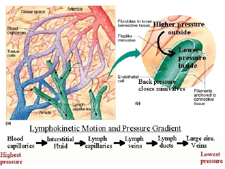
Lymphatic Capillaries • Similar to blood capillaries, with modifications: – More permeable – Loosely joined minivalves – Withstand interstitial pressure and remain open • Minivalves function as one-way gates that: – Allow interstitial fluid to enter, but not escape lymph capillaries • During inflammation, lymph capillaries absorb cell debris, pathogens, cancer cells • Lacteals – specialized lymph capillaries present in intestinal mucosa - absorb digested fat and deliver chyle to the blood
Lymphatic Vessels & Transport • Have 3 tunics (as veins) • Have thinner walls, with more internal valves • Collecting vessels in the skin travel along superficial veins • Deep vessels travel along arteries • Nutrients are supplied from branching vasa vasorum (network of small arterioles, capillaries, and venules that supply the outer tissue of large blood vessels) § The lymphatic system lacks a pumping organ § Vessels are low-pressure conduits § Uses the same methods as veins to propel lymph: § § Pulsations of nearby arteries Contractions of smooth muscle in the walls of lymphatics Lymphatics & the Breast
Lymphatic Trunks • Lymphatic trunks are formed by union of largest collecting ducts • Major trunks include: – Paired lumbar, bronchomediastinal, subclavian, and jugular trunks – A single intestinal trunk • Lymph is delivered into 1 of 2 large trunks – Right lymphatic duct – drains right upper arm and the right side of head and thorax – Thoracic duct – arises from cisterna chyli and drains rest of body
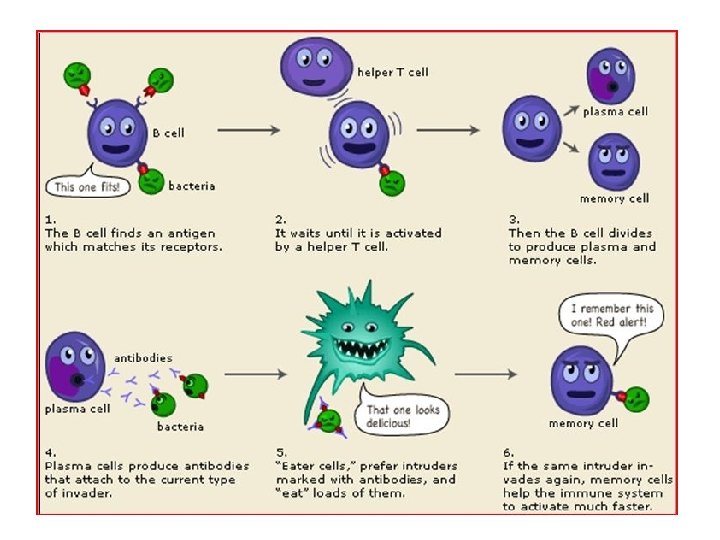
Lymphoid Cells • Lymphocytes are THE main cells involved in immune response • 2 main kinds: T cells and B cells, they protect body against antigens • Antigen – anything the body perceives as foreign: bacteria, viruses, mismatched RBCs or cancer cells • T cells: manage immune response; attack foreign cells • B cells: produce plasma cells, which secrete antibodies => Antibodies immobilize antigens Other lymphoid cells: • Macrophages – phagocytize foreign substances and activate T cells • Dendritic cells – spiny-looking cells, functions similar to macrophages • Reticular cells – supports other cell types in lymphoid organs
Lymphoid Tissue § Scattered reticular tissue elements in every body organ Lymph Nodes • Principal lymphoid organs of the body • Within connective tissue and along lymphatic vessels • Aggregations of nodes occur near body surface in inguinal, axillary, and cervical regions of the body • 2 basic functions: – Filtration – macrophages destroy microorganisms and debris – Immune system activation – monitor and attack antigens
Structure of a Lymph Node • Nodes are bean-shaped and surrounded by a capsule • Trabeculae extends inward from capsule and divide node into compartments • Nodes have 2 histologically distinct regions: a cortex and a medulla • Cortex contains follicles with dividing B cells • Cortex houses T cells
Lymph nodes 2 Regions: • Cortex – B cells – T cells • Medulla – Lymph Sinuses & macrophages Lymph Nodes Animation
Lymphoid Organs • • • Lymph Nodes Spleen Thymus Tonsils Peyer’s patches MALT
Spleen • White pulp – B and T cells carry out immune function. • Red pulp – Removes aged and defective RBCs – Stores breakdown products of RBCs • Erythrocyte production in fetus • Stores blood platelets • Has regenerative properties
Thymus • Secretes thymopoietin, thmosins to make T -cells immunocompetent • Bilobed organ • Trabeculae divide lobe into lobules. • Thymic corpuscles
Tonsils & Adenoids • Trap bacteria which work their way into the follicles where they are destroyed • This helps develop memory
Appendix • Possibly works with the Peyer's patches to help defend against invaders from the digestive system
Aggregates of Lymphoid Follicles • Peyer’s patches – isolated clusters, similar to tonsils – In the wall of the distal portion of the small intestine – Similar structures are found in the appendix • Peyer’s patches and the appendix: – Destroy bacteria – Generate “memory” lymphocytes for long-term immunity
MALT • Mucosa-associated lymphatic tissue – Peyer’s patches, tonsils, and appendix (digestive tract) – Lymphoid nodules in the walls of the bronchi (respiratory tract) • MALT protects the digestive and respiratory systems from foreign matter
Causes of Edema • Edema Accumulation of interstitial fluid • Blockage of lymphatic system • Increased pressure in veins • Lack of albumin – Decreases fluid returning to blood capillaries by osmosis • Inflammation
Homeoimbalances of the Lymphatic System – Autoimmune Lymphoproliferative Syndrome (ALPS) – Lymphatic Filariasis – Mesenteric Lymphadenitis – Swollen Lymph Nodes – Castleman Disease – Adenoids – Splenomegaly – Hodgkin's disease – Kawasaki disease
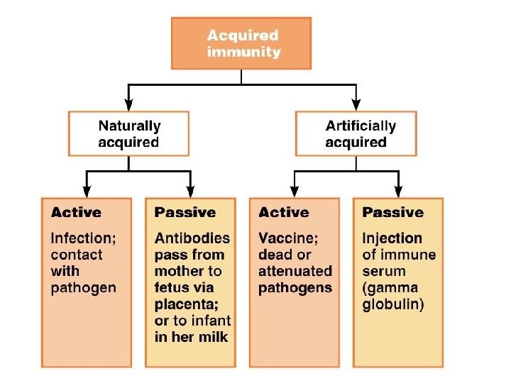
Immunity: 2 Defense Systems • Innate (nonspecific) system responds quickly and consists of: – First line of defense – skin and mucosae prevent entry of microorganisms – Second line of defense – antimicrobial proteins, phagocytes • Inhibit spread of invaders throughout the body • Inflammation is its most important mechanism • Adaptive (specific) defense system – Third line of defense – mounts attack against foreign substances • Has memory, antigen-specific, and antigen-mediated immunity • Works in conjunction with the innate system • Recognizes specific foreign substances – Immobilizes, neutralizes, or destroys foreign substances
First line of defense: Surface membrane barriers • Skin and mucous membrane – Layered epidermis and shedding of epithelial cells – Sebum inhibits growth of bacteria and fungi – Mucous traps microbes, dust and pollutants. • • • Lacrimal apparatus Saliva Vaginal secretions Flow of urine Defecation and vomiting Gastric juices destroy bacteria and their toxins
First line of defense: Surface membrane barriers • Skin and mucous membrane – Layered epidermis and shedding of epithelial cells – Sebum inhibits growth of bacteria and fungi – Mucous traps microbes, dust and pollutants. • • • Lacrimal apparatus Saliva Vaginal secretions Flow of urine Defecation and vomiting Gastric juices destroy bacteria and their toxins
Second line of defense: chemical and cellular defenses • Antimicrobial proteins – Interferon – Complement – Transferrins • Natural killer cells • Phagocytes – Neutrophils – Dendritic cells – Macrophages • Wandering • Fixed – Eosinophils
Interferons • Produced by lymphocytes, macrophages and fibroblasts. • Interfere with translation of viral proteins • Degrade viral RNA • Activate macrophages and NK cells • Interferon Animation
Complement Cascade Animation
Phagocytes • Macrophages are the chief phagocytic cells – Free macrophages wander in search of cellular debris • Kupffer cells (liver) and microglia (brain) are “fixed” macrophages • Neutrophils become phagocytic when encountering infectious material • Eosinophils are weakly phagocytic against parasitic worms • Mast cells bind and ingest a wide range of bacteria Mechanism • Pseudopods engulf the antigen into a phagosome • Invaders are digested by proteolytic enzymes • Indigestible and residual material is removed by exocytosis Figure 21. 2 a
Phagocytosis
Phagocyte Mobilization
Fever • Abnormally high body temperature in response to invading microorganisms • Body’s thermostat is reset upwards in response to pyrogens, chemicals secreted by leukocytes and macrophages exposed to bacteria and other foreign substances • High fevers are dangerous because they can denature enzymes • Moderate fever can be beneficial, as it causes: – Liver and spleen to sequester iron and zinc – Increases metabolic rate, which speeds up tissue repair
Inflammatory response Stages Inflammation Animation • Release of Chemical Alarms • Vasodilatation & Permeability of BV • Emigration of phagocytes: Dispose cellular debris & pathogens • Sets the stage for repair • Prevent spread of damaging chemicals & pathogens Signs of inflammation – – – Redness Heat Swelling Pain Impairment of function
Comparison of Immune Cells
Adaptive Resistance • Specificity—recognition of particular antigens • Memory—remembers previously encountered antigens • Systemic—immunity is not restricted to the initial infection site • Immune responses – Antibody-mediated or humoral immune responses (late 1800 s) – Cell-mediated immune responses (mid 1900 s)
Antigens and antigen receptors • Antigens can be entire microbes, parts of microbes or chemical components of pollen, egg white, blood cells, …….
Antibodies “immunoglobulins” • Four looping polypeptide chains linked together through disulfide bonds. • Heavy chains are identical and have a hinge • Light chains are half as long. • Variable region is the antigen binding site • Constant region forms the stem of the antibody and determines its class • Do not destroy antigen; inactivate and tag it for destruction • form an antigen-antibody (immune) complex
Antibody Action • Defensive mechanisms used by antibodies: – Complement fixation – antibodies bound to cells change shape and expose complement binding sites – Complement activation – uses a positive feedback cycle to promote phagocytosis – Neutralization – antibodies block binding sites on viruses – Precipitation – soluble molecules are cross-linked into large insoluble complexes
Immunoglobulin classes • Ig. D is attached to B-cell plasma membrane • Ig. M is released during primary response. Indicates current infection. • Ig. G is the most aboundant. Can cross placenta & blood vessel walls. • Ig. A found in body secretions prevents attachment to body surfaces. • Ig. E causes release of histamine (allergies) by attaching to mast cells & basophils.
Immunological memory • Primary immune response • Secondary immune response
Lymphocytes • Immature lymphocytes released from bone marrow are essentially identical • Whether a lymphocyte matures into a B cell or a T cell depends on where in the body it becomes immunocompetent – B cells mature in the bone marrow – T cells mature in the thymus
Key: Red bone marrow Immature lymphocytes Circulation in blood 1 Thymus 2 Immunocompetent, but still naive, lymphocyte migrates via blood 3 Activated Immunocompetent B and T cells recirculate in blood and lymph 1 Bone marrow 2 Lymph nodes, spleen, and other lymphoid tissues 3 = Site of lymphocyte origin = Site of development of immunocompetence as B or T cells; primary lymphoid organs = Site of antigen challenge, activation, and final diff erentiation of B and T cells 1 Lymphocytes destined to become T cells migrate to the thymus and develop immunocompetence there. B cells develop immunocompetence in red bone marrow. 2 After leaving the thymus or bone marrow as naïve immunocompetent cells, lymphocytes “seed” the lymph nodes, spleen, and other lymphoid tissues where the antigen challenge occurs. 3 Antigen-activated immunocompetent lymphocytes circulate continuously in the bloodstream and lymph and throughout the lymphoid organs of the body. Figure 20. 8
T Lymphocytes • CD 4 T cell - also known as a T Helper (Th) cell • CD 8 T cell - also known as a Cytotoxic T (Tc) cell
B Lymphocytes Clonal Selection • Production of clones initiated by antigen binding • Plasma cells secrete antibodies • Memory cells are long lived
Humoral & Cell-Mediated Immunity • Humoral = antibody mediated immunity – Involves B cells – Antibodies circulate through “humors” inactive and mark invaders for destruction • Cell-mediated = cellular immunity – Involves T cells – Attack targets directly or release chemical mediators to enhance inflammation/ activate other WBCs
Homeostatic imbalances : Immunodeficiencies • Abnormally behaving immune cells • Severe combined immunodeficiency (SCID) syndromes – Congenital conditions • Acquired immune deficiency syndromes – Hodgkin’s Disease – HIV – AIDS
Homeostatic imbalances : Autoimmune disease – Tend to be more prevalent in women • Type I diabetes—destroys pancreatic beta cells • Multiple sclerosis—destroys myelin sheaths • Myasthenia gravis—impairs communication between nerve and muscle • Lupus erythematosus—systemic disease of skin, kidneys, heart, and lungs • Rheumatoid arthritis—destruction of joints
Organ transplants • Autografts—grafts from the same person to another body site • Isografts—grafts between genetically identical individuals • Allografts—grafts among the same species • Xenografts—grafts taken from another animal species
Hypersensitivities Hypersensitivity Reactions in the Skin
Hypersensitivities Delayed complex Acute Subacute Immediate cytotoxic Immune
Type I Hypersensitivity Animation Type II Hypersensitivity
Type III Hypersensitivity
Animations • Flash animation of a NK cell interacting with a normal body cell. • Flash animation of a NK cell interacting with a virus-infected cell or tumor cell not expressing MHC-I molecules. • Flash animation of apoptosis by NK cells. • HIV Replication