LYMPHATIC SYSTEM AND ITS DISEASES Dr Farhanul Huda


")


. Becomes clinically evident after birth")

.")


 Clinical features Subclinical (latent) There is excess interstitial fluid and histological abnormalities")


/creatinine levels, and urinalysis results should be checked if")


- Slides: 62
LYMPHATIC SYSTEM AND ITS DISEASES Dr. Farhanul Huda Associate Professor Dept. of Surgery AIIMS, Rishikesh
ANATOMY
A 1 -way system that returns lymph via vessels to the CVS. Consists of: Fluid, known as lymph Vessels that transport lymph Organs that contain lymphoid tissue (eg, lymph nodes, spleen, and thymus)
Organ Function Lymph Contains nutrients, oxygen, hormones, and fatty acids, as well as toxins and cellular waste products, that are transported to and from cellular tissues Lymphatic vessels Transport lymph from peripheral tissues to the veins of the cardiovascular system Lymph nodes Monitors the composition of lymph, the location of pathogen engulfment and eradication, the immunologic response, and the regulation site Spleen Monitors the composition of blood components, the location of pathogen engulfment and eradication, the immunologic response, and the regulation site Thymus Serves as the site of T-lymphocyte maturation, development, and control
Functions are : Restoration of excess interstitial fluid and proteins to the blood Absorption of fats and fat-soluble vitamins from the digestive system Defense against invading organisms
LYMPH Fluid derived from plasma. Contains nutrients, oxygen, and hormones, as well as toxins and cellular waste. This fluid is removed by lymphatic vessels that pass through lymph nodes. As the lymph passes through the lymph nodes, lymphocytes and monocytes enter it.
LYMPHATIC VESSELS Are blind-ended tubes with thin endothelial walls. Coalesce to form larger meshlike vessels. Eventually form 2 lymphatic ducts: the right lymphatic duct and the thoracic duct. Have 1 -way valves to prevent any backflow.
LYMPH NODES Bean-shaped structures Filter the lymph before it rejoins the blood stream. Approximately 600 -700, predominantly in the neck, axillae, groin, mediastinum, and mesenteries of the GI tract. Constitute a main line of defense by hosting 2 types of immune protective cell lines, T and B lymphocytes.
2 distinct regions, the cortex and the medulla. The cortex contains follicles, which are collections of lymphocytes. Center of the follicles is called germinal centers that has B-lymphocytes while the remaining cells of the cortex are T-lymphocytes. Vessels entering the lymph nodes are called afferent lymphatic vessels and those exiting are called efferent lymphatic vessels
LYMPHADENOPATHY
ETIOLOGY Five broad etiologic categories : An immune response to infective agents (eg, bacteria, virus, fungus) Inflammatory cells in infections involving the lymph node Metastasis Malignancy of lymphocytes or macrophages (eg, leukemia, lymphoma) Storage disorders
PRESENTATION Duration Associated symptoms Past illnesses, infections, local trauma, or bites. Constitutional symptoms. If recurrent infections, HIV must be considered. Family and social history
PHYSICAL EXAMINATION Complete general examination. The skin and the soft tissue drained by the enlarge node should be carefully examined. The character of the lymph node should be noted. Whether the lymphadenopathy is a local or a general phenomenon.
GENERALIZED LYMPHADENOPATHY: Upper respiratory tract infections (rhinovirus, adenovirus, influenzavirus, parainfluenza virus, respiratory syncytial virus) Epstein-Barr virus (EBV) Cytomegalovirus (CMV) Varicella-zoster virus Herpes simplex virus Paramyxovirus Coxsackieviruses A and B Echovirus Enterovirus Human herpesvirus-6 Human immunodeficiency virus
WORK UP
LABORATORY STUDIES Perform the least invasive test that provides the most information. CBC. Serum LDH- to determine the turnover rate of cells in the case of leukemia or lymphoma. Tuberculin skin test. Monospot and titers for EBV, CMV, catscratch disease, or toxoplasmosis
IMAGING STUDIES Chest radiography to assess for bacterial pneumonias or tuberculosis, and hilar adenopathy in the case of malignancy. Ultrasonography. CT scan. 18 F-FDG PET. FNAC BIOPSY
LYMPHEDEMA
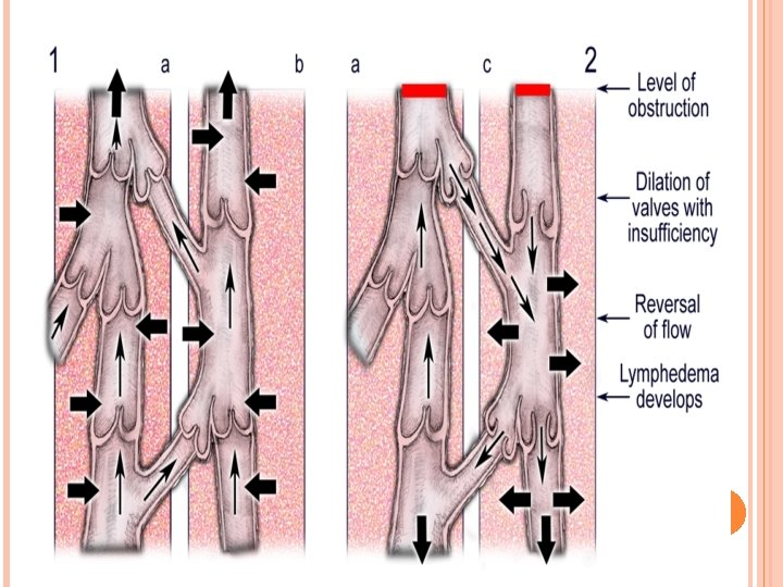
• Lymphatic obstruction • Dec. lymphatic transport • Stagnation of HMW proteins
• High protein edema • High oncotic pressure in the interstitium favors the accumulation of additional water. • Massive dilatation of lymphatic vessels • Inflammation • Shrunken and hard lymph nodes
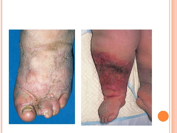
DERMATOLOGIC PATHOLOGY The overlying skin becomes thickened and displays the peau d'orange appearance. Chronic lymphedema causes fissuring and impairment of the epidermis, allowing bacteria to enter and grow, and leading to lymphorrhea, the leakage of lymph. With chronic lymphedema, the development of verrucous, cobblestone plaques, a condition known as elephantiasis nostra verrucosa (ENV), can occur.
ETIOLOGY In primary lymphedema, there is congenital hypoplasia or aplasia of the peripheral lymphatics or valvular incompetence. In secondary lymphedema, the lymphatic drainage is blocked due to: Recurrent attacks of lymphangitis Malignancy Obesity Surgery
PRIMARY LYMPHEDEMA
A developmental abnormality of the lymphatic system. 3 main types, distinguished by their age of onset: 1. Congenital lymphedema (Milroy disease) 2. Lymphedema praecox (Meige disease) 3. Lymphedema tarda Involve the lower extremities almost exclusively.
CONGENITAL LYMPHEDEMA 10 -25% of all primary lymphedema cases. Autosomal-dominant. Anaplastic lymphatic channels. Manifests at birth or later, up to age 1 year. F: M= 2: 1. LL: UL= 3: 1 The edema is most commonly pitting and non painful. 2/3 rd of patients have bilateral lymphedema. May improve spontaneously with increasing age.
LYMPHEDEMA PRAECOX Most common form of primary lymphedema (6580%). Becomes clinically evident after birth and before age 35 years. F: M= 4: 1 About 70% of cases are UL. A hypoplastic pattern, with the lymphatics reduced in caliber and number.
LYMPHEDEMA TARDA Manifests later in life, usually in persons older than 35 years. Caused by a defect in the lymphatic valves, resulting in incompetent valve function. Accounts for only 10% of cases.
ASSOCIATED CONDITIONS Distichiasis lymphedema syndrome is lymphedema associated with distichiasis (double row of eyelashes). Vertebral abnormalities, spinal arachnoid cysts, hemangiomas, cleft palate, ptosis, short stature, webbed neck, strabismus, thoracic duct abnormalities, and microphthalmia. Associated with yellow nail syndrome.
ASSOCIATED CONDITIONS Other genetic syndromes and cutaneous conditions associated with primary lymphedema include the following: Turner syndrome Noonan syndrome Klinefelter syndrome Neurofibromatosis type 1 Hemangiomas Xanthomatosis Congenital absence of nails
SECONDARY LYMPHEDEMA
SECONDARY LYMPHEDEMA Caused by an acquired defect in the lymphatic system. Associated with obesity, infection, neoplasm, trauma, and therapeutic modalities.
SECONDARY LYMPHEDEMA 1. Filariasis The most common cause of secondary lymphedema. Caused by a mosquito-borne nematode infection with the parasite Wucheria bancrofti. Commonly occurring in developing countries around the world. Results in permanent lymphedema of the limb.
SECONDARY LYMPHEDEMA 2. Malignancy and cancer treatment Obstruction from metastatic cancer or primary lymphoma or secondary to radical lymph node dissection and excision. The most commonly affected area is the axillary region after mastectomy Lymphedema can also be seen after regional dissection of pelvic, para-aortic, and neck lymph nodes.
SECONDARY LYMPHEDEMA Other causes Trauma Varicose vein surgery Congestive heart failure Portal hypertension Peripheral vascular surgery Lipectomy Burns Burn scar excision Insect bites Extrinsic pressure
CLINICAL PRESENTATION
HISTORY Chronic swelling of an extremity. First noticed by the patient as an asymmetry or increased circumference of an extremity. As swelling slowly progresses, patients may have difficulty fitting into clothing. May cause fatigue due to the size and weight of the extremity, embarrassment in public, and severe impairment of daily activities. Recurrent bacterial or fungal infections. Fevers, chills, and generalized weakness. History of recurrent episodes of cellulitis, lymphangitis, fissuring, ulcerations, and/or verrucous changes.
HISTORY In primary lymphedema, the history of onset is more typical. Associated with other anomalies and genetic disorders. In secondary lymphedema, the associated history is based on the primary etiology. If due to filariasis, the history should include travel or habitation in an endemic area. Other patients should have a clear history of a neoplasm obstructing the lymphatic system, recurrent episodes of lymphangitis and/or cellulitis, obesity, trauma, or lymphedema resulting after surgery and/or radiation therapy. A recent history of varicose vein surgery also is reported.
PHYSICAL EXAMINATION The earliest symptom is nontender, pitting edema progressing to a nonpitting one Radial enlargement of the area. Involvement of the distal extremities is followed by proximal advancement. Erythema and peau d'orange of skin.
Grade (Brunner) Clinical features Subclinical (latent) There is excess interstitial fluid and histological abnormalities in lymphatics and lymph nodes, but no clinically apparent lymphoedema I Oedema pits on pressure and swelling largely or completely disappears on elevation and bed rest II Oedema does not pit and does not significantly reduce upon elevation III Oedema is associated with irreversible skin changes, i. e. fibrosis, papillae
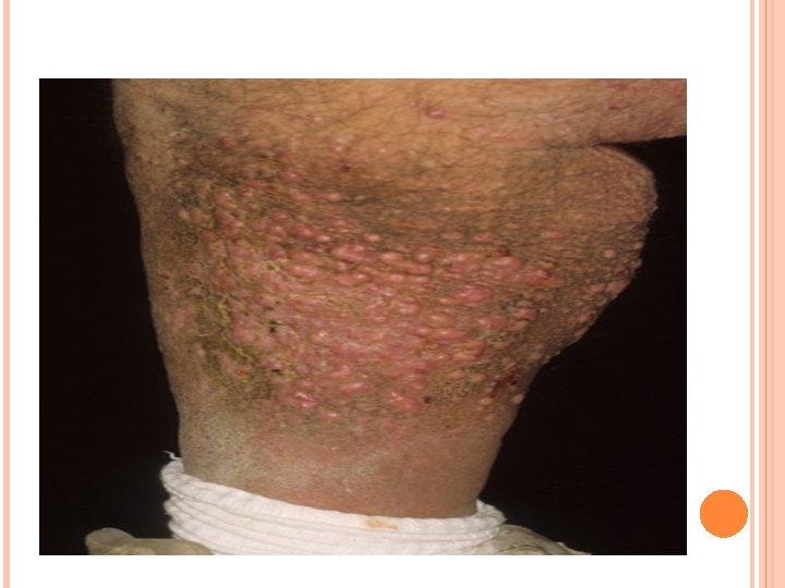
ELEPHANTIASIS NOSTRA VERRUCOSA ENV is an area of cobble-stoned, hyperkeratotic, papillomatous plaques most commonly seen on the shins. The plaques can be weepy or oozing a clear or yellow fluid. Fissuring, ulcerations, skin breakdown. Superinfection is common and can manifest as impetigo with yellow crusts.
EXAMINATION A positive Stemmer sign (inability to pinch the dorsal aspect of skin between the first and second toes) Patients with congenital lymphedema may also present with recurrent cellulitis, papillomatosis, large caliber leg veins, and upsloping "ski-jump" toenails.
DIFFERENTIALS Deep vein thrombosis Hypoalbuminemia Renal failure Lipedema Postoperative complications Baker cyst Idiopathic edema Congestive heart failure Idiopathic edema Neurofibromatosis Sclerema neonatorum
Features of lipoedema that help differentiate it from lymphoedema ■ Occurs almost exclusively in women ■ Onset nearly always coincides with puberty ■ Nearly always bilateral and symmetrical ■ Involvement of trunk ■ The feet are not involved, leading to an inverse shouldering effect at the malleoli ■ No pitting ■ No response to elevation or compression ■ No skin changes of lymphoedema (negative Stemmer’s sign) ■ MRI shows subcutanteous fat but no fluid accumulation
WORKUP OF LYMPHEDEMA
Liver function, blood urea nitrogen (BUN)/creatinine levels, and urinalysis results should be checked if a renal or hepatic etiology is suspected. Specific markers should be checked if a neoplasm is suspected. CBC with differential should be checked if an infectious etiology is being considered. An indication for CT scanning or MRI is suspicion of malignancy. MRI is useful to show lymph trunk anatomy and causes of obstructive secondary lymphedema.
Ultrasonography to evaluate the lymphatic and venous systems. Lymphangiography is now rarely used because of the potential adverse effects. Fluorescence microlymphography demonstrates a lack of microlymphatics. Lymphoscintigraphy.
TREATMENT OF LYMPHEDEMA
The goal is to restore function, reduce physical and psychologic suffering, and prevent the development of infection. Initiate therapy as early as possible before extensive, irreversible fibrosclerotic changes occur in the interstitium. Strict compliance is essential. The majority of compliant patients can be treated successfully with conservative measures
1. HYGIENE AND SKIN CARE Appropriate skin care to prevent recurrent cellulitis or lymphangitis. Meticulous hygiene to remove keratinous debris and bacteria. Cleanse the skin regularly and dry thoroughly. Regular inspection is necessary to identify any open wounds or developing cellulitis.
2. PHYSICAL THERAPY AND COMPRESSION The first-line treatment. Aimed at improving lymphedema with manual lymphatic drainage, massage, and exercise. It advocates the use of compression stockings (at a minimum of 40 mm Hg), multilayer bandaging, or pneumatic pumps. Encourage patients to lose weight, avoid minor trauma, and avoid constrictive clothing that might have a tourniquet effect. Encourage elevation of the affected extremity whenever possible, particularly at night.
3. SURGERY Surgical treatment is palliative, not curative, and it does not obviate the need for continued medical therapy. Surgical treatment is reserved for patients who do not improve with conservative measures or for cases in which the extremity is so large that it impairs daily activities and prevents successful conservative management. Surgical procedures are classified as physiologic or excisional.
PHYSIOLOGIC SURGERY Physiologic procedures attempt to improve lymphatic drainage. Multiple techniques have been described, including omental transposition, buried dermal flaps, enteromesenteric bridging, lymphangioplasty, and microvascular lympholymphatic anastomosis.
EXCISIONAL SURGERY Excisional techniques remove the affected tissues, thus reducing the lymphedema-related load. The Charles procedure is a radical excisional technique. This procedure involves the total excision of all skin and subcutaneous tissue from the affected extremity. The underlying fascia is then grafted, using the skin that has been excised. This technique is extreme and is reserved for only the most severe cases. Complications include ulceration, hyperkeratosis, keloid formation, hyperpigmentation, weeping dermatitis, and severe cosmetic deformity. A variant of the Charles procedure, total superficial lymphangiectomy, involves debulking of the entire limb.
COMPRESSION THERAPY Patients should use compression garments continuously during the day. They should also have graduated compression that increases from distal to proximal on the affected extremity. Intermittent pneumatic pump compression therapy provides sequential, active compression from distal to proximal, effectively milking the lymph from the extremity. This treatment is most appropriately used prior to fibrosclerotic evolution, which it assists in preventing. Contraindications to intermittent pneumatic pump compression therapy include congestive heart failure, deep vein thrombosis, and active infection.
MLD Manual lymphatic drainage according to the Vodder and/or Leduc techniques. Compression garments are essential between treatments. Manual massage of the affected extremity; this recruits collateral vessels, allowing the accumulated lymph to be drained into neighboring regions with normally functioning lymphatics.
PHARMACOLOGIC THERAPY Cellulitis At the earliest signs of infection, institute topical or systemic antifungal or antimicrobial therapy to prevent the development of sepsis. Filariasis has been treated with DEC and albendazole Benzopyrones These drugs bind to accumulated interstitial proteins, inducing macrophage phagocytosis and proteolysis. The resulting protein fragments pass more readily into the venous capillaries and are removed by the vascular system. The benzopyrones aid in decreasing excess edematous fluid, softening the limb, decreasing skin temperature, and reducing the number of secondary infections. Of note, however, is that hepatotoxicity has been associated with coumarin therapy.
PHARMACOLOGIC THERAPY Retinoids Help normalize keratinization and decrease inflammatory and fibrotic changes. Topical agents Topical emollients and keratolytics have been recommended to improve secondary epidermal changes.
What every patient with lymphoedema should receive ■ An explanation of why the limb is swollen and the underlying cause ■ Guidance on skin hygiene and care and the avoidance of acute infective episodes ■ Anti-fungal prophylactic therapy to prevent athlete’s foot ■ Rapid access to antibiotic therapy if necessary, hospital admission for acute infective episodes ■ Appropriate instructions regarding exercise therapy ■ Manual lymphatic drainage (MLD) ■ Multilayer lymphoedema bandaging (MLLB) ■ Compression garments and, if appropriate, specialised footwear ■ Advice on diet ■ Access to support services and networks