Lymphatic System and Immunity Lymphatic System the other


,")


 →")
 →")


 - immunogens 1. This is any chemical substance that is not")
 1. These are produced by plasma cells and are specific for")


- Slides: 62
Lymphatic System and Immunity
Lymphatic System - the other circulatory system
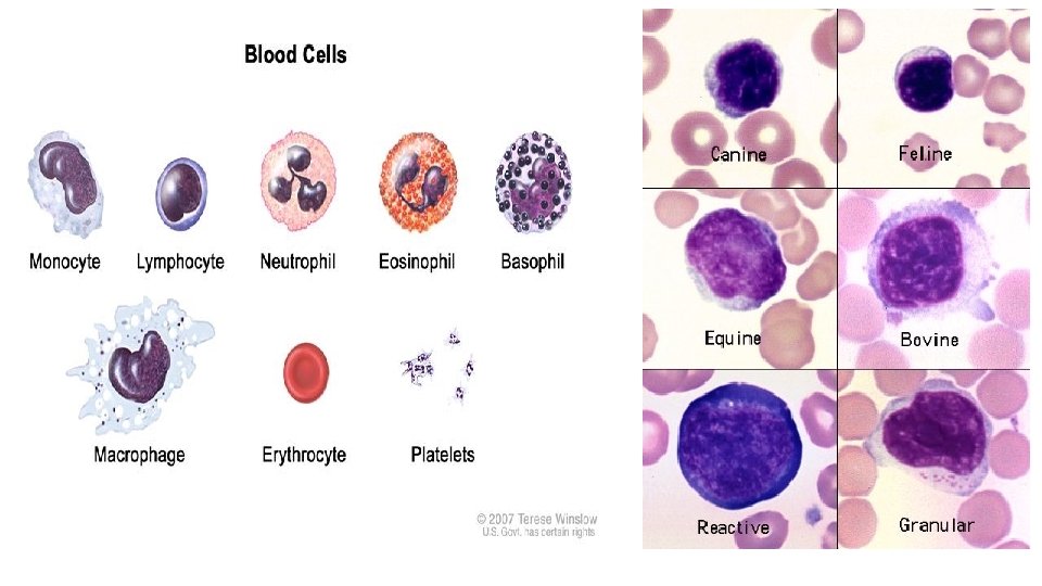
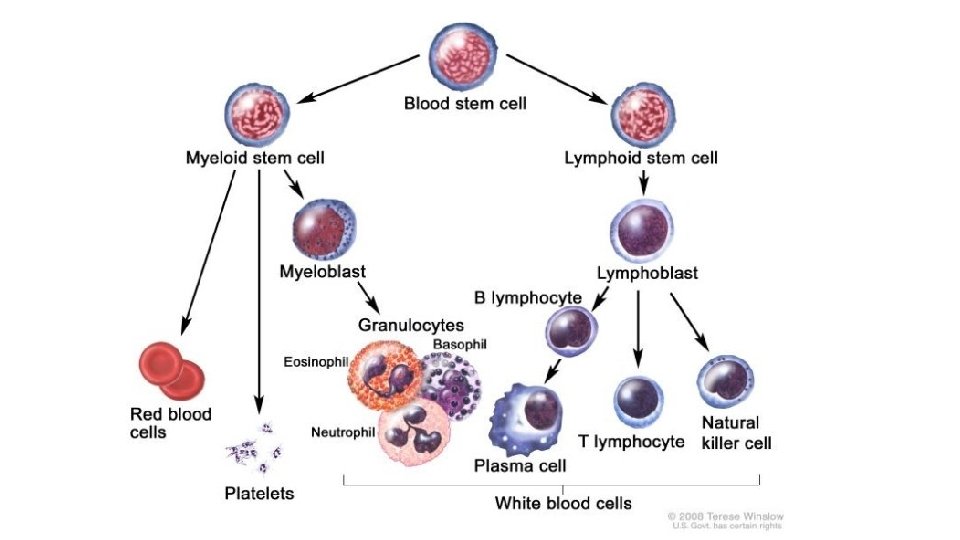
A. Structures - lymph fluid, lymph vessels, lymph tissue, red bone marrow - makes the WBC, lymphocytes - makes B and T cells
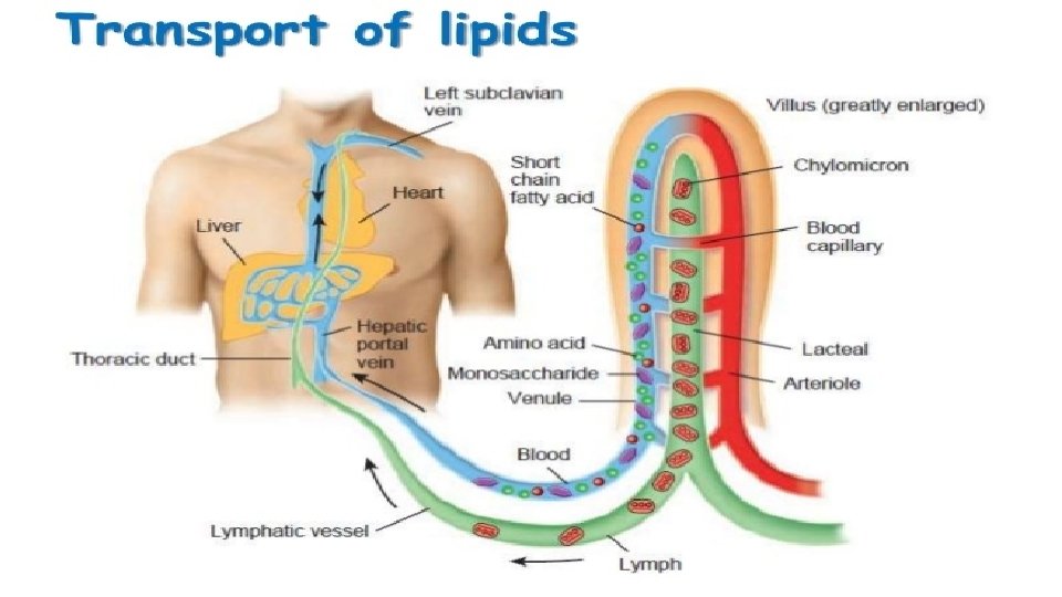
B. Functions 1. Drains interstitial fluid returns to circulatory system 2. Transports lipids, vitamins from GI tract to blood
B. Functions 1. Drains interstitial fluid returns to circulatory system 2. Transports lipids, vitamins from GI tract to blood 3. Protection - uses lymphocytes, lymph organs
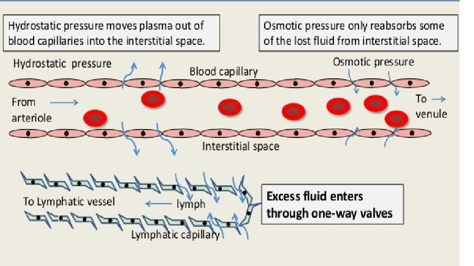
C. Lymph vs. interstitial fluid 1. Same chemical composition (like plasma - less protein), different location. 2. Lymph is in lymph vessels, interstitial is between cell
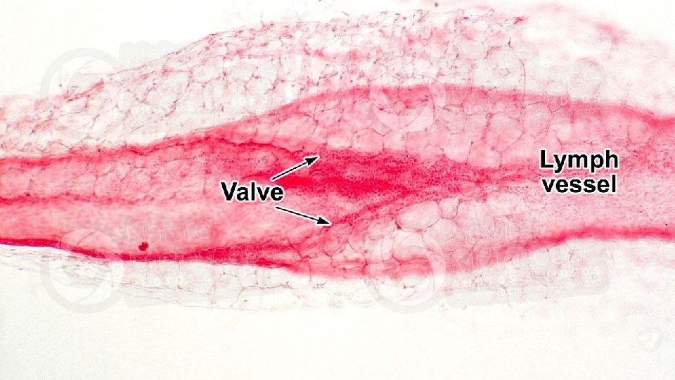
D. Lymphatic vessels 1. Lymphatic capillaries between blood capillaries 2. Interstitial fluid flows into lymphatic capillaries due to a pressure gradient and then, due to an overlapping of the capillary walls, cannot escape
D. Lymphatic vessels 1. Lymphatic capillaries between blood capillaries 2. Interstitial fluid flows into lymphatic capillaries due to a pressure gradient and then, due to an overlapping of the capillary walls, cannot escape 3. Lymphatic capillaries contain valves like veins
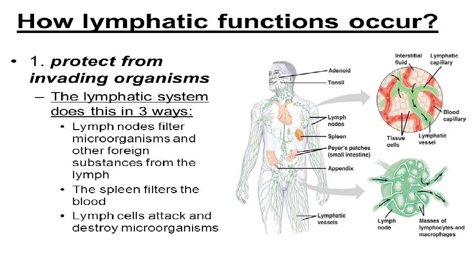
E. Lymphatic tissue 1. Lymph nodes 2. Tonsils 3. Spleen 4. Thymus
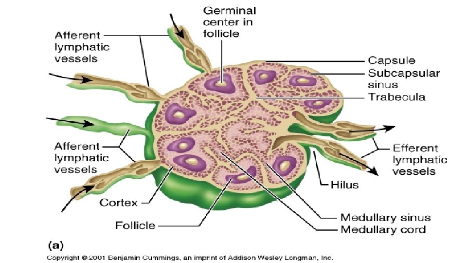
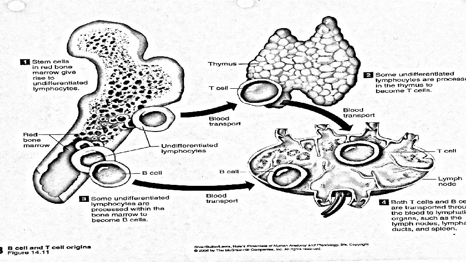
E. Lymphatic tissue 1. Lymph nodes a. Oval shape surrounded by a capsule and divided interiorly into regions (follicles) producing T/B cells and macrophages
E. Lymphatic tissue 1. Lymph nodes a. Oval shape surrounded by a capsule and divided interiorly into regions (follicles) producing T/B cells and macrophages b. Lymph nodes filter the lymph being returned to the blood. Fibers trap substances and the T/B cells or macrophages destroy them c. Lymph flows into the node through afferent vessels and leaves through efferent vessels
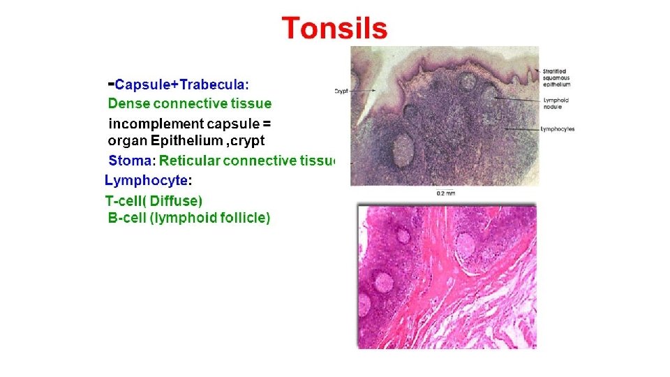
E. Lymphatic tissue 2. Tonsils a. Lymphatic nodes near pharynx b. Positioned to interpret foreign substances inhaled or swallowed. c. Contain T/B cells
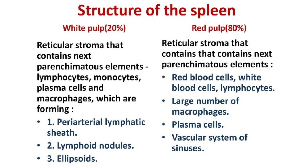
E. Lymphatic tissue 3. Spleen a. Lies between stomach and diaphragm contains plasma cells, RBC, macrophages and leukocytes
E. Lymphatic tissue 3. Spleen a. Lies between stomach and diaphragm contains plasma cells, RBC, macrophages and leukocytes b. Does not filter lymph. Site of B cell proliferation into antibody producing plasma cells
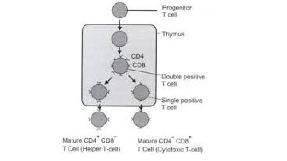
E. Lymphatic tissue 4. Thymus a. Behind sternum, between lungs contains T cells, macrophages b. Grows until puberty, then atrophies c. Produces and distributes T cells
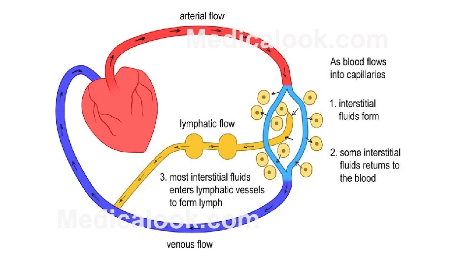
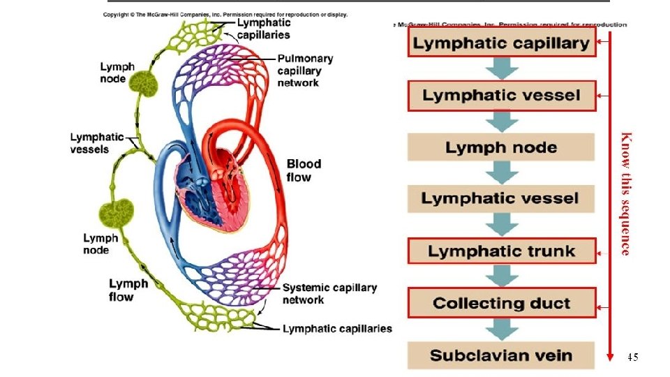
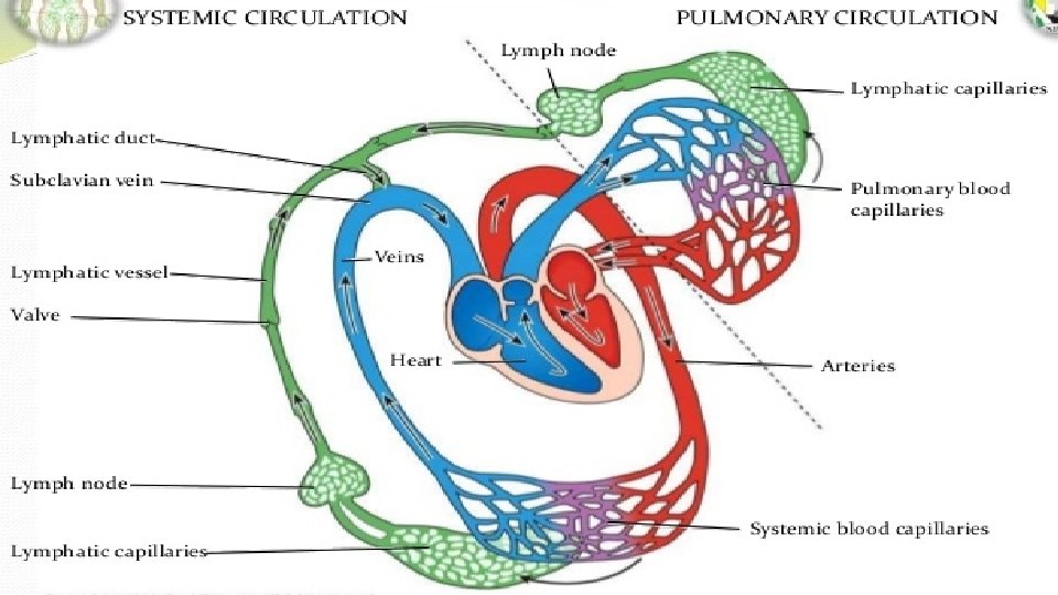
F. Lymph circulation
F. Lymph circulation 1. heart → artery → capillary → cells (interstitial fluid) → lymphatic capillaries → lymphatic vessels → lymph node → lymph trunks → thoracic duct (lymph from top left side/all lower body) → Left jugular/subclavian OR right lymphatic duct (right top side) → right jugular/subclavian → superior vena cava → heart
F. Lymph circulation 1. heart → artery → capillary → cells (interstitial fluid) → lymphatic capillaries → lymphatic vessels → lymph node → lymph trunks → thoracic duct (lymph from top left side/all lower body) → Left jugular/subclavian OR right lymphatic duct (right top side) → right jugular/subclavian → superior vena cava → heart 2. Flow is maintained by skeletal muscle movement and/or respiratory movement
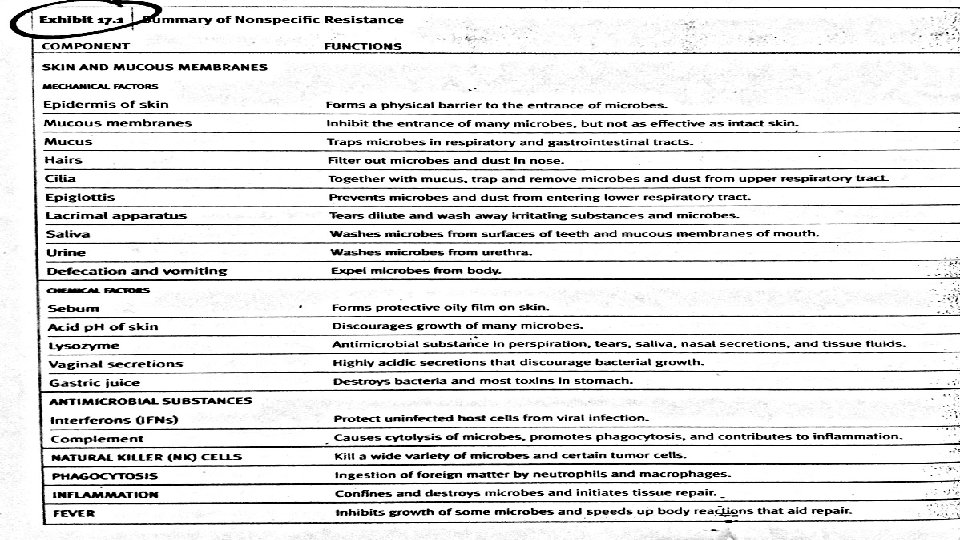
G. Nonspecific resistance to disease 1. One function of the lymphatic system is protection against pathogens (disease producers). The ability to do so is called resistance. Nonspecific resistance is a general response by the body to counteract many pathogens.
Immunity - The response of the body to destroy a specific pathogen.
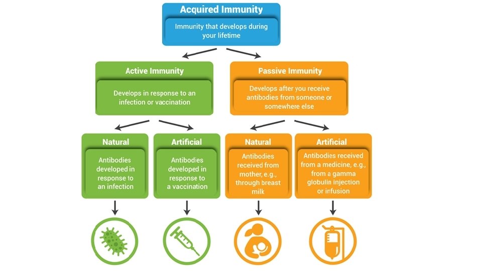
A. Acquired immunity - immunity granted to the body by prior contact with that pathogen 1. Naturally acquired active immunity person becomes sick and survives. 2. N. acquired passive immunity - immunity is passed from one person to another Ex - mother to child through placenta or breast milk. 3. A. acquired active immunity - from vaccination with the pathogen 4. Artificially acquired passive immunity - injection of antibodies
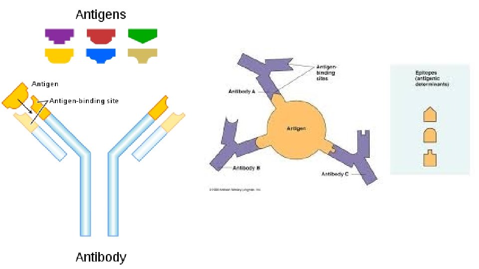
B. Antigens (Ag) - immunogens 1. This is any chemical substance that is not recognized by the body as self. It will lead to the production of specific antibodies and will react specifically with them.
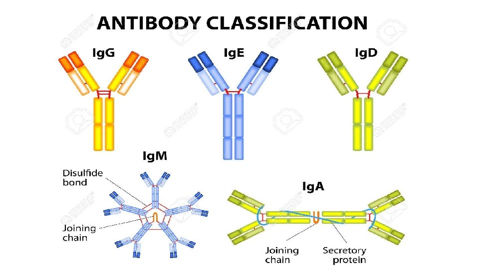
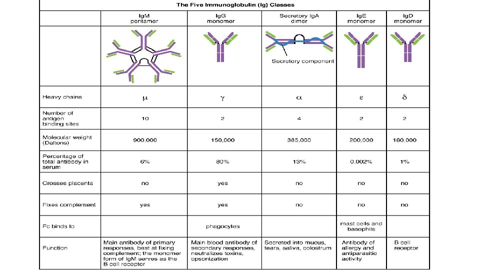
C. Antibodies (Abs) 1. These are produced by plasma cells and are specific for an antigen like a lock and key. 2. There are 5 classes of antibodies or immunoglobulins.
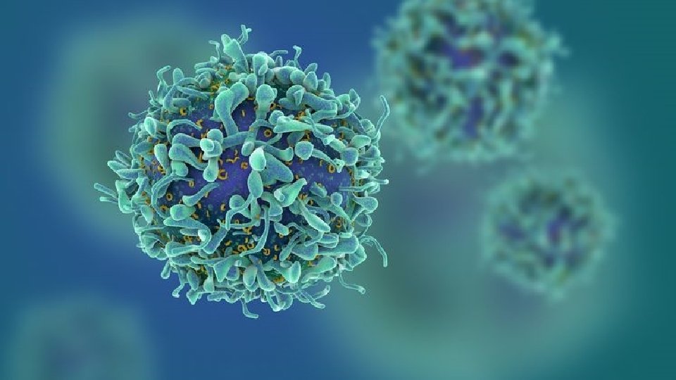
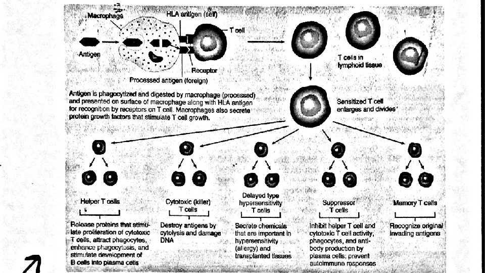
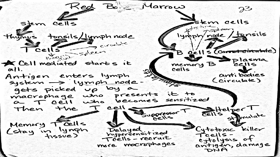
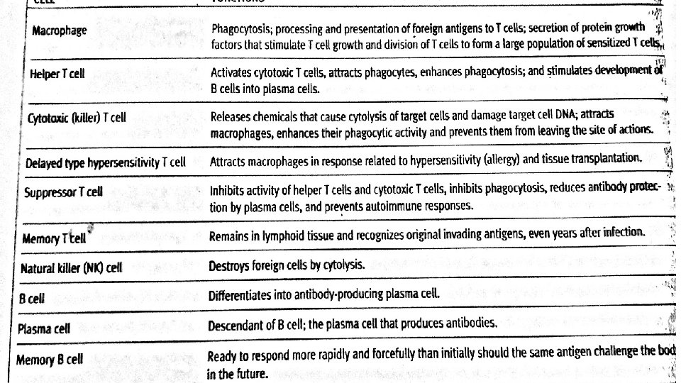
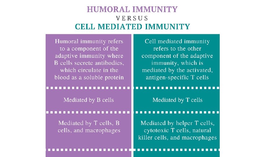
D. Methods of destroying antigens 1. Cell mediated immunity - uses T cells which directly attack and destroy. Mostly used for antigens like fungus, parasites, etc (not bacteria/viruses)
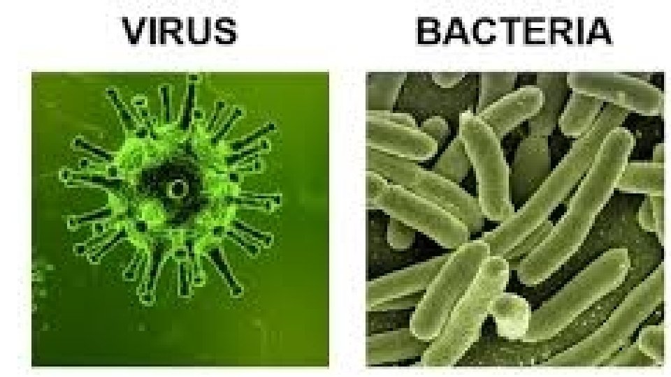
2. Antibody mediated immunity - uses B cells which produce antibodies. Used against bacteria and viruses
D. Methods of destroying antigens 1. Cell mediated immunity uses T cells which directly attack and destroy. Mostly used for antigens like fungus, parasites, etc (not bacteria/viruses) 2. Antibody mediated immunity - uses B cells which produce antibodies. Used against bacteria and viruses 3. The cells used by each are produced by lymph nodes, spleen, GI tract, red bone marrow
E. Cell mediated immunity
F. Antibody mediated immunity 1. Sensitized T cells travel to the tonsils, lymph nodes and spleen to activate the immobile B cells. The B cells produce the antibodies which are then free to circulate and kill and the memory B cells.
Antibody mediated immunity Cell mediated immunity
Antibody mediated immunity Cell mediated immunity
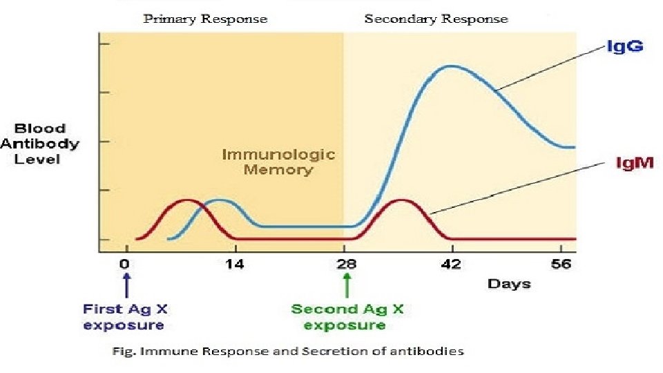
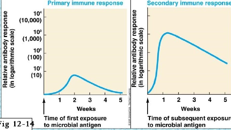
F. Antibody mediated immunity 1. Sensitized T cells travel to the tonsils, lymph nodes and spleen to activate the immobile B cells. The B cells produce the antibodies which are then free to circulate and kill and the memory B cells. 2. There is a primary response (1 st time infected - slow) and a secondary response (subsequent infections – quicker).
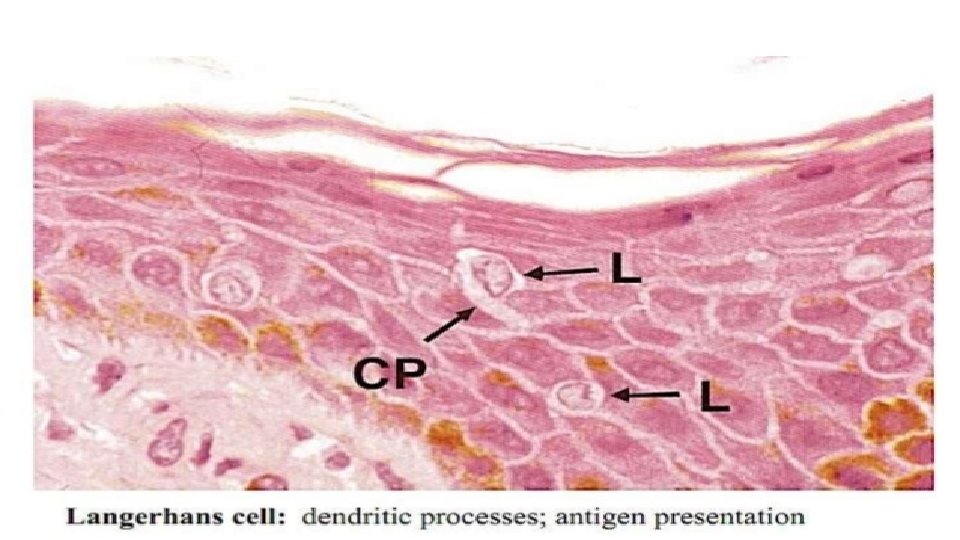
G. Skin 1. Langerhans cells in the skin will bind to the antigen and then present it to a helper T cell