Lymphatic System and Immunity Chapter 16 Functions of


 • Found throughout body except: –")


- Slides: 32
Lymphatic System and Immunity Chapter 16
Functions of Lymphatic System 1. Draining interstitial fluid 2. Transporting dietary lipids 3. Protection
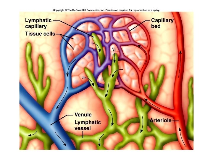
Lymphatic Vessels • Begin as closed ended lymph capillaries in tissue spaces between cells • NOT A CIRCULATING FLUID • Interstitial fluid drains into lymphatic capillaries, forming lymph. • Lymph capillaries merge to form lymphatic vessels
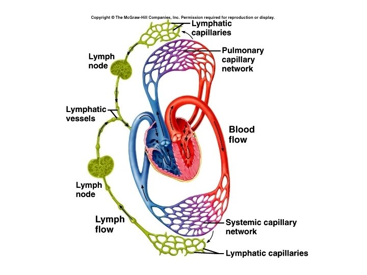
Lymphatic vessels carry lymph into and out of lymph nodes • • and finally back to the vascular system.
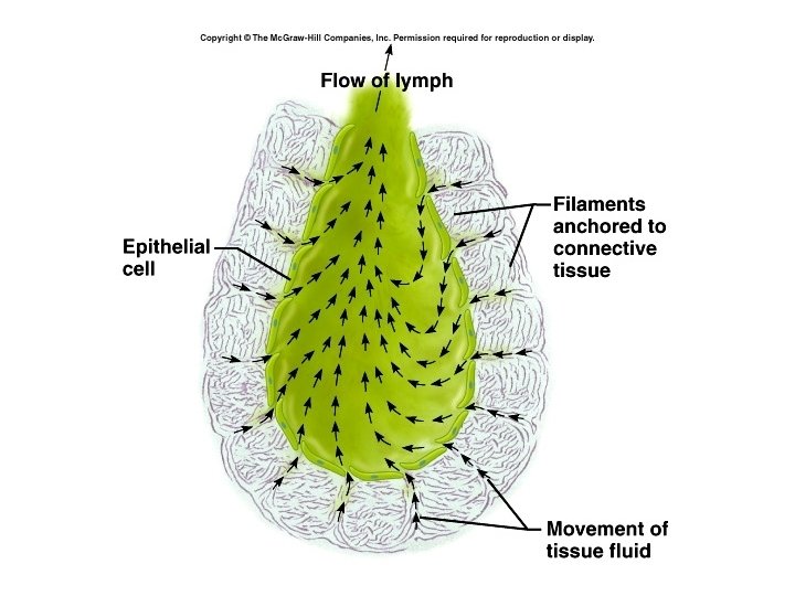
Lymphatic capillaries • Made of a single layer of squamous epithelial cells • Slightly larger than blood capillaries • Cells overlap and act as one-way valves • Opened by pressure of interstitial fluid • Anchoring filaments attach cells to surrounding tissue
Lymphatic vessels • Resemble veins (same 3 layers) • Found throughout body except: – Avascular tissues – Central nervous system – Splenic pulp – Bone marrow
Lymphatic vessels join to form lymphatic trunks. Lymphatic trunks join to form : Thoracic duct (3/4 of body) Right lymphatic duct (drains right arm, and right side of head, neck and upper torso) These empty into subclavian veins at junction with internal jugular vein.
Formation of lymph: Fluid leaves capillaries by diffusion and filtration Escaped proteins If lymph flow blocked = tissue swelling or edema Specialized lymphatic capillaries in vili of small intestine transport lipids - they are called lacteals, and the fluid is called chyle.
Lymphatic Organs • • • Red bone marrow Thymus gland Lymph nodes Lymph nodules Spleen Primary organs Secondary organs
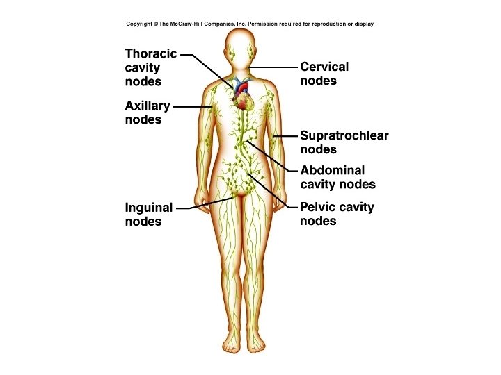
Lymph Nodes • • • Lymph is filtered through lymph nodes Found in clusters “Waste water treatment plants” Vary in size Principal groupings in cervical, axillary and inguinal regions. • Provide biological filtration • Site of cancer growth and metastasis
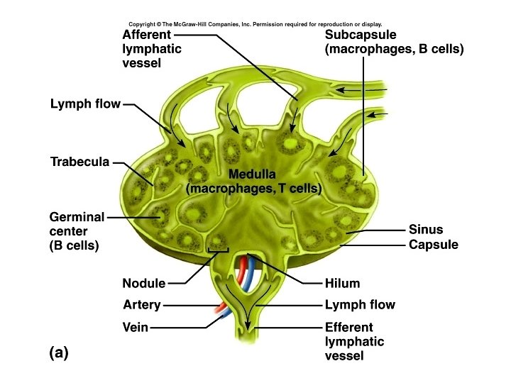
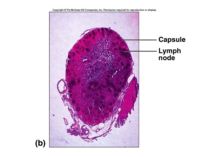
• Vessels enter node on convex side • Lymph passes through irregular channels called sinuses • Leaves node through one or two efferent vessels at the hilum or hilus • Capsule, cortex and medulla • Cortex contains lymph nodules • Follicular dendritic cells • Germinal centers – B cells proliferate
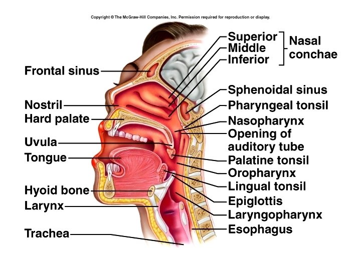
Lymph nodules are also found singly or in groups throughout the mucous membranes of the respiratory, urinary, reproductive and digestive tracts. MALT – mucosa associated lymphoid tissue Peyer’s patches in ileum Tonsils Some in appendix
Tonsils – lymphoid tissue under the mucous membranes of the throat palatine tonsils pharyngeal tonsil – adenoid lingual tonsils First line of defense Tonsillectomy
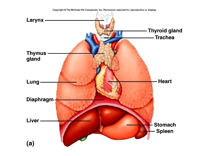
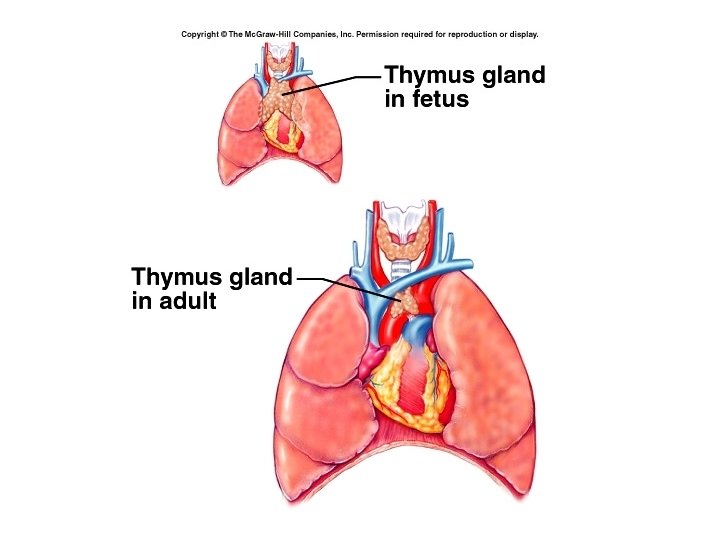
Thymus gland • in mediastinum above the heart • largest at age 10 -12 then begins to atrophy • Pre - T cells come from bone marrow and develop into T cells • T cells then go to other lymphatic tissues • Thymus produces hormone thymosin - aids maturation of T cells elsewhere in body
Spleen • • Largest lymphoid organ In upper left quadrant of abdomen Has a hilum and a capsule Sinuses contain blood instead of lymph
White pulp: little islands, mostly B cells Red pulp: Venous sinuses Splenic cords – RBCs, macrophages, lymphocytes, plasma cells and granulocytes