Lymph nodes examination Wail Alamoudi Classification Head neck

 LL (inguinal")


 3. subscapular axillary")


 2. vertical group (Along femoral vessels)")


- Slides: 24
Lymph nodes examination Wail Alamoudi
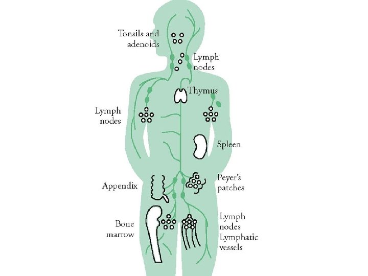
Classification • • Head & neck and clavicle UL (axillary , epitrochlear) LL (inguinal , femoral and popliteal) Abdomen ( paraaortic , liver and spleen )
Head & neck and clavicle • There approximately 300 lymph nodes in the neck
Head & neck and clavicle Inspection : Open mouth and see the 1. Tonsils 2. Adenoids (pharyngeal tonsils)
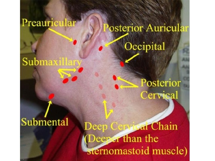
Palpation • Perauricular • Posterior auricular • Occipital • Submandibular • Submental • Tonsillar • • Anterior cervical Posterior cervical Deep cervical Supraclavicular
Examination of the Neck • Perauricular: in front of the tragus of ear • Posterior auricular: behind ear • Occipital: at base of skull
Examination of the Neck • Submandibular • Submental • Tonsillar: at angle of mandible
• Anterior cervical: superficial to the sternocleidomastoid • Posterior cervical: along anterior edge of the trapezius • Deep cervical : lies below the sternomastoid and cannot be palpated without getting underneath the muscle • Supraclavicular
• The deep cervical chain of lymph nodes – Inform the patient that this procedure will cause some discomfort. – Hook your fingers under the anterior edge of the sternomastoid muscle. – Ask the patient to bend their neck toward the side you are examining. – Move the muscle backward and palpate for the deep nodes underneath
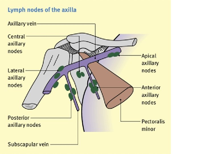
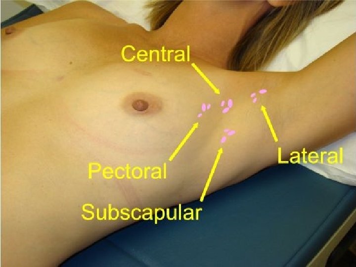
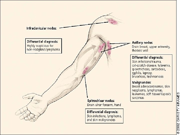
Axillary LN
1. central lymph nodes 2. pectoral axillary lymph nodes (or "anterior") 3. subscapular axillary lymph nodes (or "posterior") 4. apical lymph nodes (or "medial" or "subclavicular") 5. brachial lymph nodes (or "lateral")
Epitrochlear LN
Inguinal LN
Inguinal LN 1. horizontal group (Along inguinal ligament) 2. vertical group (Along femoral vessels)
1. Superomedial superficial inguinal 2. Superolateral superficial inguinal 3. Inferior superficial inguinal 4. Deep inguinal lymph nodes
Wail Alamoudi 0504649662
Examination of Lymph Nodes • Small, mobile, discrete, nontender nodes are common and termed shotty • Nodes are abnormal if greater than 1 cm and/or present greater than one month • Hard nodes suggest malignancy • Tender nodes suggest infection • Rubbery nodes suggest lymphoma
• in abnormal nodes, describe in terms of – location – size – delimination (discrete or matted together) – mobile or fixed – consistency (soft, hard, firm) – tenderness
• scalene lymph nodes are located deep in the neck near the cervical vertebra.