Lymph Glands lymphoglandulae After initial maturation in the
 After initial maturation in the primary immune organs, \"virgin\" B and")
 The site of T cell homing is the paracortex. Normal lymphocytes")


- Slides: 13
Lymph Glands (lymphoglandulae) After initial maturation in the primary immune organs, "virgin" B and T lymphocytes are released into the peripheral blood and home to specific sites within the lymph node (and the other secondary organs), controlled by incompletely understood homing receptors. The sites of B cell homing include: 1. The primary and secondary follicles of the lymph node cortex-the sites of antigen presentation to B cells, and subsequent proliferation and differentiation in response to same. 2. The medullary cords, where plasma cells aggregate, and release their immunoglobulins into the efferent lymph.
Lymph Glands (lymphoglandulae) The site of T cell homing is the paracortex. Normal lymphocytes recirculate, passing from blood into and through the lymph nodes, and then into efferent lymphatics, surveilling for the presence of the antigen for which they have a unique and specific receptor on their surface If this antigen is not present, the lymphocytes leave the node. Virgin lymphocytes have a finite lifespan, numbered in weeks, unless they come in contact with antigen.
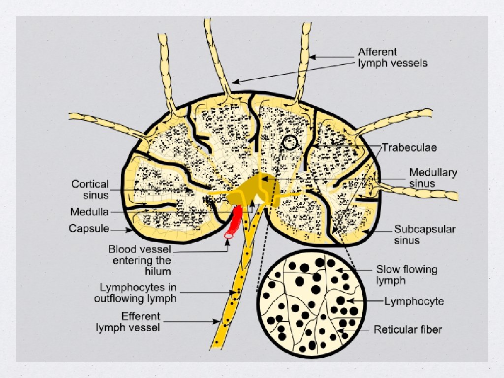
Lymphatic circulation Lymph circulates to the lymph node via afferent lymphatic vessels and drains into the node just beneath the capsule in a space called the subcapsular sinus. The subcapsular sinus drains into trabecular sinuses and finally into medullary sinuses. The sinus space is criss-crossed by the pseudopods of macrophages which act to trap foreign particles and filter the lymph. The medullary sinuses converge at the hilum and lymph then leaves the lymph node via the efferent lymphatic vessel towards either a more central lymph node or ultimately for drainage into a central venous subclavian blood vessel, most via Virchow's node and Ductus Thoracicus.
The Lymph Found only in the closed lymphatic vessels. It’s transparent, colorless, or slightly yellow, watery fluid of specific gravity about 1. 015; it closely resembles the blood plasma, but is more dilute Lymph should be distinguished from “tissue fluid” which is found outside the lymphatic vessels in the tissue spaces.
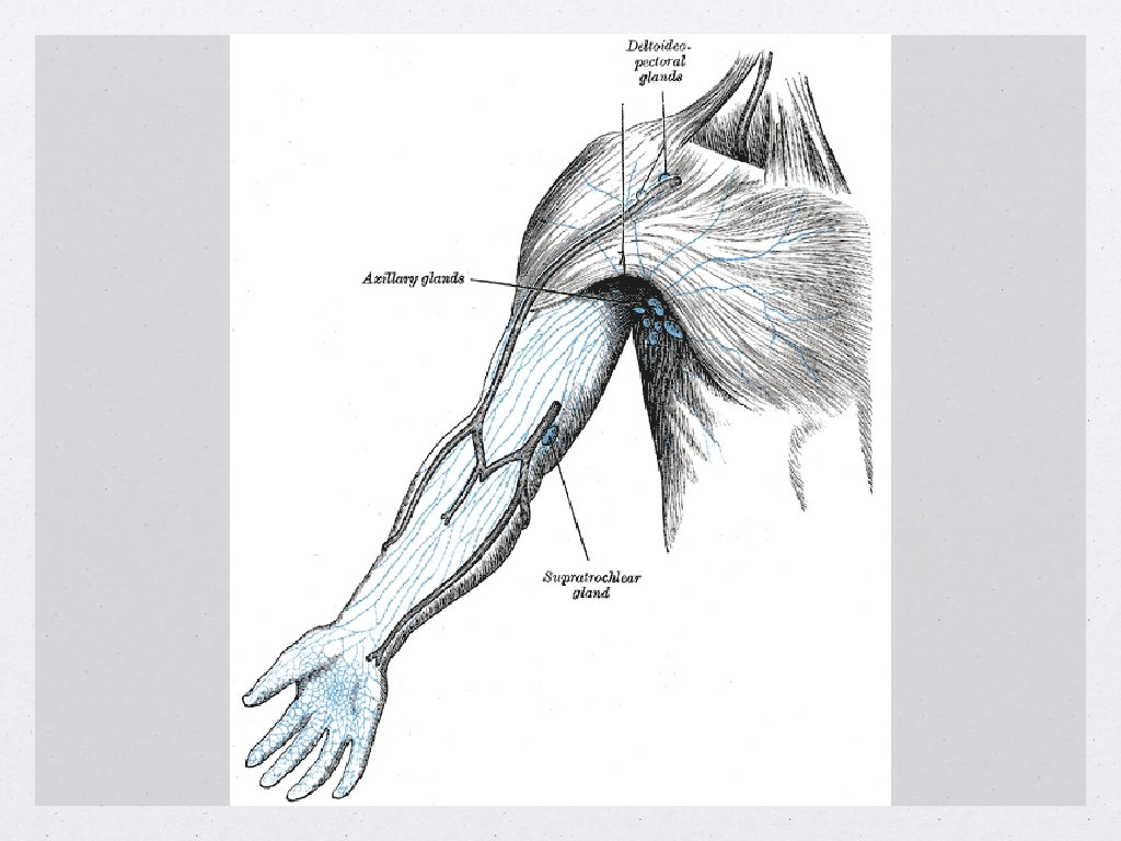
Lymph nodes of the arm These drain the whole of the arm, and are divided into two groups, superficial and deep. Deep lymph glands of the arm: These comprise the axillary glands, which are 20 -30 individual glands and can be subdivided into: ◦ ◦ ◦ • Lateral glands Anterior or pectoral glands Posterior or subscapular glands Central or intermediate glands Medial or subclavicular glands Superficial lymph glands of the arm: ◦ Supratrochlear glands: Situated above the medial epicondyle of the humerus, medial to the basilic vein, they drain the C 7 and C 8 dermatomes. ◦ Deltoideopectoral glands: Situated between the pectoralis major and deltoid muscles inferior to the clavicle.
Lymph Nodes of The Leg The superficial inguinal lymph nodes: They receive as afferents lymphatic vessels from the following: • Integument of the penis • Scrotum • Perineum • Buttock • Abdominal wall below the level of the umbilicus • Vulva • Anus (below the pectinate line) • The lower extremity (foot, leg and thigh) They are divided into three groups: • Supramedial or Superomedial • Superolateral • Inferior
1. Supromedial superficial inguinal 2. Superolateral superficial inguinal 3. Inferior superficial inguinal 4. Deep inguinal lymph nodes
Lymph Nodes of The Leg The deep inguinal lymph nodes: - The deep inguinal lymph nodes drain superiorly to the external iliac lymph nodes, then to the pelvic lymph nodes and on to the para-aortic lymph nodes.
1. Supromedial superficial inguinal 2. Superolateral superficial inguinal 3. Inferior superficial inguinal 4. Deep inguinal lymph nodes
THANKS Dr. Ryan AL. Ghanemi