Lung volumes and Capacities Functional residual capacity Helium


- Slides: 23
Lung volumes and Capacities, Functional residual capacity Helium dilution method By Prof. Dr. Samia Jawed
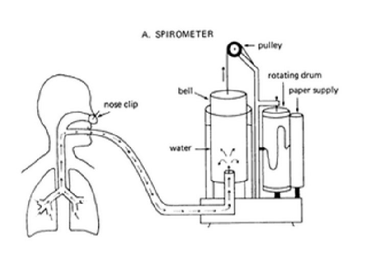
Student spirometer.
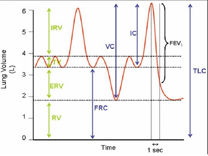
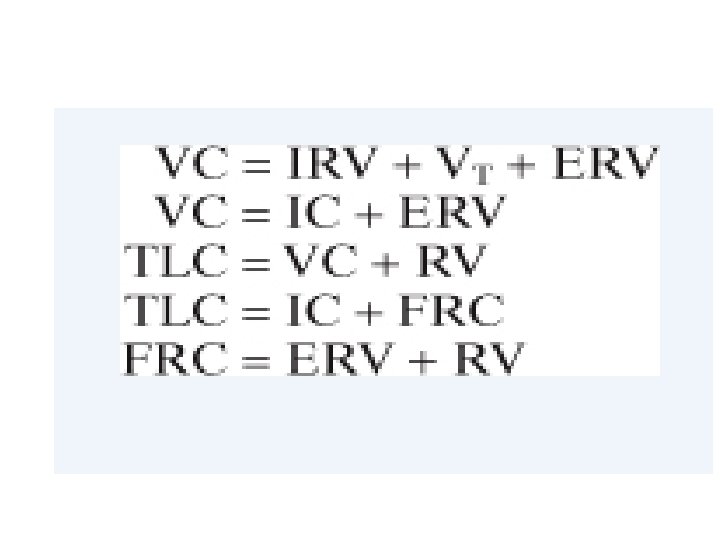
• THE GRAPHICAL RELATIONSHIP AMONG VARIOUS LUNG VOLUME AND CAPACITIES.
Spirogram
• What are the advantages of residual volume, FRC, dead space air?
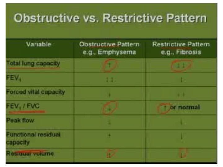
• Clinical measurements of specific volumes and capacities provide insights into lung function and origin of disease processes.
• These tests are screening and not diagnostic.
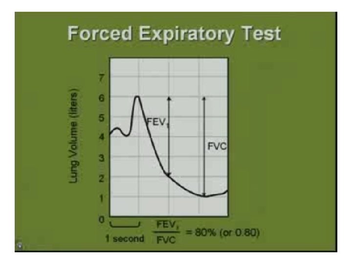
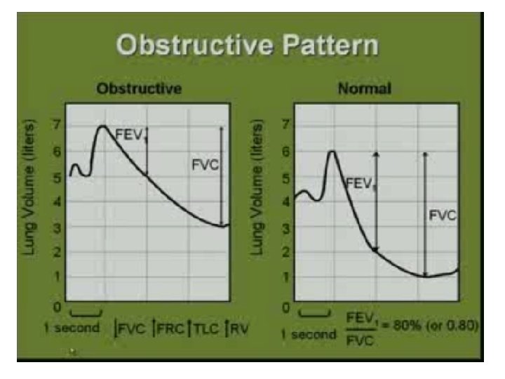
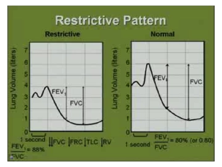
• The VC is the maximum volume of air that an individual can move in a single breath. • The most useful assessment of VC is to expire as quickly and forcefully as possible. • This way we get timed vital capacity or forced vital capacity. (FVC). • During the FVC maneuver , volume of air exhaled in the first second is FEV 1.
• From 0 -20 years vital capacity increases • From 20 -60 years it remains stable if there is no pathology • With more aging elasticity of lungs decreases and residual volume increases • Pathology affects vital capacity • Like neuromuscular disorders, lower motor neuron diseases, myasthenia gravis, kyphosis, scoliosis,
• Lung infections, collapse, pleural effusion, bronchial Asthma, emphysema, chronic Brochitis etc. • Males have more VC. Why? • VC decreases in pregnancy and while lying down.
Flow volume curves in different conditions
Helium dilution method
FRC and its measurement.
• What is the difference between hyperventilation, hyperpnoea, tachypnoea?