
Lung nodule detection in lowdose and highresolution CT
- Slides: 20
Lung nodule detection in low-dose and high-resolution CT scans Alessandra Retico INFN Pisa P. Delogu 12, M. E. Fantacci 12, I. Gori 3, A. Preite Martinez 1, A. Retico 2, A. Tata 2 1 University of Pisa 2 INFN Pisa 3 Bracco Imaging S. p. A. Milano Frontier Science 2005 – Milano, 12 -17 September A. Retico - INFN Pisa
Lung cancer • Lung cancer is the main cause of cancer death in the male population and the second one in the female population • The 5 -year survival rate is 60 -70% for early-stage cancers (stage IA). Most cancers are detected in advanced stages. The overall 5 -year survival rate is only 10 -15% No improvement has occurred in the last 20 years • It has been demonstrated that screening programs with chest X-ray do not lead to a reduction of the mortality rate • Computed Tomography (CT) has proved to be more sensitive in detecting small, early-stage cancers • The efficacy of CT-based screenings in reducing mortality rate for lung cancer has not been proved yet. Trial screening programs are being carried out in the US, Canada, Japan and Europe Frontier Science 2005 A. Retico - INFN Pisa
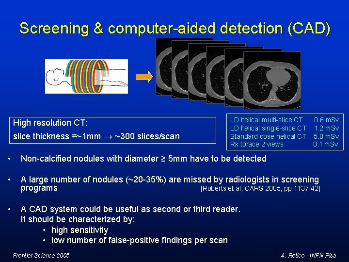
Screening & computer-aided detection (CAD) High resolution CT: slice thickness =~1 mm → ~300 slices/scan LD helical multi-slice CT 0. 6 m. Sv LD helical single-slice CT 1. 2 m. Sv Standard dose helical CT 5. 0 m. Sv Rx torace 2 views 0. 1 m. Sv • Non-calcified nodules with diameter ≥ 5 mm have to be detected • A large number of nodules (~20 -35%) are missed by radiologists in screening programs [Roberts et al, CARS 2005, pp 1137 -42] • A CAD system could be useful as second or third reader. It should be characterized by: • high sensitivity • low number of false-positive findings per scan Frontier Science 2005 A. Retico - INFN Pisa
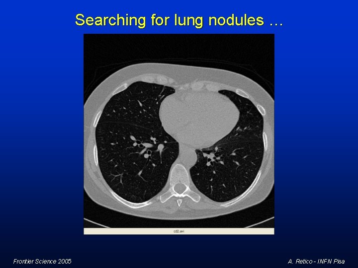
Searching for lung nodules … Frontier Science 2005 A. Retico - INFN Pisa
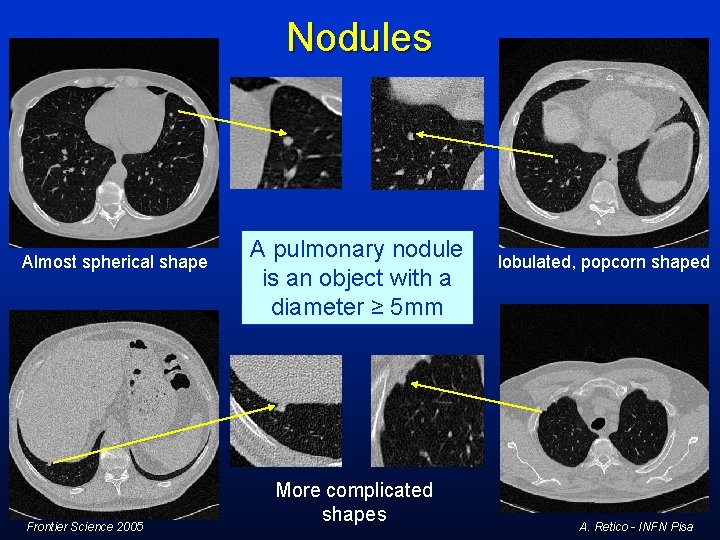
Nodules Almost spherical shape Frontier Science 2005 A pulmonary nodule is an object with a diameter ≥ 5 mm More complicated shapes lobulated, popcorn shaped A. Retico - INFN Pisa
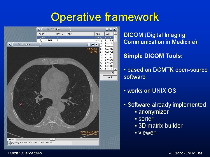
Operative framework DICOM (Digital Imaging Communication in Medicine) Simple DICOM Tools: • based on DCMTK open-source software • works on UNIX OS • Software already implemented: § anonymizer § sorter § 3 D matrix builder § viewer Frontier Science 2005 A. Retico - INFN Pisa
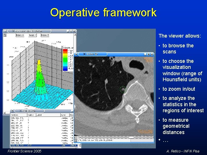
Operative framework The viewer allows: • to browse the scans • to choose the visualization window (range of Hounsfield units) • to zoom in/out • to analyze the statistics in the regions of interest • to measure geometrical distances • … Frontier Science 2005 A. Retico - INFN Pisa
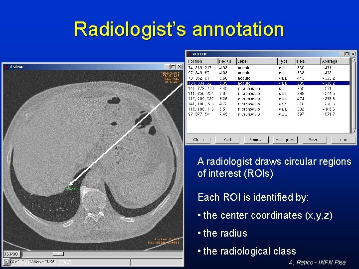
Radiologist’s annotation A radiologist draws circular regions of interest (ROIs) Each ROI is identified by: • the center coordinates (x, y, z) • the radius • the radiological class Frontier Science 2005 A. Retico - INFN Pisa
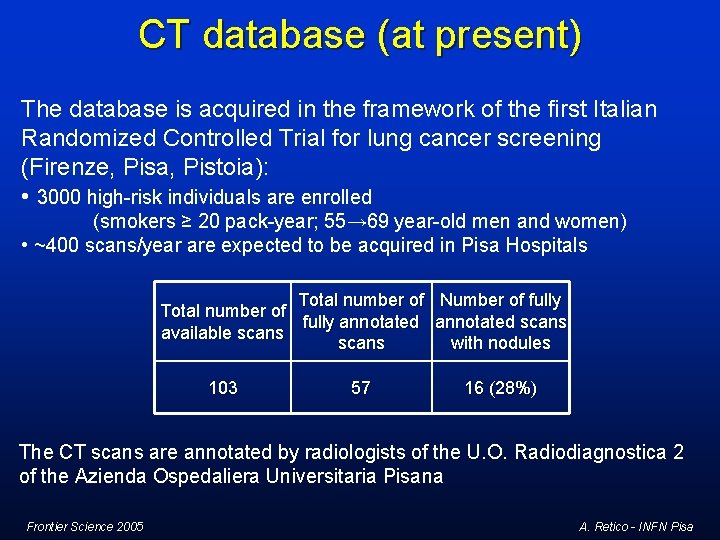
CT database (at present) The database is acquired in the framework of the first Italian Randomized Controlled Trial for lung cancer screening (Firenze, Pisa, Pistoia): • 3000 high-risk individuals are enrolled (smokers ≥ 20 pack-year; 55→ 69 year-old men and women) • ~400 scans/year are expected to be acquired in Pisa Hospitals Total number of Number of fully Total number of fully annotated scans available scans with nodules 103 57 16 (28%) The CT scans are annotated by radiologists of the U. O. Radiodiagnostica 2 of the Azienda Ospedaliera Universitaria Pisana Frontier Science 2005 A. Retico - INFN Pisa
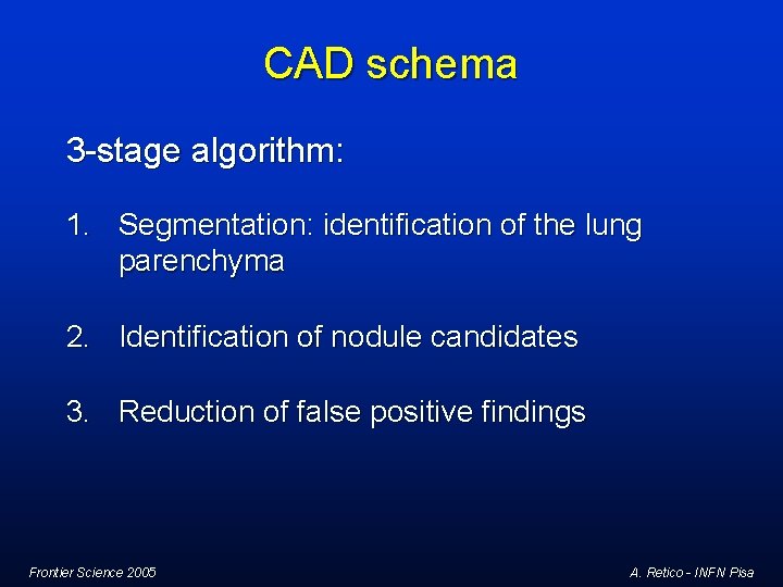
CAD schema 3 -stage algorithm: 1. Segmentation: identification of the lung parenchyma 2. Identification of nodule candidates 3. Reduction of false positive findings Frontier Science 2005 A. Retico - INFN Pisa
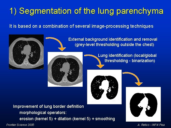
1) Segmentation of the lung parenchyma It is based on a combination of several image-processing techniques External background identification and removal (grey-level thresholding outside the chest) Lung identification (local/global thresholding - binarization) Improvement of lung border definition morphological operators: erosion (kernel 5) + dilation (kernel 5) + smoothing Frontier Science 2005 A. Retico - INFN Pisa
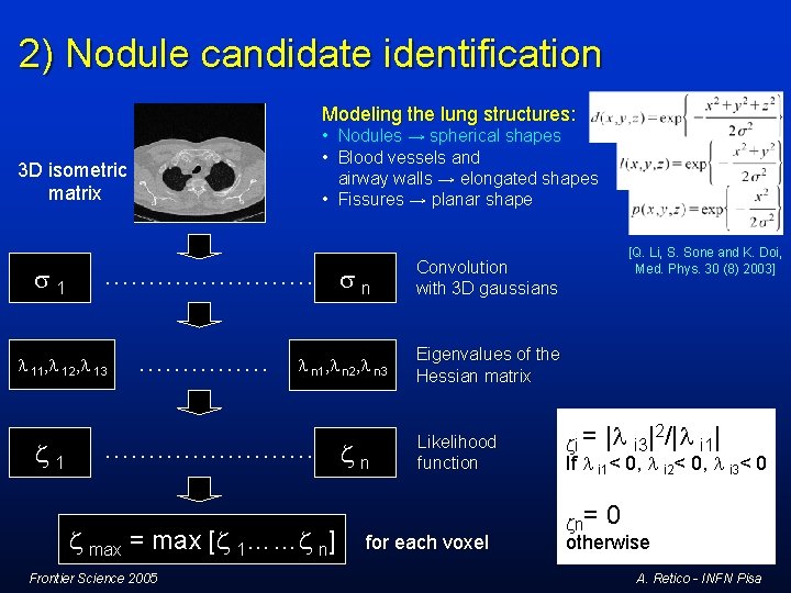
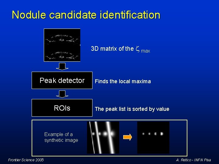
2) Nodule candidate identification Modeling the lung structures: • Nodules → spherical shapes • Blood vessels and airway walls → elongated shapes • Fissures → planar shape 3 D isometric matrix s 1 ………… s n l 11, l 12, l 13 z 1 …………… l n 1, l n 2, l n 3 ………… z max = max [z 1……z n] Frontier Science 2005 zn [Q. Li, S. Sone and K. Doi, Med. Phys. 30 (8) 2003] Convolution with 3 D gaussians Eigenvalues of the Hessian matrix Likelihood function for each voxel zi = |l i 3|2/|l i 1| zn= 0 If l i 1< 0, l i 2< 0, l i 3< 0 otherwise A. Retico - INFN Pisa
Nodule candidate identification 3 D matrix of the z Peak detector ROIs max Finds the local maxima The peak list is sorted by value Example of a synthetic image Frontier Science 2005 A. Retico - INFN Pisa
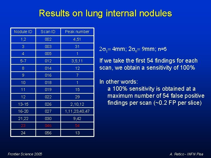
Results on lung internal nodules Nodule ID Scan ID Peak number 1, 2 002 4, 51 3 003 31 4 005 1 5 -7 012 3, 5, 11 8 014 12 9 016 7 10 018 1 11 019 15 12 022 29 13 -15 026 2, 10, 12 16 -20 027 1, 11, 23, 40, 47 21, 22 030 9, 42 23 046 54 24 056 13 Frontier Science 2005 2 s 1= 4 mm; 2 sn= 9 mm; n=5 If we take the first 54 findings for each scan, we obtain a sensitivity of 100% In other words: a 100% sensitivity is obtained at a maximum number of 54 false positive findings per scan (~0. 2 FP per slice) A. Retico - INFN Pisa
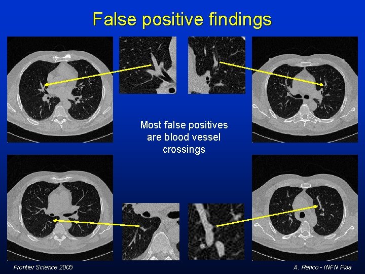
False positive findings Most false positives are blood vessel crossings Frontier Science 2005 A. Retico - INFN Pisa
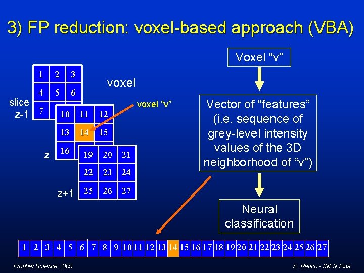
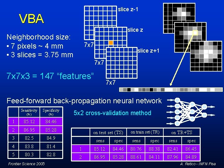
3) FP reduction: voxel-based approach (VBA) Voxel “v” 1 2 3 4 5 6 slice z-1 7 voxel 8 10 9 11 z voxel “v” 12 13 14 16 17 19 18 20 22 15 23 21 24 Vector of “features” (i. e. sequence of grey-level intensity values of the 3 D neighborhood of “v”) z+1 25 26 27 Neural classification 1 2 3 4 5 6 7 8 9 10 11 12 13 14 15 16 17 18 19 20 21 22 23 24 25 26 27 Frontier Science 2005 A. Retico - INFN Pisa
slice z-1 VBA slice z Neighborhood size: • 7 pixels ~ 4 mm • 3 slices = 3. 75 mm 7 x 7 slice z+1 7 x 7 x 3 = 147 “features” 7 x 7 Feed-forward back-propagation neural network Sensitivity Specificity (%) 1 85. 12 84. 46 2 86. 95 85. 28 3 82. 5 84. 9 4 83. 8 81. 4 5 80. 3 82. 8 Frontier Science 2005 5 x 2 cross-validation method on test set (TS) on train set (TR) on TR+TS sens spec 1 85. 12 84. 46 80. 76 88. 38 82. 41 86. 45 2 86. 95 85. 28 88. 61 84. 11 87. 96 84. 89 A. Retico - INFN Pisa
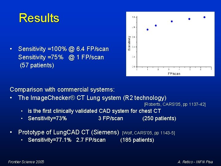
Results • Sensitivity =100% @ 6. 4 FP/scan Sensitivity =75% @ 1 FP/scan (57 patients) Comparison with commercial systems: • The Image. Checker® CT Lung system (R 2 technology) [Roberts, CARS’ 05, pp 1137 -42] • is the first clinically validated CAD system for chest CT • Sensitivity=73% 3 FP/scan (250 patients) • Prototype of Lung. CAD CT (Siemens) • Sensitivity=77. 1% 2. 7 FP/scan Frontier Science 2005 [Wolf, CARS’ 05, pp 1143 -5] (185 patients) A. Retico - INFN Pisa
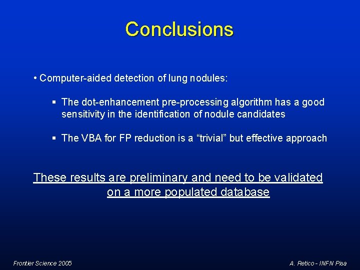
Conclusions • Computer-aided detection of lung nodules: § The dot-enhancement pre-processing algorithm has a good sensitivity in the identification of nodule candidates § The VBA for FP reduction is a “trivial” but effective approach These results are preliminary and need to be validated on a more populated database Frontier Science 2005 A. Retico - INFN Pisa
Thanks for your attention! This work has been carried out in the framework of the INFN (Istituto Nazionale di Fisica Nucleare)-funded MAGIC-5 (Medical Applications in a GRID Infrastructure Connection) project. We would like to acknowledge: Dr. L. Battolla, Dr. F. Falaschi and Dr. C. Spinelli of the “Unità Operativa Radiodiagnostica 2” of the “Azienda Ospedaliera Universitaria Pisana” and Prof. D. Caramella and Dr. T. Tarantino of the Diagnostic and Interventional Radiology Division of the “Dipartimento di Oncologia, Trapianti e Nuove Tecnologie in Medicina” of the Pisa University who have made available the full annotated CT scans. E-mail: retico@df. unipi. it Frontier Science 2005 A. Retico - INFN Pisa