Lung diseases November 22 2011 Respiration system pathophysiology

Lung diseases November 22, 2011

Respiration system pathophysiology - Disease can be divided into 3 groups: Ventilation Difussion Defect in regulation of breathing Defect of ventilation Perfusion Obstructive diseases Restrictive diseases - They can be manifested along or in combinations Defect of diffusion Defect in oxygene transport Defect of perfusion

o Lung Diseases are primarily placed into two categories n 1. Obstructive Lung Diseases Asthma p COPD p n n n Emphysema Chronic Bronchitis 2. Restrictive Lung Diseases Asbestosis p Pulmonary Fibrosis p

Obstructive lung diseases o They are characterized by airway obstruction that is worse with expiration. o Either more force (i. e. , use of accessory muscles of expiration) is required to expire a given volume of air or emptying of the lungs is slowed or both. o The unifying symptom of obstructive disease is dyspnea, the unifying sign is wheezing. o The most common obstructive diseases are asthma, chronic bronchitis and emphysema. o Because many individuals have both bronchitis and emphysema, they are often called COPD

Airway obstruction caused by emphysema, chronic bronchitis, and asthma Normal lung Emphysema Bronchitis Asthma
 Ø Asthma is a chronic inflammatory disorder of the airways")
Asthma bronchiale (GINA 2006) Ø Asthma is a chronic inflammatory disorder of the airways in which many cells and cellular elements play a role. Ø The chronic inflammation causes an associated increase in airway hyperresponsiveness that leads to recurrent episodes of wheezing, breathlessness, chest tightness, and coughing, particularly at night or in the early morning. Ø These episodes are usually associated with widespread but variable airway obstruction that is often reversible either spontaneously or with treatment.

Factors that contribute to airflow limitation in asthma
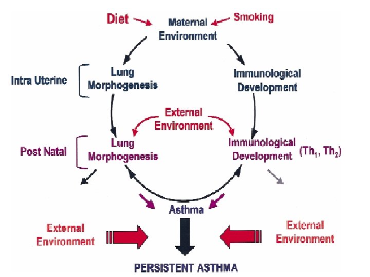

The hyper-responsive airways in asthma respond to a wide-range of provoking factors

Proportions of asthmatic children sensitised to the common allergens

Types of asthma Allergic asthma Non-allergic asthma Ig. E-mediated asthma Ig. E non-mediated asthma

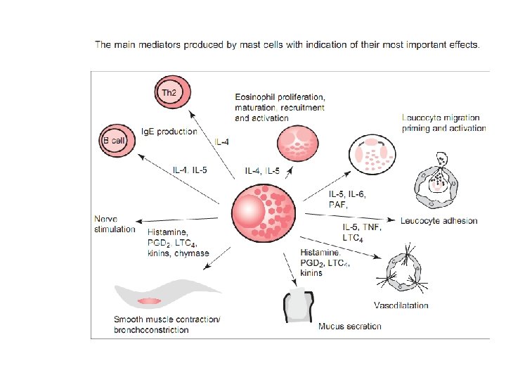
Pathogenesis of allergic asthma

Asthma response Early phase Late phase
")
Pathogenesis of ASA (non atopic asthma)

Paradigma of asthma pathogenesis

Polygenic nature of asthma

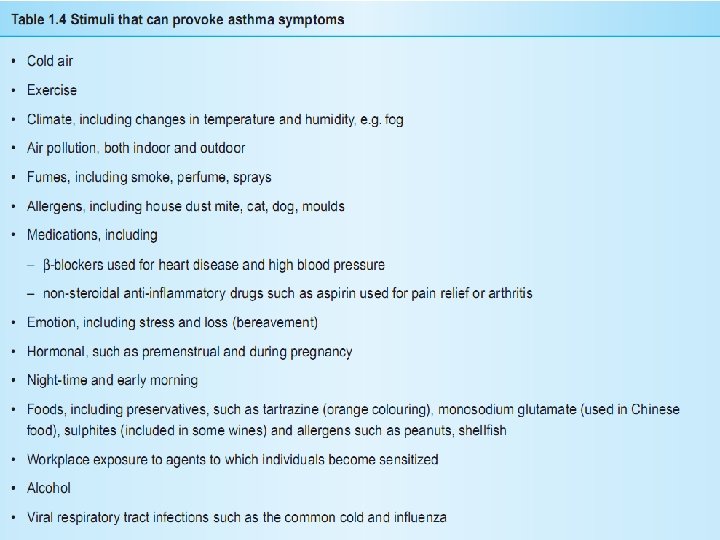
Common precipitants of asthma exacerbations • Respiratory virus infections • Allergens • Air pollution • Excercise and hyperventilation • Changes in wheather • Occupational factors • Foods, additives, drugs • Endocrine factors • Stress

Asthma – clinical manifestations q During full remision n Individuals are asymptomatic and pulmonary function tests are normal. q During partial remision There are no clinical symptoms but pulmonary function tests are abnormal q During attacks n n Individuals are dyspneic and respiratory effort is marked Breath sounds are decreased except for considerable wheezing, dyspnea, non-productive coughing, tachycardia and tachypnea occur

Asthma - pulmonary function o Spirometry shows decreases in expiratory flow rate, forced expiratory volume (FEV), and forced vital capacity (FVC) o FRC and total lung capacity (TLC) are increased. o Blood gas analysis shows hypoxemia with early respiratory alkalosis or late respiratory acidosis.

Classification of Asthma Severity: Clinical Features Before Treatment Days with symptoms Nights with symptoms PEF % of personal best peak flow Step 4 Severe persistent Continual Frequent <= 60% Step 3 Moderate Daily >= 5 times per month 3 -6 times per week 3 -4 times per month >= 80% <= 2 times per week <= 2 times per month >= 80% Step 2 Mild persistent Step 1 Mild intermittent >60% - < 80% persistent

Treatments Goals: To reverse of acute attacks § To control recurrent attacks § To reduce bronchial inflammation and the associated hyperreactivity § + elimination of allergens (if it is possible) § Drugs: Allergen´s immunotherapy § Bronchodilator (Beta agonists, Anticholinergic agents, Theophylline) § Immunosuppressant (corticosteroids) § Others (Leukotriene modifiers, antihistamine, e. g. ) §
 using allergen extracts has been administered in many")
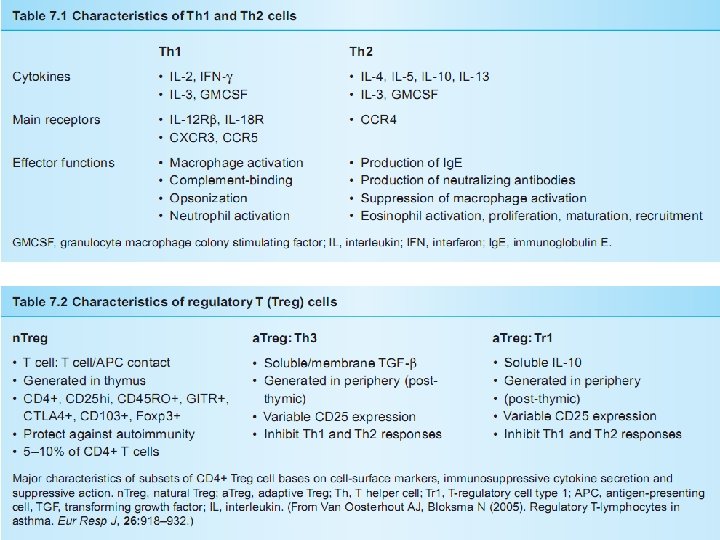
Allergen-specific immunotherapy o Specific immunotherapy (SIT) using allergen extracts has been administered in many countries for the treatment of allergic diseases. o Mechanisms of action: - Although the mechanisms of action of SIT have not been fully defined, some studies suggest that SIT may shift the immune system´s balance from Th 2 to Th 1 cells, with increased production of interleukin (IL-12) and interferon gamma (IFNgamma). SIT also increases the anti-inflammatory cytokine IL-10.
 o salmeterol,")
Bronchodilator Beta 2 agonists - selective 2 agonists o albuterol (short acting) o salmeterol, formoterol (long lasting)

Anticholinergic agents

Methylxanthine o Inhibits phosphodiesterase and therefore increase c. AMP o Reduce intracellular calcium o Cause membrane hyperpolarisation to prevent activity of smooth muscle Eg. Theophylline (similar to caffeine) o Decrease of infiltration of eosinophils into epithelium

Corticosteroids o Inhibit the attraction of inflammatory cells to the site of allergic reaction o Block leukotriene synthesis o Inhibit cytokine production and adhesion protein activation o Reverse 2 receptor down-regulation

The mechanisms of action of corticosteroids Krejsek et al. , 2004

Leukotriene mediators - Inhibitors of 5 -lipoxygenázy - Antagonists of cysteinyl LT receptors SINGULAIR® eg. SINGULAIR® (montelukast sodium): leukotriene D 4 receptor antagonist

Anti- Ig. E and others….
 COPD is defined as pathologic lung changes consistent with")
Chronic obstructive pulmonary disease (COPD) COPD is defined as pathologic lung changes consistent with emphysema or chronic bronchitis. o It is syndrome characterized by abnormal tests of expiratory airflow that do not change markedly over time, and without a reversible response to pharmacological agents. o 5 -20% of adult population o Most frequently in men o The fifth leading cause of death o

The complex, heterogenous overlapping of the three primary diagnoses include under diseases of air flow limitation is present on the next picture:

1. Chronic bronchitis is defined as hypersecretion of mucus and chronic productive cough that continues for at least 3 months of years for at least 2 consecutive years. Incidence is increased in smokers (up to twentyfold) and even more so in workers exposed to air pollution. It is a major health problem for the elderly population. Repeated infections are common.

Chronic bronchitis - etiology o It is primarily caused by cigarette smoke, both active and passive smoking have been implicated o Other risk factors: - profesional exposition - air pollution - repeated infections of airways - genetics

Chronic bronchitis - morphology o Inspired irritants not only increase mucus production but also increase the size and number of mucous glands and goblet cells in airway epithelium o The mucus produced is thicker and more tenacious than normal. This sticky mucus coating makes it much more likely that bacteria, such as H. influenze and S. pneumoniae, will become embedded in the airway secretions, there they reproduce rapidly. o Ciliary function is impaired, reducing mucus clearance further. The lung´s defense mechanisms are tehrefore compromised, increasing susceptibility to pulmonary infection and injury. o The bronchial walls become inflamed and thickened from edema and accumulation of inflammatory cells.

Initially chronic bronchitis affects only the larger bronchi, but eventually all airways are involved. o The thick mucus and hypertrophied bronchial smooth muscle obstruct the airways and lead to closure, particularly during expiration, when the airways are narrowed. o The airways collapse early in expiration, trapping gas in the distal portions of the lung. o Obstruction eventually leads to ventilation-perfusion mismatch, hypoventilation (increased Pa. CO 2) and hypoxemia. o
")
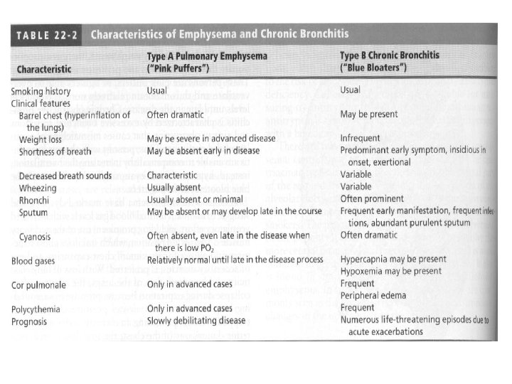
Chronic bronchitis – clinical manifestations o Individuals usually have a productive cough („smoker´s cough“) and evidence of airway obstruction is shown by spirometry o Bronchitis patients are often described as „blue bloaters“ due to their tendency to exhibit both hypoxemia/hypercapnia and right heart failure with peripheral edema in spite of only moderate obstructive changes on pulmonary functional tests. o Acute episodes (e. g. after infection) result in marked hypoxemia that leads to polycytemia and cyanosis (blueness) associated with an increase in pulmonary artery pressure, impairing right ventricular function, and significant jugular venous distension and ankle edema (bloated)

Chronic bronchitis – evaluation and treatment o Diagnosis is made on the basis of physical examination, chest radiograph, pulmonary function tests and blood gas analyses. o The best „treatment“ is prevention, because pathological changes are not reversible. o If the individuals stops smoking, disease progression can be halted o Therapy: - bronchodilators - expectorans - chest physical therapy - steroids - antibiotics

Chronic bronchitis: low-flow oxygen therapy o It is administered with care to individuals with severe hypoxemia o o o and CO 2 retention Because of the chronic elevation of Pa. CO 2, the central chemoreceptors no longer act as the primary stimulus for breathing. This role is taken over by the peripheral chemoreceptors, which are sensitive to changes in Pa. O 2. Peripheral chemoreceptors do not stimulate breathing if the Pa. O 2 is much more than 60 mm. Hg. Therefore, if oxygen therapy causes Pa. O 2 to exceed 60 mm. Hg, the stimulus to breathe is lost, Pa. CO 2 increases, and apnea results. If inadequate oxygenation cannot be achieved without resulting in respiratory depression, the individual must be mechanically ventilated)
 accompanied by")
2. Emphysema n It is abnormal permanent enlargement of gasexchange airways (acini) accompanied by destruction of alveolar walls and without obvious fibrosis. n In emphysema, obstruction results from changes in lung tissues, rather than mucus production and inflammation, as in chronic bronchitis. n The major mechanism of airflow limitation is loss of elastic recoil.

Healthy lung Emphysema lung

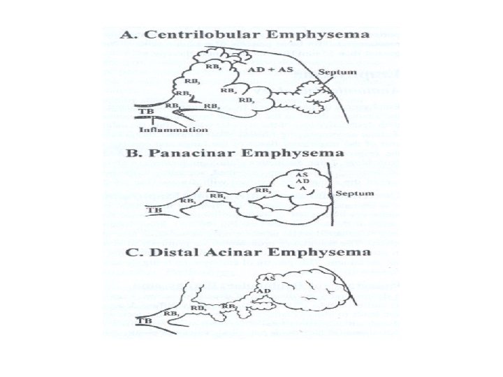
Types of emphysema o Three distinctive types of alveolar destruction have been described, according to the portion of the acinus first involved with disease: 1) Centrilobular (centriacinar): - septal destruction occurs in the respiratory bronchioles and alveolar ducts, usually in the upper lobes of the lung. The alveolar sac (alveoli distal to the respiratory bronchiole) remains intact. It tends to occur in smokers with chronic bronchitis. 2) Panacinar (panlobular): - It involves the entire acinus with damage more randomly distributed and involving the lower lobes of the lung. It tends to occur in patients with 1 -antitrypsin deficiency. 3) Distal acinar (subpleural): - It is typically seen in a young adult with a history of a spontaneous pneumothorax.

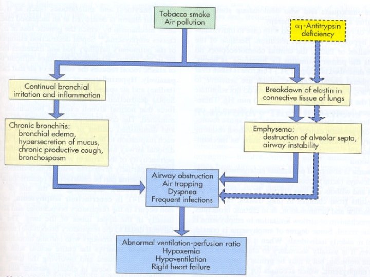
Types of emphysema o Primary emphysema: - it is commonly linked to an inherited deficiency of the enzyme 1 -antitrypsin that is a major component of 1 globulin, a plasma protein. - Normally it inhibits the action of many proteolytic enzymes. - Individuals with deficiency of this enzyme (AR) have an increased likelihood of developing emphysema because proteolysis in lung tissues is not inhibited. o Secondary emphysema: - It is also caused by an inability of the body to inhibit proteolytic enzymes in the lung. It results from an insult to the lungs from inhaled toxins, such as cigarette smoke and air pollution.

Pathophysiology of emphysema o Emphysema begins with destruction of alveolar septa o It is postulated that inhaled oxidants, such as those in cigarette smoke and air pollution, tip the normal balance of elastases (proteolytic enzymes) and antielastases (such as 1 -antitrypsin) such that elastin is destroyed at an increased rate o Expiration becomes difficult because loss of elastic recoil reduces the volume of air that can be expired passively. o Hyperinflation of alveoli causes large air spaces (bullae) and air spaces adjacent to pleura (blebs) to develop. o The combination of increased RV in the alveoli and diminished caliber of the bronchioles causes part of each inspiration to be trapped in the acinus.

Mechanisms of air trapping in emphysema o Damaged or destroyed alveolar walls no longer support and hold open the airways, and alveoli lose their property of passive elastic recoil. o Both of the se factors contribute to collapse during expiration.

Emphysema – clinical manifestations o Patients with emphysema are able to maintain a higher alveolar minute ventilation than those with chronic bronchitis. Thus they tend to have a higher Pa. O 2 and lower Pa. CO 2 and have classically been referred to as „pink puffers“ o Physical examination often reveals a thin, tachypneic patient using accessory muscles and pursed lips to facilitate respiration. The thorax is barrel-shaped due to hyperinflation. o There is little cough and very little sputum production (in „pure“ emphysema)

Emphysema – evaluation o Pulmonary function tests: - indicate obstruction to gas flow during expiration - airway collapse and air trapping lead to a decrease in FVC and FEV 1 and an increase in FRC, RV, and TLC. - diffusing capacity is decreased because destruction of the alveolocapillary membrane o Arterial blood gas measurements are usually normal until latge in the disease

Emphysema – approach to therapy o Smoking cessation is the most important o o o intervention Inhaled anticholinergic agets 2 -adrenergic agonists Steroids Low-flow oxygen therapy in selected individuals Lung transplant can be considered
 o It is AR inherited disorder that results from defective epithelial")
Cystic fibrosis (mucoviscidosis) o It is AR inherited disorder that results from defective epithelial ion transport o On simplistic level, CF is associated with abnormal secretions Cl- that may cause obstructive problems within the respiratory, digestive and reproductive tracts. o The CF gene has been localized on chromosome 7 its mutation result in the abnormal expression of the protein cystic fibrosis transmembrane regulator (CFTR) = chloride channel present on the surface of many cells (airways, bile ducts, pancreas, sweat ducts, vas deferens)

Pathogenesis of cystic fibrosis lung diseases

Cystic fibrosis – clinical manifestations o The most common manifestations are respiratory and gastrointestinal. o Respiratory symptoms include: persistent cough or wheeze and recurrent or severe pneumonia Physical signs include barrel chest and digital clubbing. o Gastrointestinal manifestations include: meconium ileus at birth, failure to thrive, and malabsorptive symptoms, such as frequent loose and oily stools o Male with CF are typically infertile (98%) o May be liver disease or diabetes mellitus

Cystic fibrosis – evaluation and treatment o The standard method of diagnosis is the sweat test, which will reveal sweat chloride concentration in excess of 60 m. Eql/L. o Genotyping for CFTR mutation (above 800 variations) o Treatment: - chest physical therapy - bronchodilators - antibiotics - pancreatic enzymes, vitamins

Restrictive lung diseases The lung volumes are reduced either because of: 1. Alteration in lung parenchyma. 2. Diseases of the pleura, chest wall or neuromuscular apparatus. Physiologically restrictive lung diseases are defined by reduced total lung capacity, vital capacity and functional residual capacity, but with preserved air flow.

Restrictive lung diseases may be divided into the following groups: o Intrinsic lung diseases (diseases of the lung parenchyma) o Extrinsic disorders (extra-parenchymal diseases)

Intrinsic Lung Diseases These diseases cause either: o Inflammation and/or scarring of lung tissue (interstitial lung disease) or o Fill the air spaces with exudate and debris (pneumonitis). o These diseases are classified further according to the etiological factor.

Extrinsic Disorders The chest wall, pleura and respiratory muscles are the components of respiratory pump. Disorders of these structures will cause lung restriction and impair ventilatory function. These are grouped as: o Non-muscular diseases of the chest wall. o Neuromuscular disorders.

Interstitial lung diseases o There a large number of diseases that affect the interstitium of the lung it is connective tissue present between the alveolar epithelium and capillary endothelium o Some of these diseases have known etiology, e. g. occupational diseases o Others are diseases of unknown etiology - most frequent of these are idiopatic pulmonary fibrosis (diffuse interstitial fibrosis), pulmonary fibrosis associated with collagen-vascular diseases, and sarcoidosis.
 o Wegener’s granulomatosis")
Granulomatous Lung Diseases o Infections o Sarcoidosis o Hypersensitivity pneumonitis (EAA) o Wegener’s granulomatosis (WG) o Reaction to tumours o Foreign body o Pneumoconiosis (Berrylium, o o o o o Aluminium, Cobalt) Drug reactions Drug abusers Necrotising sarcoidal granulomatosis (NSG) Eosinophilic pneumonia Bronchocentric granulomatosis (BCG) Churg Strauss syndrome Lymphoid interstitial pneumonia (LIP) Sjogren’s disease Amyloidosis Incidental
 collection of mature mononuclear phagocytes (macrophages and/or")
What is a granuloma? “a compact (organised) collection of mature mononuclear phagocytes (macrophages and/or epithelioid cells) which may or may not be accompanied by accessory features such as necrosis or infiltration of inflammatory leucocytes” Adams, 1983 Key features. A granuloma is: Discrete Avascular Comprises epithelioid histiocytes EJ Mark, 2004

Granulomas and granulomatous inflammation: synonymous or different? o Granuloma is well defined (sarcoidal or tuberculoid type) o Granulomatous inflammation n Diffuse process, ill-defined n Palisading histiocytes in zones

The granulomas o Necrotising or non-necrotising? o Is the necrosis n Caseous n Abscess-like n Degeneration / fibrinoid necrosis o ‘Distinct and compact’ or ‘Soft and diffuse’?

Necrotising granulomas Caseous necrosis Abscess-like necrosis Fungi TB Histoplasmosis Coccidioidomycosis Pneumocystis Candida Aspergillus Phycomycosis Blastomycosis Crypococcosis Bacteria Nocardia, Actinomyces Viruses
 o Is there associated interstitial")
Non-necrotising granulomas (mostly in the context of diffuse disease) o Is there associated interstitial pneumonitis? o Nature of the granulomas? o Distribution of disease?

Non-necrotising granulomas: Interstitial inflammation ABSENT If there are o Tight well formed granulomas o Evidence of multisystem disease o ‘Lymphatic’ distribution Consider Sarcoidosis Berylliosis Aluminium If not o Random distribution? Airways? Vessels? o Try viewing under polarised light o History of inhalation or injection? o Food, dust, haemosiderin, amyloid,

Non-necrotising granulomas: Interstitial inflammation PRESENT If there are o Inflammation and granulomas centriacinar o Granulomas often ‘soft’ o Foamy macrophages, cholesterol clefts, COP-like features Consider Hypersensitivity Pneumonitis (EAA) If not o Random distribution? o Check history o Other pathological features Drug reaction Aspiration pneumonia Foreign material? Eosinophilic pneumonia

Diffuse Interstitial Pulmonary Fibrosis o Synonyms: idiopathic pulmonary fibrosis, interstitial pneumonia, cryptogenic fibrosing alveolitis. o o o Pathology Thickening of interstitium. Initially, infiltration with lymphocytes and plasma cells. Later fibroblasts lay down thick collagen bundles. These changes occur irregularly within the lung. Eventually alveolar architecture is destroyed – honeycomb lung

Etiology Unknown, may be immunological reaction. o o o Clinical Features Uncommon disease, affects adults in late middle age. Progressive exertional dyspnea, later at rest. Non-productive cough. Physical examination shows finger clubbing, fine inspiratory crackles throughout both lungs. Patient may develop respiratory failure terminally. The disease progresses insidiously, median survival 4 -6 years.

Pulmonary Function o Spirometry reveals a restrictive pattern. FVC is reduced, but FEV 1/FVC supernormal. o All lung volumes – TLC, FRC, RV – are reduced. o Pressure volume curve of the lung is displaced downward and flattened. o Arterial Pa. O 2 and Pa. CO 2 are reduced, p. H normal. o On exercise Pa. O 2 decreases dramatically. o Physiologic dead space and physiologic shunt and VQ mismatch are increased. o Diffuse impairment contributes to hypoxemia on exercise. o There is marked reduction in diffusing capacity due to thickening of blood gas barrier and VQ mismatch.

Sarcoidosis o A disease characterized by the presence of granulomatous tissue. o This is a systemic disease which involves eyes, brain, heart, lungs, bones and kidneys, skin, liver and spleen. o On pathology a non-caseating granuloma composed of histiocytes, giant cells and lymphocytes. o In advanced lung disease fibrotic changes are seen.

Etiology o Unknown, likely immunological basis . Clinical Fetures Four stages are identified: o Stage 0: No obvious intrathoracic involvement o Stage 1: Bilateral hilar lymphadenopathy, often accompanied by arthritis, uveitis and erythema nodosum. o Stage 2: Pulmonary parenchyma is also involved, changes in mid and upper zones. o Stage 3: Pulmonary infiltrates and fibrosis without adenopathy.

Pulmonary Function o No impairment occurs in stages 0 and 1. o In stages 2 and 3 restrictive changes are seen. Treatment and Prognosis o 85% of these patients improve spontaneously, but 15% may develop progressive fibrosis and respiratory failure. o Treatment is other observation, but in symptomatic patients or deteriorating PFT’s – treatment recommended. o Prednisone 0. 5 - 1 mg/kg initially, then tapered and continued for 6 months to 1 year.
 o Stopping the occupational")
Therapy o It depends on etiology (if it is known) o Stopping the occupational exposure o Antibiotics o Diseases of unknown etiology (sarcoidosis, idiop. pulmonary fibrosis) corticosteroids o Oxygen therapy
 in the lung o The normal lung")
Pulmonary edema It is excess water (fluid) in the lung o The normal lung contains very little water or fluid. It is kept dry by lymphatic drainage and a balance among capillary hydrostatic pressure, capillary oncotic pressure, and capillary permeability o In addition, surfactant lining the alveoli repels water, keeping fluid from entering the alveoli.

Pulmonary edema - pathogenesis
 High pressure (hydrostatic, cardiogenic) edema - It is associated")
Classification of pulmonary edema 1) High pressure (hydrostatic, cardiogenic) edema - It is associated with elevated capillary hydrostatic pressure 2) Low pressure (high permeability, noncardiogenic) edema - It refer to conditions in which hydraulic filtration coefficint is elevated and osmotic reflection coefficient is reduced interstitial edema x alveolar edema

Effects of pulmonary edema o Pulmonary vascular pressure and volume In cardiogenic edema the increase in left atrial pressure is reflected passively in a retrograde direction to the pulmonary veins, capillaries, and arteries. This increase in pulmonary vascular pressure produces an increase in pulmonary blood volume. In permeability edema the passive increase in vascular volume is absent but the fundamental process of lung injury releases substances which may produce pulmonary vasoconstriction leadint to increased pulmonary artery pressure despite normal left atrial pressure.

o Pulmonary blood flow redistribution Cardiogenic edema is associated with a redistribution of blood flow in the lungs such that the lung bases, which normally receive the highest blood flow, experience a decrease in blood flow while the apices, which normally receive hte least amount of flow, experience an increase in blood flow. Perfusion redistribution becomes relevant in gas exchange. Perfusion of the pulmonary capillaries in an edema-filled alveolus has the effect of a right-to-left shunt since venous blood which is not exposed to alveolar air is admixed with oxygenated blood from nonedematous alveoli. Vasodilator therapy for congestive heart failure, while improving cardiac function, usually increases the severity of hypoxemia by reversing pulmonary blood flow redistribution.

o Lung compliance Interstitial edema produces a reduction in lung compliance which increases the elastic work the muscles must do to achieve a given tidal volume. Furthermore, even small amounts of edema fluid interfere with surfactant function, leading to increased surface tension, alveolar instability, and alveolar collapse. In cardiogenic edema the increase in pulmonary blood volume causes a further increase in lung stiffness.
 There are several factors increasing airway resistance: 1) 2) 3)")
o Airway resistance (AR) There are several factors increasing airway resistance: 1) 2) 3) 4) A reduction in lung volume produces an increase in airway resistance Edema in the bronchovascular sheath produces compression of small airways Fluid in the airways combined with edema of the bronchial mucosa narrows the lumen and increase AR. Reflex bronchospasm which occurs in some patients with congestive heart failure – „cardiac asthma“

o Oxygenation Alveolar edema produces a right-to-left shunt, which has the same effect on arterial PO 2 as an anatomic shunt. o Acid-base balance - mild forms of pulmonary edema stimulate interstitial „J“ receptors in the lung, leading to hyperventilation and respiratory alkalosis. More severe forms increasing the work of breathing lead to relative hypoventilation and respiratory acidosis. In cardiogenic edema while the metabolism of the respiratory muscles is increased, cardiac dysfunction leads to decreased blood flow, resulting in reduced tissue PO 2, anaerobic metabolism, and metabolic acidosis. -

Therapy The treatment is based on pathophysiologic consequences and on pathogenic mechanisms: o Oxygen and respiratory support o Acid-base balance o Reduce pulmonary capillary pressure (increase plasma oncotic pressure)
- Slides: 90