
LUMINESCENCE SPECTROSCOPY OF WIDEGAP SOLIDS Eduard Aleksanyan Post


- Slides: 37
LUMINESCENCE SPECTROSCOPY OF WIDE-GAP SOLIDS Eduard Aleksanyan Post. Doc at the Institute of Physics, University of Tartu 29. 08. 2013, Yerevan
Education • • 1990 – 1997 Secondary School #20, Yerevan 1997 – 2000 Anania Shirakaci Lycey, Yerevan 2000 – 2004 Yerevan State University, Department of Physics Bachelor of Science 2004 – 2006 Yerevan State University, Department of Physics, Master’s School 2006 – 2009 Yerevan Physics Institute, Department of Solid State Physics, Ph. D student 2007 – 2008 Institute of Physics, University of Tartu, Department of ionic crystals, Tartu, Estonia, Ph. D student 2009 – 2011 Yerevan Physics Institute, Department of Solid State Physics, Researcher 2011 - 2014 Institute of Physics, University of Tartu, Department of ionic crystals, Tartu, Estonia, Post. Doc
OUTLINE Ø Luminescence Spectroscopy Ø Synchrotron Radiation o o o SUPERLUMI BW 3 MAX-III Ø Intrinsic (e-h pairs or excitons) and extrinsic (impurity ions and defects) luminescence Ø Scintillators, phosphors, LEDs Ø Conclusion
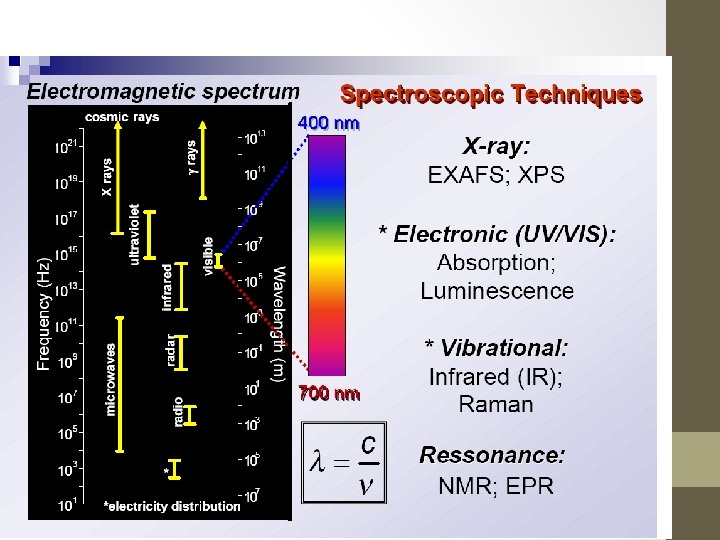
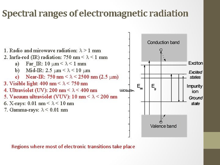
Spectral ranges of electromagnetic radiation 1. Radio and mirowave radiation: > 1 mm 2. Inrfa-red (IR) radiation: 750 nm < < 1 mm a) Far_IR: 10 m < < 1 mm b) Mid-IR: 2. 5 m < < 10 m c) Near-IR: 750 nm < < 2500 nm (2. 5 m) 3. Visible light: 400 nm < < 750 nm 4. Ultraviolet (UV): 200 nm < < 400 nm 5. Vacuum ultraviolet (VUV): 10 nm < < 200 nm 6. X-rays: 0. 01 nm < < 10 nm 7. Gamma-rays: < 0. 01 nm Regions where most of electronic transitions take place
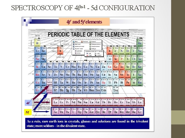
SPECTROSCOPY OF 4 fn-1 - 5 d CONFIGURATION
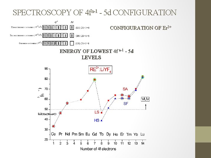
SPECTROSCOPY OF 4 fn-1 - 5 d CONFIGURATION OF Er 3+ ENERGY OF LOWEST 4 f n-1 - 5 d LEVELS
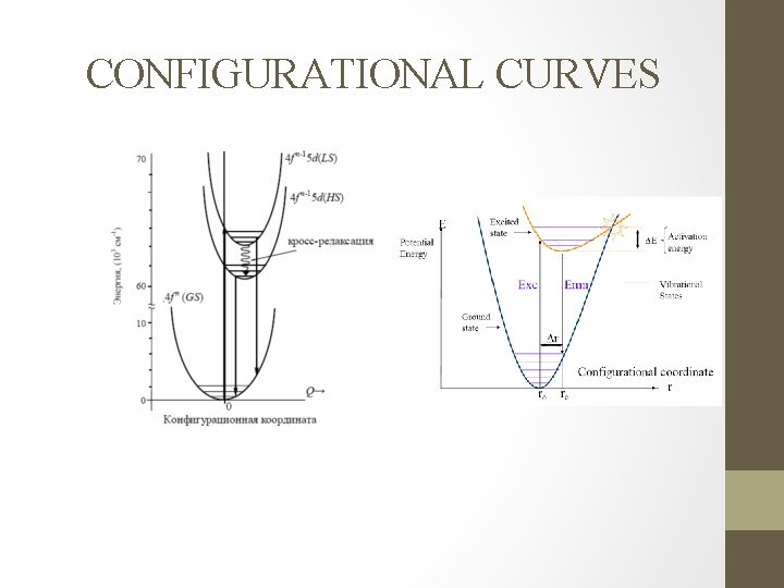
CONFIGURATIONAL CURVES
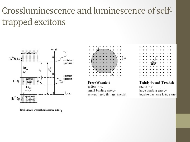
Crossluminescence and luminescence of selftrapped excitons
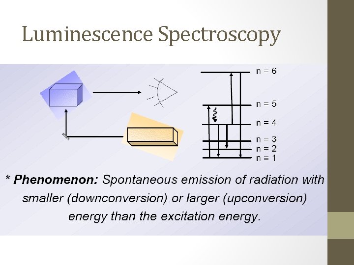
Luminescence Spectroscopy
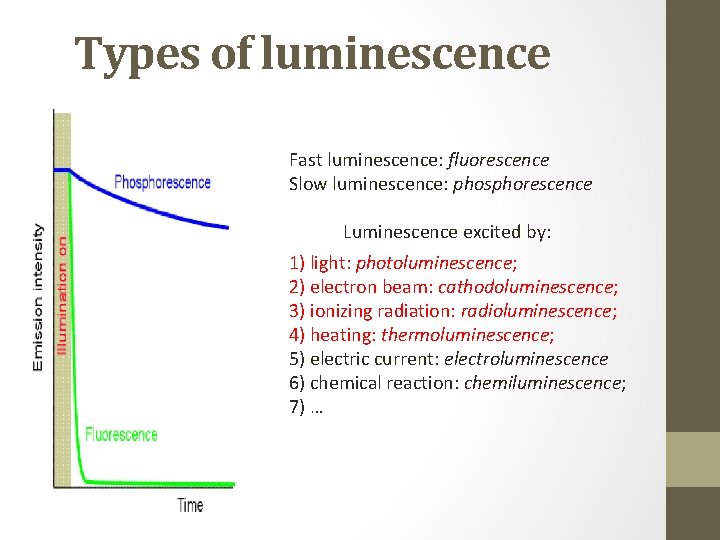
Types of luminescence Fast luminescence: fluorescence Slow luminescence: phosphorescence Luminescence excited by: 1) light: photoluminescence; 2) electron beam: cathodoluminescence; 3) ionizing radiation: radioluminescence; 4) heating: thermoluminescence; 5) electric current: electroluminescence 6) chemical reaction: chemiluminescence; 7) …
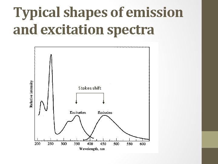
Typical shapes of emission and excitation spectra Stokes shift
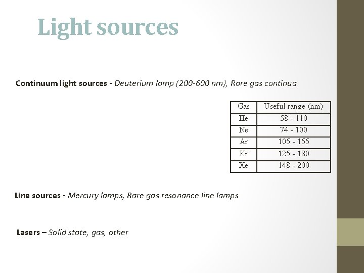
Light sources Continuum light sources - Deuterium lamp (200 -600 nm), Rare gas continua Gas He Ne Ar Kr Xe Line sources - Mercury lamps, Rare gas resonance line lamps Lasers – Solid state, gas, other Useful range (nm) 58 - 110 74 - 100 105 - 155 125 - 180 148 - 200
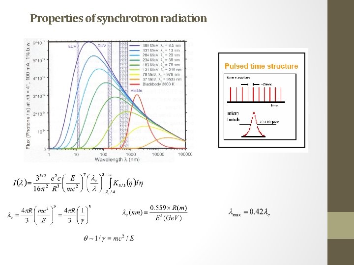
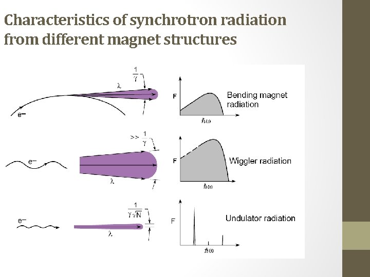
Synchrotron radiation Characteristics of Synchrotron Radiation: • Synchrotron radiation is highly collimated (within angle ~1/ = mec 2/E) • High brightness: synchrotron radiation is extremely intense (hundreds of thousands of times higher than conventional X-ray tubes). • Wide energy spectrum: synchrotron radiation is emitted within a wide range of energies, allowing a beam of any energy to be produced. • Synchrotron radiation is highly polarized. • It is emitted in very short pulses, typically less than a nano-second. • It’s spectral characteristics are calculable. Synchrotron radiation – a perfect tool for material science
Properties of synchrotron radiation
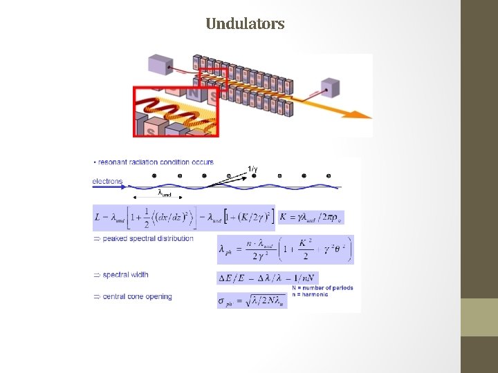
Undulators
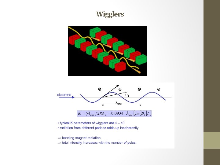
Wigglers
Characteristics of synchrotron radiation from different magnet structures
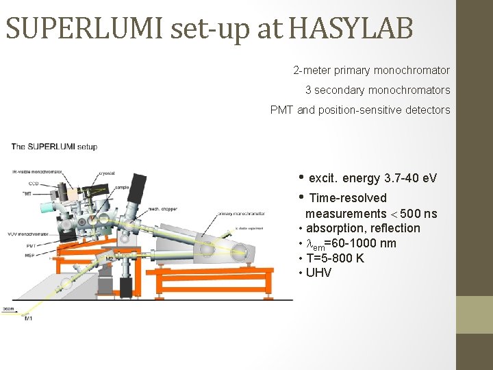
SUPERLUMI set-up at HASYLAB 2 -meter primary monochromator 3 secondary monochromators PMT and position-sensitive detectors • excit. energy 3. 7 -40 e. V • Time-resolved measurements 500 ns • absorption, reflection • em=60 -1000 nm • T=5 -800 K • UHV
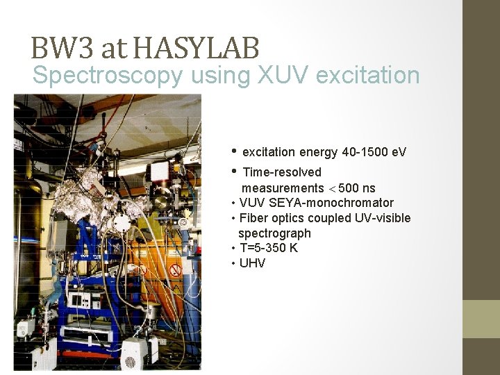
BW 3 at HASYLAB Spectroscopy using XUV excitation • excitation energy 40 -1500 e. V • Time-resolved measurements 500 ns • VUV SEYA-monochromator • Fiber optics coupled UV-visible spectrograph • T=5 -350 K • UHV
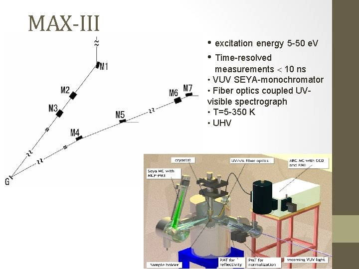
MAX-III • excitation energy 5 -50 e. V • Time-resolved measurements 10 ns • VUV SEYA-monochromator • Fiber optics coupled UVvisible spectrograph • T=5 -350 K • UHV
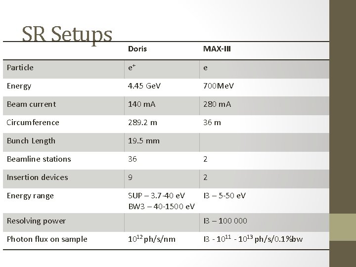
SR Setups Doris MAX-III Particle e+ e Energy 4. 45 Ge. V 700 Me. V Beam current 140 m. A 280 m. A Circumference 289. 2 m 36 m Bunch Length 19. 5 mm Beamline stations 36 2 Insertion devices 9 2 Energy range SUP – 3. 7 -40 e. V I 3 – 5 -50 e. V BW 3 – 40 -1500 e. V Resolving power Photon flux on sample I 3 – 100 000 1012 ph/s/nm I 3 - 1011 - 1013 ph/s/0. 1%bw
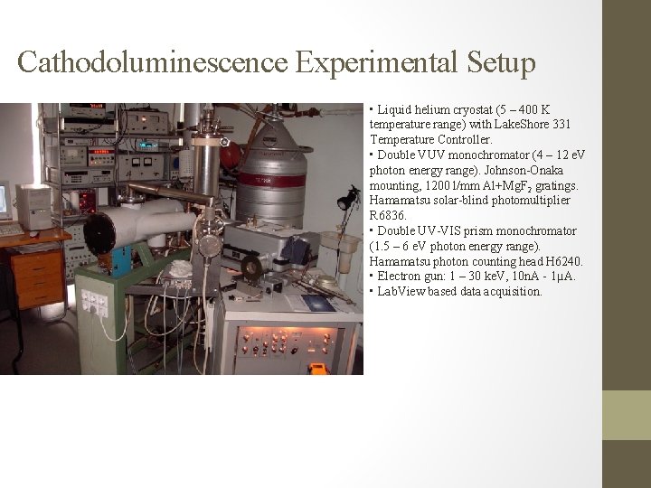
Cathodoluminescence Experimental Setup • Liquid helium cryostat (5 – 400 K temperature range) with Lake. Shore 331 Temperature Controller. • Double VUV monochromator (4 – 12 e. V photon energy range). Johnson-Onaka mounting, 1200 l/mm Al+Mg. F 2 gratings. Hamamatsu solar-blind photomultiplier R 6836. • Double UV-VIS prism monochromator (1. 5 – 6 e. V photon energy range). Hamamatsu photon counting head H 6240. • Electron gun: 1 – 30 ke. V, 10 n. A - 1µA. • Lab. View based data acquisition.
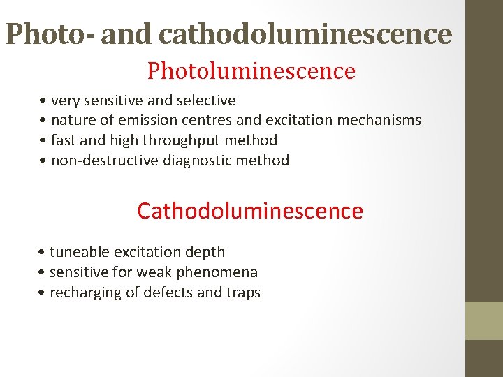
Photo- and cathodoluminescence Photoluminescence • very sensitive and selective • nature of emission centres and excitation mechanisms • fast and high throughput method • non-destructive diagnostic method Cathodoluminescence • tuneable excitation depth • sensitive for weak phenomena • recharging of defects and traps
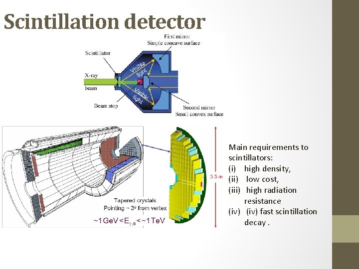
Scintillation detector Main requirements to scintillators: (i) high density, (ii) low cost, (iii) high radiation resistance (iv) fast scintillation decay.
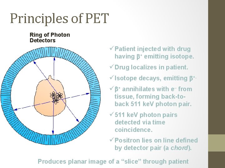
Principles of PET Ring of Photon Detectors ü Patient injected with drug having + emitting isotope. ü Drug localizes in patient. ü Isotope decays, emitting +. ü + annihilates with e– from tissue, forming back-toback 511 ke. V photon pair. ü 511 ke. V photon pairs detected via time coincidence. ü Positron lies on line defined by detector pair (a chord). Produces planar image of a “slice” through patient
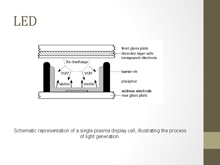
LED Schematic representation of a single plasma display cell, illustrating the process of light generation.
Quantum splitting (quantum cutting) schemes We need phosphor with Q > 100%
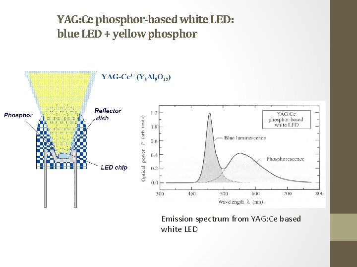
YAG: Ce phosphor-based white LED: blue LED + yellow phosphor Emission spectrum from YAG: Ce based white LED
Dosimeters Mechanism of thermoluminescence
MOBILITAS Postdoctoral Research Grant DEVELOPMENT OF NOVEL SCINTILLATORS BASED ON THIN NANOCRYSTALLINE FILMS SUPERVISOR – Marco Kirm 23. 09. 2011 – 22. 09. 2014
Objects of research • Pure and RE doped Hf. O 2 and Zr. O 2 1. Defects and traps in pure hafnia and zirconia 2. Intrinsic excitations 3. RE (Ce 3+, Pr 3+, Gd 3+, Eu 3+ etc. ) in hafnia and zirconia. 4. Methods – ALD, PLD, sol-gel chemistry • High-k material – Hf. O 2 - 16 -50, Zi. O 2 -18 -38, Si. O 2 – 3. 9 • High density - Hf. O 2 – 11. 78 g/cm 3, Zi. O 2 - 6. 21 g/cm 3 • Possible control of crystallographic phases • Data from localized volume
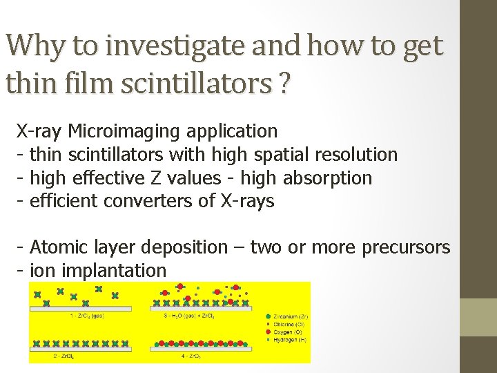
Why to investigate and how to get thin film scintillators ? X-ray Microimaging application - thin scintillators with high spatial resolution - high effective Z values - high absorption - efficient converters of X-rays - Atomic layer deposition – two or more precursors - ion implantation
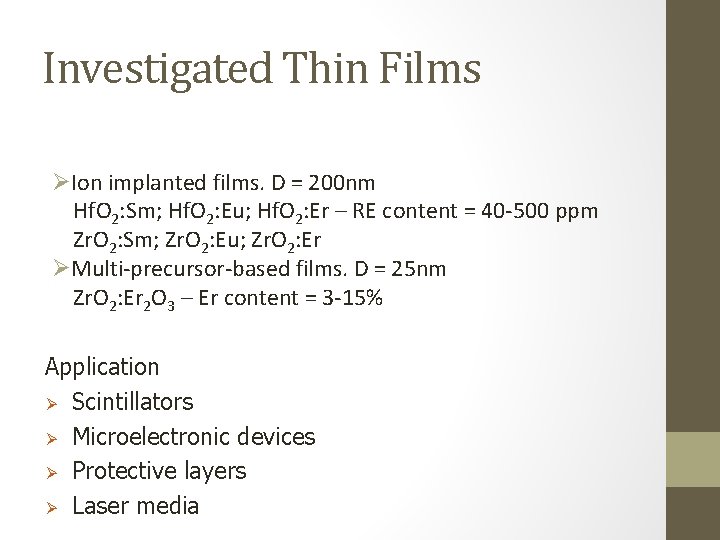
Investigated Thin Films ØIon implanted films. D = 200 nm Hf. O 2: Sm; Hf. O 2: Eu; Hf. O 2: Er – RE content = 40 -500 ppm Zr. O 2: Sm; Zr. O 2: Eu; Zr. O 2: Er ØMulti-precursor-based films. D = 25 nm Zr. O 2: Er 2 O 3 – Er content = 3 -15% Application Ø Scintillators Ø Microelectronic devices Ø Protective layers Ø Laser media
Conferences and Publications • • • • • Harutunyan, V. V. ; Saakyan, A. A. ; Aleksanyan, E. M. ; Grigoryan, R. P. ; Hakobyan, G. R. Creation of Aluminium Nanolayer on a Corundum Monocrystals Surface by High-Energy Electron Irradiation. International Conference “New technologies for development of heterosemiconductors for device applications. 2006, 65. Lixin Ning, Peter A. Tanner, Vachagan V. Harutunyan, Eduard Aleksanyan, Vladimir N. Makhov, Marco Kirm. Luminescence and excitation spectra of YAG: Nd 3+ excited by synchrotron radiation. Journal of Luminescence, Volume 127, Issue 2, 2007, 397 - 403. Aleksanyan Eduard, Harutunyan Vachagan, Kink Margarita, Kink Rein, Kirm Marco, Maksimov Yuri, Makhov Vladimir, Ouvarova Tatiana. Upconverted VUV 5 d - 4 f luminescence of Er 3+ doped into Li. YF 4 and Ba. Y 2 F 8 crystals under Ar. F-laser excitation. The 15 th International Conference on Luminescence and Optical Spectroscopy of Condensed Matter. Lyon, 7 -11 July, 2008, 525. Aleksanyan Eduard, Harutunyan Vachagan, Kostanyan Radik, Feldbach Eduard, Kirm Marco, Liblik Peeter, Makhov Vladimir, Vielhauer Sebastian. Luminescence and excitation spectra due to inter- and intraconfigurational transitions of Er 3+ in YAG: Er. The 15 th International Conference on Luminescence and Optical Spectroscopy of Condensed Matter. Lyon, 7 -11 July, 2008, 336. Aleksanyan Eduard, Harutunyan Vachagan, Kostanyan Radik, Feldbach Eduard, Kirm Marco, Liblik Peeter, Makhov Vladimir, Vielhauer Sebastian. 5 d – 4 f luminescence of Er 3+ in YAG: Er 3+. International Baltic Sea Region conference “Functional materials and nanotechnologies 2008”, April 1 -4, Riga, 2008, 38. V. N. Makhov, A. Lushchik, Ch. B. Lushchik, M. Kirm, E. Vasil’chenko, S. Vielhauer, V. V. Harutunyan, E. Aleksanyan. Luminescence and radiation defects in electron-irradiated Al 2 O 3 and Al 2 O 3: Cr. Nuclear Instruments & Methods in Physics Research B, Volume 266, Issues 12 -13, 2008, 2949 - 2952. Алексанян, Э. М. ВУФ люминесценция ионов Er 3+ в кристаллах Li. YF 4 и Ba. Y 2 F 8. Известия НАН Армении. Физика // Journal of Contemporary Physics, 44(2), 2009, 113 - 118. E. M. Aleksanyan. VUV Luminescence of Er 3+ Ions in Li. YF 4 and Ba. Y 2 F 8 Crystals. Journal of Contemporary Physics (Armenian Academy of Sciences), 2009, Vol. 44, No. 2, pp. 75– 79. Eduard Aleksanyan, Vachagan Harutunyan, Radik Kostanyan, Eduard Feldbach, Marco Kirm, Peeter Liblik, Vladimir N. Makhov, Sebastian Vielhauer. 5 d– 4 f luminescence of Er 3+ in YAG: Er 3+. Optical Materials, Volume 31, Issue 6, 2009, 1038 - 1041. Eduard Aleksanyan, Vachagan Harutunyan, Margarita Kink, Rein Kink, Marco Kirma, Yuri Maksimov, Vladimir N. Makhov, Tatiana V. Ouvarova. Upconverted 5 d– 4 f luminescence from Er 3+ and Nd 3+ ions doped into fluoride hosts excited by Ar. F and Kr. F excimer lasers. Optics Communications, Volue 283, Issue 1, 2010, 49 - 53. E. Aleksanyan, M. Kirm, E. Feldbach, K. Kukli, S. Lange, I. Sildos, A. Tamm. Luminescence Properties of RE 3+ Doped Hafnia and Zirconia Thin Films Grown by Atomic Layer Deposition Method. International Conference “Functional materials and nanotechnologies 2012”, April 17 -20, Riga, 2012, 50. E. Aleksanyan, M. Kirm, S. Vielhauer, V. Harutyunyan. Investigation of Luminescence Processes in YAG Single Crystals Irradiated by 50 Me. V Electron Beam. Lumdetr 2012 - 8 th International Conference on Luminescent Detectors and Transformers of Ionizing Radiation. September 10 -14, 2012, Halle (Saale), Germany, P-Tue-56. Eduard Aleksanyan, Marco Kirm, Sebastian Vielhauer, Vachagan Harutyunyan. Investigation of Luminescence Processes in YAG Single Crystals Irradiated by 50 Me. V Electron Beam. Radiation Measurements 2013, In Press, Accepted Manuscript, DOI: 10. 1016/j. radmeas. 2013. 01. 036. V. Nagirnyi, E. Aleksanyan, G. Corradi, M. Danilkin, E. Feldbach, M. Kerikmäe, A. Kotlov, A. Lust, K. Polgár, A. Ratas, I. Romet, V. Seeman. Recombination luminescence in Li 2 B 4 O 7 doped with manganese and copper. Radiation Measurements 2013, In Press, Accepted Manuscript, DOI: 10. 1016/j. radmeas. 2013. 02. 005. Eduard Aleksanyan, Marco Kirm, Eduard Feldbach, Vitali Nagirnyi, Aarne Maaroos, Hugo Mändar. Luminescence Properties of Hafnia and Zirconia Nanopowders Prepared by Solution Combustion Synthesis. International Conference “Functional materials and nanotechnologies 2013”, April 21 -24, Tartu, Estonia, 2013, OR-25. N. V. Vasil’eva, D. A. Spassky, I. V. Randoshkin, E. M. Aleksanyan, S. Vielhauer, V. O. Sokolov, V. G. Plotnichenko, V. N. Kolobanov, A. V. Khakhalin. Optical Spectroscopy of Gd 3(Alx. Ga 1 -x)5 O 12: Ce 3+ Epitaxial Films. International Conference “Functional materials and nanotechnologies 2013”, April 21 -24, Tartu, Estonia, 2013, OR-1. Vachagan Harutunyan, Vladimir Makhov, Eduard Aleksanyan. X-ray Excited Emission of YAG and YAG: Nd 3+ Single Crystals. International Conference “Functional materials and nanotechnologies 2013”, April 21 -24, Tartu, Estonia, 2013, PO-177. Sebastian Vielhauer, Eduard Aleksanyan, Eduard Feldbach, Marco Kirm, Henri Mägi, Vitali Nagirnyi, Ergo Nõmmiste and Sergey Omelkov. VUV Photoluminescence Spectroscopy Setup MAX III. International Conference “Functional materials and nanotechnologies 2013”, April 21 -24, Tartu, Estonia, 2013, PO-187.
Thank You For Your Attention !
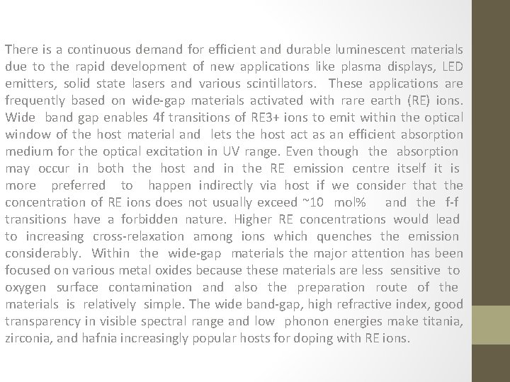
There is a continuous demand for efficient and durable luminescent materials due to the rapid development of new applications like plasma displays, LED emitters, solid state lasers and various scintillators. These applications are frequently based on wide-gap materials activated with rare earth (RE) ions. Wide band gap enables 4 f transitions of RE 3+ ions to emit within the optical window of the host material and lets the host act as an efficient absorption medium for the optical excitation in UV range. Even though the absorption may occur in both the host and in the RE emission centre itself it is more preferred to happen indirectly via host if we consider that the concentration of RE ions does not usually exceed ~10 mol% and the f-f transitions have a forbidden nature. Higher RE concentrations would lead to increasing cross-relaxation among ions which quenches the emission considerably. Within the wide-gap materials the major attention has been focused on various metal oxides because these materials are less sensitive to oxygen surface contamination and also the preparation route of the materials is relatively simple. The wide band-gap, high refractive index, good transparency in visible spectral range and low phonon energies make titania, zirconia, and hafnia increasingly popular hosts for doping with RE ions.