Luciferins and luciferases The common piddock A fluorescent


 / luciferase (enzyme) reaction (Dubois 1885 -7) First carried out on")

 luciferase Firefly luciferase produces light by")


- Slides: 37
Luciferins and luciferases
The common piddock A fluorescent clam that drills its way into rock
The luciferin (substrate) / luciferase (enzyme) reaction (Dubois 1885 -7) First carried out on the common piddock, then on firefly extracts
Understanding luciferase
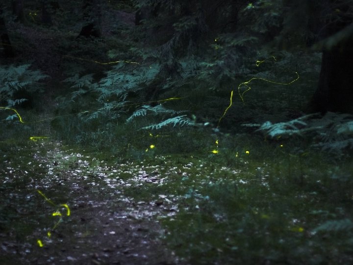
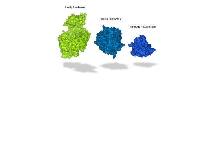
Luciferase: • • Photinus pyralis Firefly (Photinus pyralis) luciferase Firefly luciferase produces light by ATP-dependent oxidation • Bioluminescence or light emission is determined by a luminometer
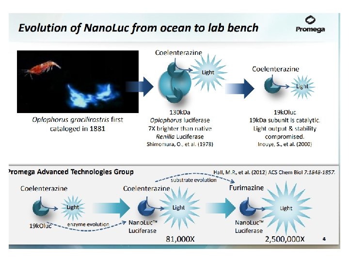
Luciferin chemistry There are many different luciferins, each specific luciferin has its own special luciferase. Terrestrial organisms Bacteria Vargulin Marine organisms Firefly Dinoflagellate Emission maxima of most luminous Coelenterazine marine organisms 450 -490 nm Best Wavelenghth for light trasmission in ocean water Maximal sensitivity of visual pigments of most marine organisms
Renilla reniformis è un celenterato coloniale
Gaussia Princeps è un copepode mesopelagico si trova nelle acque temperate e tropicali di tutto il mondo
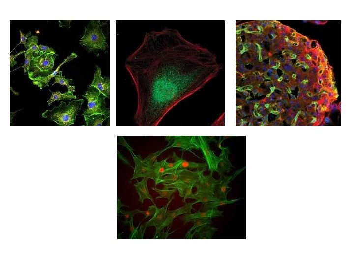
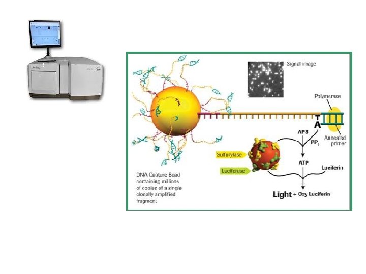
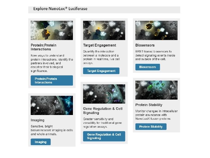
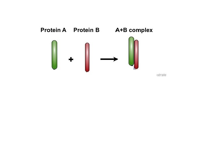
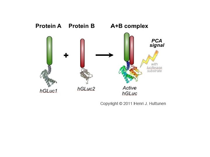
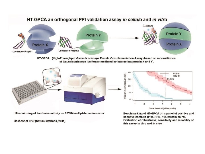
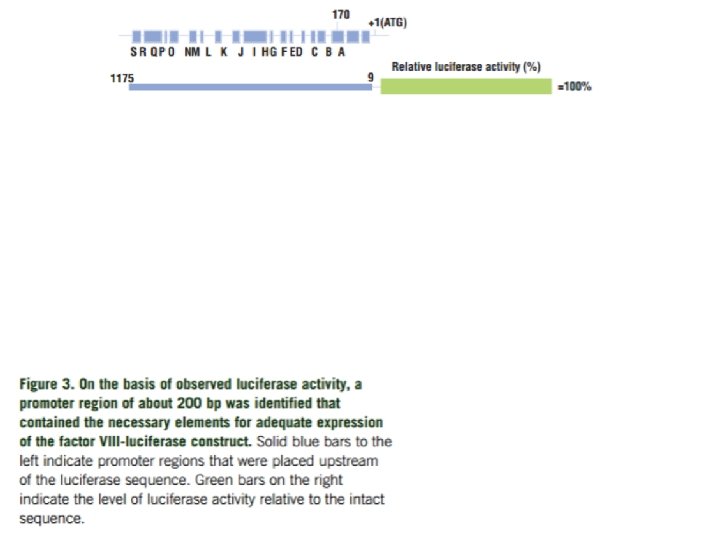
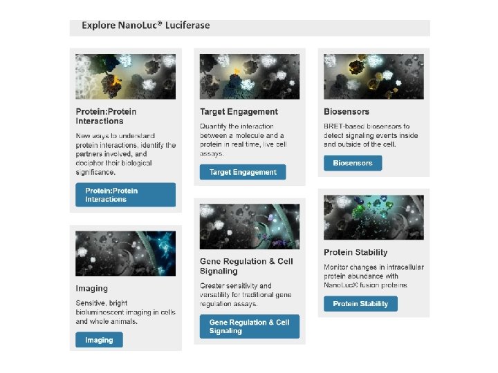
Biotechnological uses of luciferase In PCA to monitor protein interactions To test for anti-oxidants Pyrosequencing (454 sequencing) As a reporter gene To assess promoter strength and activity In vivo imaging Cancer biology
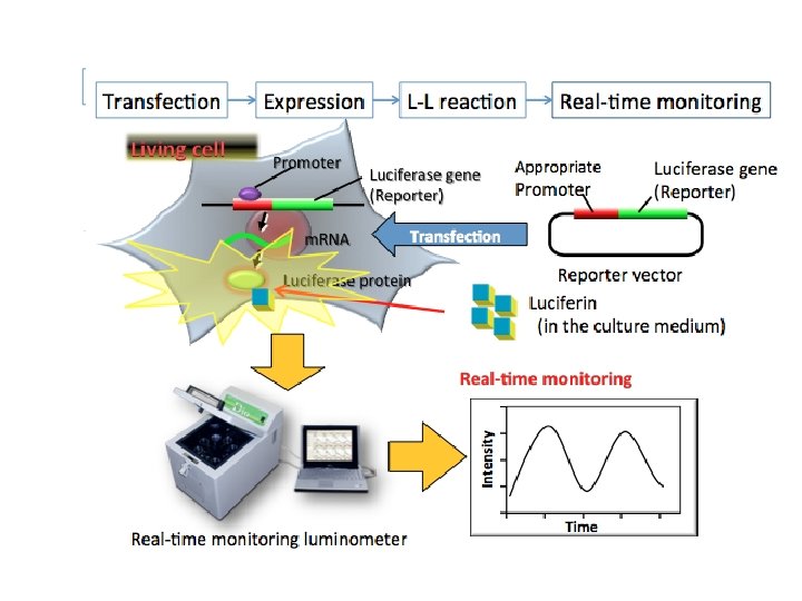
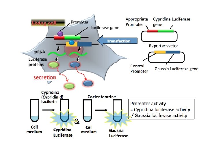
Reporter Gene Assays are powerful tools for studying gene regulation and serving as indicators of specific cellular signals. By cloning a promoter or 3´-UTR of interest into a plasmid containing the Luciferase reporter gene, it is possible to study elements in these DNA sequences that regulate gene expression. Alternatively, by inserting defined elements known to bind to different classes of transcription factors or enhancers into these vectors, assays measuring cell signaling pathways that activate these factors can be built.
The luciferase reporter assay is commonly used as a tool to study gene expression at the transcriptional level. It is widely used because it is convenient, relatively inexpensive, and gives quantitative measurements instantaneously. It has broad applications across various fields of cell and molecular biology – wherever you want to measure or track expression of a cloned gene.
Dual-Luciferase Reporter Assay System
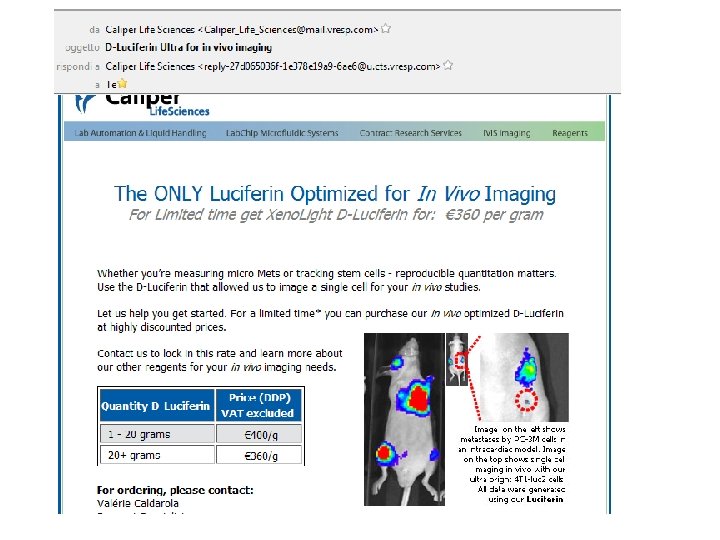
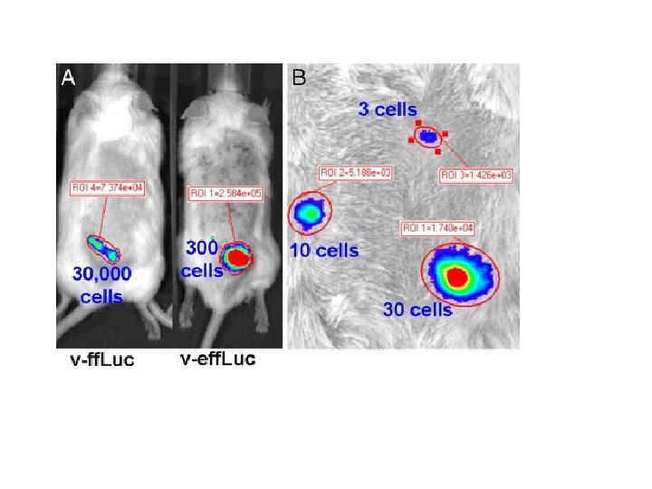
In vivo imaging of luciferase expression in mice: CMV promoter
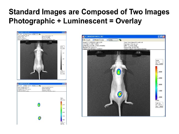
Since the luciferase gene can be stably transfected into cells under the control of most any promoter, signal will not be lost following dilution by cell division. Luciferase assays in mammalian systems are particularly sensitive because they are not subject to high background as a result of tissue autofluorescence Bioluminescent imaging is dependent on highly sensitive detectors. Typically a charged-coupled device (CCD) camera is used to image luciferase signal in whole animals. CCD cameras can be cooled to reduce thermal noise, and therefore background. Additionally, CCD systems detect the entire visible spectrum and near infrared wavelengths, allowing them to detect the light that is not absorbed by mammalian tissues.
The substrate luciferin was injected into the intraperitoneal cavity at a dose of 150 mg/kg body weight (30 mg/ml luciferin) approximately 5 min before imaging.
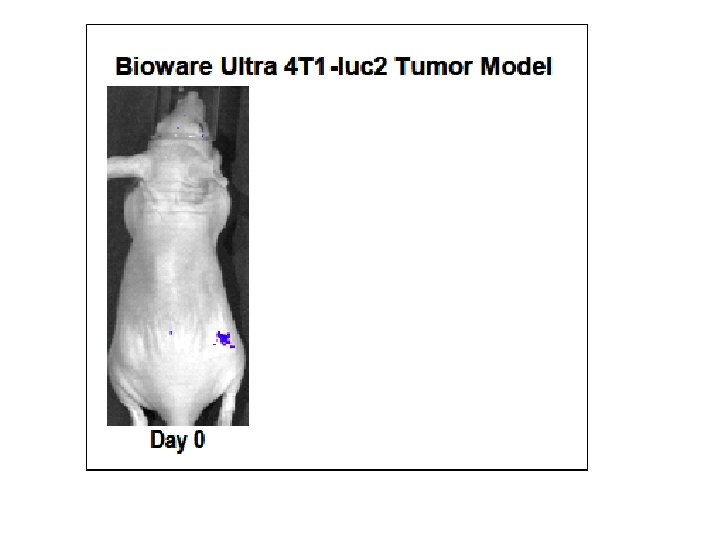
Bone metastasis Melanoma Prostate model Breast cancer