LPT 601 FRESH MEAT TECHNOLOGY UNIT I Structure


, precursor")


 Meat Source Protein Moisture Fat")
 (fresh weight) Name Protein Carbohydrate Fat Ash Calcium Phos. Phorus")


 75 PROTEN")


. Principles of Meat")
- Slides: 42
LPT 601: FRESH MEAT TECHNOLOGY UNIT I: Structure and chemistry of animal tissues (Part -II) Dr. R. K. Jaiswal Asstt. Prof. -cum-Jr. Scientist Dept. of Livestock Products Technology Bihar Veterinary College Bihar Animal Sciences University Patna-800014 (Bihar)
Meat § Animal tissues which are suitable for use as food § More precisely edible postmortem component originating from live animals
Meat terms Meat Science The basic study of the unique characteristics of muscle and other animal tissues that are used as meat, includes all facets of the meat industry, beginning with animal production and ending with final preparation for consumption. Meat Technology Deals with the handling, processing, packaging, preservation and marketing of meat, meat products and byproducts at industrial scale.
Muscle and Associated Tissue Types of Tissues § Muscle tissue § Nervous tissues § Epithelial tissues § Connective tissues § Adipose Tissues
Types of Muscular Tissue § Skeletal Muscle § Voluntary and striated § Smooth Muscle § Involuntary and smooth § Cardiac Muscle § Involuntary and striated
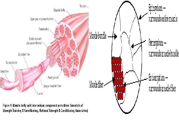
Skeletal Muscles § There are more than 600 muscles in animal body § Vary widely in shape, size and actions. § In general Skeletal muscle constitute 35 -65% of meat Muscle Epimysium Muscle bundle Muscle fiber Perimysium Endomysium & Sarcolemma Myofilaments Myofibril § Muscle fiber constitute 75 -92% of total muscle volume. § Remaining are connective tissues, blood vessels, nerve fibers and ECF
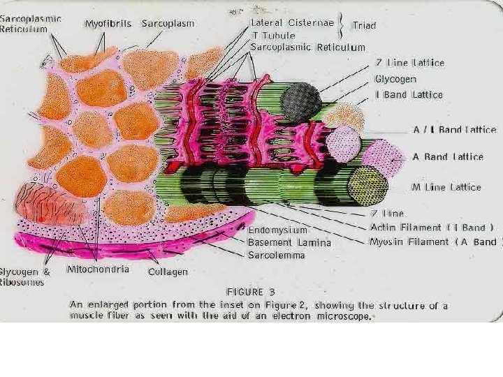
Structure of Cell
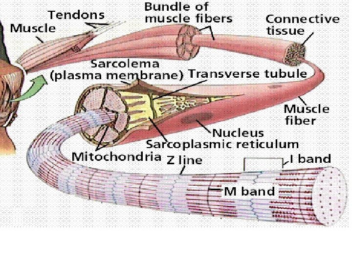
Sarcolemma • Membrane surrounding the Muscle fiber • Made up of protein and lipid material • Elastic in nature • Invaginations from Sarcolemma forming a tubular network Transverse Tubules/ T system/ T Tubules • Small mound on the surface of muscle fiber (sarcolemma) where motor nerve fiber terminate – Motor End Plate
Sarcoplasm § Cytoplasm of Muscle cell § In addition to 75 -80% water contains lipid droplets, glycogen granules, ribosomes, numerous protein, NPN compounds and inorganic constituents Nuclei • Multinucleated • At tendinous attachment more concentrated and irregularly distributed. • High in number at motor end plate • Peripheral in location in mammals whereas in fish central.
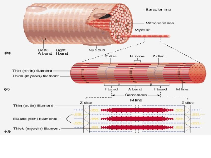
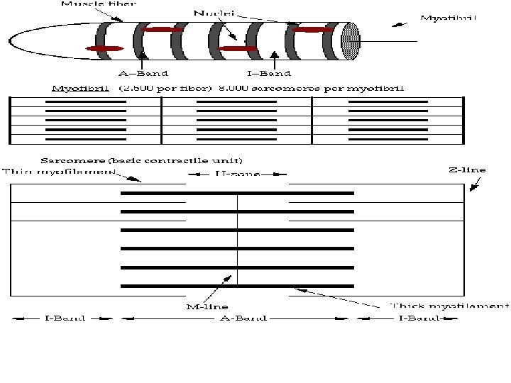
Myofibrils • Usually 1 -2 μm in diam. • Parallel to long axis • Fibers with diam. of 50 μm can accommodate 1000 -2000 myofibrils • Contractile unit is sarcomere (between two adjacent Z line) • Light and dark band • I Band (Isotropic/less dense) singly refractive, whereas A Band (Anisotropic/much dense) doubly refractive – • I band bisection by thin dense line –Z line
Myofilaments § Thick filaments of vertebrates 14 -16 nm in diam. and 1. 5 μm long (Myosin predominant/A band) § Thin filaments of vertebrates 6 -8 nm in diam. and 1. 0 μm long on either side of Z disc (Actin predominant/ I band) § Orderly arrangements of Thick and Thin filament § Thick and thin overlapping in certain region accounts for striations § In cross section of A band region Six thin filaments surround each thick filament § Each Actin filaments connect with 4 oblique Z filaments (Z line)
Sarcoplasmic reticulum • System of Tubules and cisternae/Intracellular membrane structure acting as Flattered reservoirs of Ca 2+ and corresponds to ER of other cells • Thin tubules of SR oriented in direction of myofibril. Longitudinal tubules and in H zone it forms a perforated sheet called as fenestrated collar. • At the junction of A and I band the longitudinal tubules join with lagers transversely oriented tubules called terminal cisternae. • The T tubules also runs transversely across the sarcomere at A-I junction • Connecting elements exist between terminal cisternae and T tubules. • SR and T tubules are separate, SR constitute 13% of total muscle fiber volume where as T tubules approx 0. 3%.
Mitochondria • Powerhouse of cell • Great variation in mitochondrial size and their number in muscle fibers • Abundant at periphery near the poles of nuclei and motor end plates • Additional at A-I band junction, Z disc and I bands Lysosomes • Cathepsins a group of proteolytic enz. Golgi Complex • Near the nuclei • Flattened vesicles, function as concentrating and packaging apparatus.
Sarcomere length, Dimension of I band appearance of H Zone, Pseudo H zone and M line change with contraction state.
Smooth Muscles § Innervated by the Autonomic Nervous System § Found primarily in the walls of hollow organs & tubes § In most of the case spindle-shaped cells typically arranged in sheets § Single nucleus centrally located § Cells do not have t-tubules & have very little sarcoplasmic reticulum § No Z line or M line and striation § Actin and Myosin in same proportion § Occurs single or in bundle and surrounded by network of reticular fiber
Contd. . Two types of smooth muscle: 1. Visceral or Unitary smooth muscle § Found in the walls of hollow organs (e. g. , small blood vessels, digestive tract, urinary system, & reproductive system) § Multiple fibers contract as a unit (because impulses travel easily across gap junctions from cell to cell) &, in some cases, are self-excitable (generate spontaneous action potentials & contractions) 2. Multiunit smooth muscle § § Consists of motor units that are activated by nervous stimulation Found in the walls of large blood vessels, in eye (adusting the shape of the lens to permit accommodation & the size of the pupil)
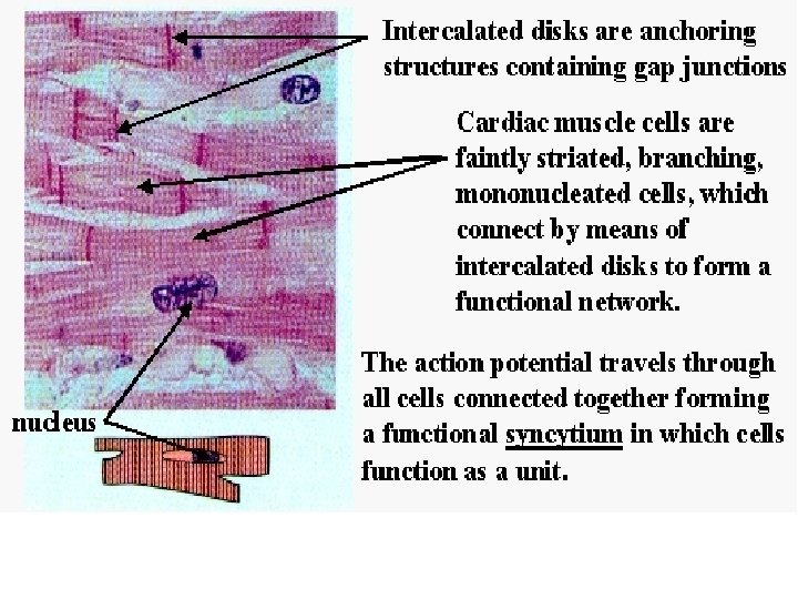
Cardiac Muscle § § § § Myocardium, the contractile layer of the heart, contains the bulk of the cardiac muscle Single centrally placed nucleus, fibers branched and shorter Sarcoplasm with numerous glycogen granules Mitochondria especially large and numerous, extensive blood circulation. Aggregates of myofilaments form fibrils of extremely variable size T Tubules at Z disc, SR less developed and terminal cisternae absent. Intercalated disc- transecting the fiber, occurs at regular interval across I bands
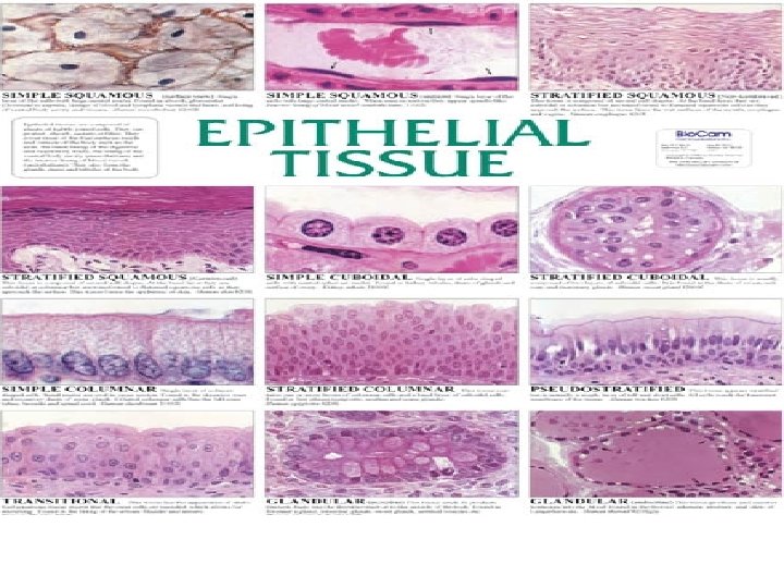
Contd. . § § § Lines the body’s surface, cavities, ducts and tubes One free surfaces a body fluid or the environment Quantitatively contributes the least to meat, but add to flavor and crispiness Associated with blood and lymph vessels and edible organs such as the kidney and liver and also in byproducts hides and skins. Classified according to shape and number of layers Protection, Secretion, Excretion, Transport, Absorption, Sense Perception.
Connective Tissues § Mesodermal origin § Functions: Support, binding, protection, insulation, Intercellular communication and exchange, Site for tissue reactions (inflammation, immune) § General characteristics: § Scattered few cells and considerable extra cellular materials. § Fibers (Protein): Arranged in matrix - as fortification/network or in dense bundles § Categories § Loose: Areolar, Adipose, mesenchymal § Dense : Reg. & Irreg. § Special: Adipose, Embryonic, Reticular, Elastic, Blood, Cartilage and Bone.
Contd. . • Throughout body • Few cells and considerable extracellular substance • Contains embedded fibers that provide structural elements, limited in adipose tissue but no fibers in blood and lymph. • Surrounding the muscle, muscle bundle and muscle fibers. Connective tissue proper • Bone, cartilage - supportive connective tissue. • Connective tissue proper = Ground substance + Cells + extra cellular fibers. • Fixed cells: Fibroblast, mesechyme cells, adipose cells • Wandering cells- Eosinophils, plasma cells, mast cells, lymph cells, free macrophages
Contd. . Ground Substance: Contain glycoproteins like mucopolysaccharides (hyaluronic acid and chondroitin sulphates), precursor of collagen and elstin etc Glycosaminoglycans (GAGs) or mucopolysaccharides – Linear polymers of repeating disaccharides of hexosamine plus a uronic acid such as glucuronic acid – Have a high negative charge and are highly hydrophilic – GAG-residues are often sulfated. – Signaling function
Contd. . Extra cellular fiber Collagen fibers § § § § Most abundant protein (20 -25%) Influence tenderness, major component of tendons and ligament but less in bone and cartilage. Amount of collagen in skeletal muscle parallel to physical activity. Glycoprotein in nature. Glycine most abundant and about 1/3 rd, Hydroxiproline (13 -14%, used for chemical assay) and proline another 1/3 rd. Structural unit for all 12 typed collagen is tropocollagen, made of three α chains (19 types) to form a triple helix Fibers are inextensible and colourless individually. Appear reddish brown in silver stain Relative insolubility and high tensile strength due to intermolecular cross linkages which increase in number and strength with age.
Contd. . Reticular fibers § Delicate and form fine networks instead of thick bundles § Silver stained sections - black threads (argyrophilia ) § Support to individual cells § Present in connective tissue surrounding liver, spleen or lymph nodes and larger vessels
Contd. . Elastic fibers § § § Less abundant and elastic in nature and rubbery in nature Present in ligamnets, arterial wall, framework of organs and muscle, Ligamentum nuchae Glycine most abundant and isodesmine and desmosine are unique amino acids. Insolubility because of non polar amino acids and its desmosine cross links. In aggregates yellow in color and contributes nothing or little to nutritive value of meat.
Connective tissue cells • • • Among the connective tissue cells only fibroblasts (true connective tissue cells), mesenchymal cells and adipose cells are of relevance Fibroblast synthesize precursor of extra cellular components of CT, namely tropocollagen, tropoelastine and ground substance. The spindle mesenchymal cells differentiate to become one of several different cell types ( fibroblasts, adipobalsts etc. ) Two types of adipose tissue (fat) (adipocytes cell)- white and brown. Brown fat remains at the time of birth, cell smaller in size but high with cytochrome in mitochondria.
Specialized connective tissue § § § Cartilage Chondrocytes embedded in gel like matrix, about 40% interlacing of collagen fibrils Hyaline (most abundant) and elastic or fibrocartilage types, based on relative amount of collagenous and elastin fibers Bone Unlike other connective tissue extracellular matrix is calcified Storage site for Ca, Mg, Na and other ions In long bone parts are Diaphysis, metaphsis, epiphysis, articular cartilage, compact zone, spongy zone, bone marrow (Red/Yellow), periosteum, endoosteum, Canaliculi, Osteocytes, osteoclasts, organic matrix (proteoglycans) and inorganic salts (Ca. PO 3) Blood and Lymph In blood (7% of BW) Plasma, Hb, Erythrocytes, Leucocytes, Platelets whereas in lymph mainly lymphocytes, and few leuco and erythrocytes.
Nervous Tissues § § § Constitute less than 1% of meat Function of it prior or during slaughter has important influence on meat quality. Neuron the bulk of nervous tissue. Neurolemma (Sheath of schwann), myelin sheath (larger fibers) Neuroplasm, Nucleus, axon, axoplasm, dendrites, axon ending, node of ranvier, schwann cell nucleus, nerve fibers and neurofibrils. Interdigitating areas of 2 neurons (Axon with dendrite)- synapses. Large nerve fibers-Myelinated and small peripheral-Unmyelinated nerve fibers.
Composition of muscle and associated tissue Carcass Composition (%) Meat Source Protein Moisture Fat Ash Beef 21 72 6 1 Chicken 20 -23 74 -76 2 1 Lamb 20 72 7 1. 5 Pork 20 72 7 1 Veal 20 75 4 1
Composition of meats (%) (fresh weight) Name Protein Carbohydrate Fat Ash Calcium Phos. Phorus Iron (mg/kg) Copper (mg/kg) Chicken 21. 1 0. 0 4. 5 1. 1 0. 012 0. 232 32. 0 3. 5 Duck 21. 4 0. 0 8. 2 1. 2 0. 010 0. 200 23. 0 5. 0 Mutton (leg) 19. 8 0. 0 12. 4 1. 2 0. 010 0. 270 30. 0 4. 0 Turkey 24. 0 0. 0 6. 7 1. 1 0. 030 0. 420 45. 0 1. 8 Goose 22. 3 0. 0 7. 1 1. 1 0. 009 0. 175 24. 0 3. 3 Ham 15. 2 0. 0 31. 0 0. 8 0. 009 0. 168 23. 0 --
Carcass Composition Flesh/Muscle: Skeletal muscle 35 -65% Bone Fat Mutton- 25% Bobby calves- 50% Veal-20 -25% Pork 12 -20% Simple lipids: esters of fatty acids with certain alcohols such as glycerol. General composition of Bones Compound or conjugate lipids: esters of fatty acids that, on hydrolysis yield such substances as phosphoric acid, amino acid, choline, carbohydrates and sulfuric acid in addition to fatty acids and an alcohol. Derived lipids: formed in the hydrolysis of simple or compound lipids
Chemical Composition of the Animal Body . Oxygen 65. 00% Carbon 18. 00% Hydrogen 10. 00% Nitrogen 3. 00% Calcium 1. 50% Phosphorus 1. 00% Potasium 35. 00% Sulfur 25. 00% Sodium 0. 15% Chlorine 0. 15% Magnesium 0. 05% Trace elements present: Aluminium, Arsenic, Barium, Boron, Bromin, Cadmium, Chromium, Cobalt, Copper, Fluorine, Iodine, Iron, Lead, Lithium, Strontium, Titanium, Vanadium, Zinc etc
Composition of Mammalian skeletal muscle Constituents Percent WATER (range 65 to 80) 75 PROTEN (range 16 to 22) 18. 5 LIPIDS (variable range 1. 5 to 13. 0) 3 NON-PROTEIN NITROGENOUS SUBSTANCES 1. 5 CARBOHYDRATES AND NONNITROGENOUS SUBSTANCES (range 0. 5 to 1. 5) 1 INORGANIC CONSTITUENTS 1 Neutral lipids (range 0. 5 to 1. 5) 1 Phospholipids 1 Cerebrosides 0. 5 Cholesterol 0. 5 Glycogen (range 0. 5 to 1. 3) 0. 8 Glucose 0. 1 Intermediate and products of cell metabolism (hexose and triose phosphates, lactic acid, citric acid, fumaric acid, succinic acid, etc. ) 0. 1
Composition of Mammalian skeletal muscle Protein Myofibrillar 9. 5 Myosin 5 Actin 2 Troponyosin 0. 8 Tropomin 0. 8 M protein 0. 4 C protein 0. 2 a-actinin 0. 2 b-actinin 0. 1 Sarcoplasmic 6 Soluble sarcoplasmic and mitochondrial enzyme 5. 5 Myoglobin 0. 3 Hemoglobin 0. 1 Cytochromes and flavoproteins 0. 1 Stroma 3 Collagen and recticulin 1. 5 Elastin 0. 1 Other insoluble proteins 1. 4
Composition of Mammalian skeletal muscle NPN NON-PROTEIN NITROGENOUS SUBSTANCES 1. 5 Creatine and Creatine phosphate 0. 5 Nucleotides [adenosine triphosphate (ATP), adenosine diphosphate (ADP), etc] 0. 3 Free amino acids 0. 3 Peptides (anserine, carnosine etc) 0. 3 Other nonprotein substances [creatinine, urea, inosine monophosphae (IMP), nicotinamide adenine dinucleotide (NAD), nicotinamide adenine dinucleotide phosphate (NADP)] 0. 1
Suggested readings 1. 2. 3. 4. 5. Forrest, J. C. (1975). Principles of Meat Science. W. H. Freeman & Co, San Francisco. Lawrie, R. A. (1990). Meat Science, 5 th edn. Pergamon Press, Oxford. “Meat Hygiene” 10 th Edition W. B. Saunders Company Ltd. London By J. F. Gracey, D. S. Collins and R. J. Huey. Principle of Meat Science 3 rd edition kendall/Hunt publlizhing company. Meat & Meat product technology 1 st edition JAYPEE Brothers New Delhi. by B D Sharma