Lower limb Muscles of lower limb muscles of

Lower limb

Muscles of lower limb § muscles of iliac region § muscles of thigh § anterior femoral muscles § midial femoral muscles § posterior femoral muscles § muscles of gluteal region § muscles of leg § muscles of foot lower limb

Muscles of iliac region M. of iliac region # iliaccus # psoas major # psoas minor Post. View

iliopsoas M. of iliac region § Origin § inner surface of upper iliac fossa § transverse processes of L 1 -L 5 § vertebral bodies of T 12 -L 4 § Insertion § lesser trochanter of the femur § Action § hip flexion § lateral rotation § Nerve : medial femoral circumflex artery § Blood : femoral nerve, L 3, 4 / ventral rami, L 1, 2, 3

Muscles of thigh anterior muscles femoral # rectus femoris # vastus lateralis # vastus intermidius # vastus midialis # sartorius Ant. View

rectus femoris muscles of thigh § Origin § anterior head: anterior inferior iliac spine § posterior head: ilium just above the acetabulum § Insertion § tibial tuberosity via patellar ligament § Action § extends knee § flexes hip § Nerve : lateral femoral circumflex artery § Blood : branches of femoral nerve, [L 2], 3, 4 v

vastus lateralis muscles of thigh § Origin § greater trochanter § lateral lip of linea aspera § lateral intermuscular septum § Insertion § tibial tuberosity via patellar ligament § Action § extends knee § can abnormally displace patella § Nerve : lateral femoral circumflex artery § Blood : branches of femoral nerve, [L 2], 3, 4 v

vastus intermidius muscles of thigh § Origin § Ant. lateral aspect of the femoral shaft § Insertion § tibial tuberosity via patellar ligament § Action § extends knee § Nerve § lateral femoral circumflex artery § Blood § branches of femoral nerve, [L 2], 3, 4 v

vastus midialisv muscles of thigh § Origin § intertrochanteric line of femur § medial aspect of linea aspera § Insertion § tibial tuberosity via patellar ligament § Action § extends knee § Nerve § descending genicular artery § femoris artery § Blood § femoral nerve, [L 2], 3, 4 v

sartorius muscles of thigh § Origin § anterior superior iliac spine § Insertion § upper medial surface of body of tibia § Action § flexes hip and knee § laterally rotates thigh if flexed at the hip § Nerve § femoral artery § descending genicular artery § Blood § femoral nerve, L 2, 3

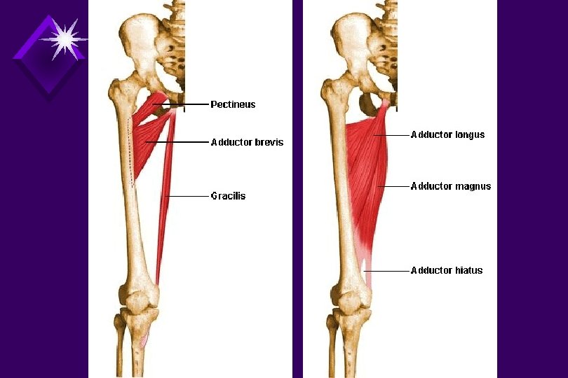
Muscles of thigh medial femoral muscles # pectineus # adductor longus # adductor brevis # adductor magnus # gracilis Ant. View(deep)

pectineus muscles of thigh § Origin § pectineal line of the pubis § superior pubic ramus § Insertion § the pectineal line of the femur § Action § flexes hip § adducts thigh § medially rotates thigh § Nerve : medial femoral circumflex artery § Blood : femoral nerve L 3, 4 obturator nerve L 2, 3, 4

adductor brevis § Origin § body & inferior ramus of pubis § Insertion § superior portion of linea aspera § Action § adducts thigh § aids in flexion of thigh § may laterally rotate thigh at the hip § Nerve § femoral artery § Blood § obturator nerve L 2, 3, 4 muscles of thigh

gracilis muscles of thigh § Origin § body of pubis & inferior pubic ramus § Insertion § medial surface of proximal tibia, inferior to tibial condyle § Action § adducts thigh § flexes knee § medially rotates tibia § Nerve § obturator artery § Blood § obturator nerve L 2, 3, 4

adductor longus muscles of thigh § Origin § anterior surface of pubis § Insertion § medial lip of linea aspera on middle half of femur § Action § adducts thigh § flexes thigh § may laterally rotate thigh at the hip § Nerve § femoral artery § Blood § obturator nerve L 2, 3, 4

adductor magnus § Origin § inferior pubic ramus § ischial tuberosity § Insertion § proximal 1/3 of linea aspera § adductor tubercle § Action § adducts the thigh § extend and laterally rotate thigh § Nerve § femoral artery § Blood § obturator nerve L 2, 3, 4 / tibial nerve muscles of thigh

Muscles of thigh posterior femoral m. # biceps femoris # semitendinosus # semimembranosus Post. View

biceps femoris muscles of thigh § Origin § ischial tuberosity § lateral lip of linea aspera & the lateral intermuscular septum § Insertion § head of fibula § maybe to the lateral tibial condyle § Action § flexor at the knee(mainly short head) § laterally rot. thigh if flexed at the knee § extends hip(long head) § Blood : gluteal artery § Nerve : tibial nerve, L 5, S 1, 2 / common peroneal nerve, L 5, S 1

semitendinosus muscles of thigh § Origin § ischial tuberosity § Insertion § medial aspect of tibial shaft § Action § extends hip § flexes knee § medially rotates tibia § Blood § gluteal artery § branches of profunda femoris § Nerve § tibial nerve of sciatic bundle, L 5, S 1, 2

Semimembranosus muscles of thigh § Origin § ischial tuberosity § Insertion § posterior medial aspect of medial tibial condyle § Action § flexes knee § extends hip § medially rotates tibia § pulls medial meniscus posterior during flexion § Blood § gluteal artery § branches of profunda femoris § Nerve : tibial nerve of sciatic bundle, L 5, S 1, 2

Muscles of thigh muscles of gluteal region # gluteus maximus # gluteus medius # gluteus minimus # tensor fascia lata Lateral View
")
gluteus maximus muscles of thigh § Origin § outer rim of ilium (medial aspect) § dorsal surface of sacrum and coccyx § sacrotuberous ligament § Insertion § IT band (primary insertion) § gluteal tuberosity of femur § Action § powerful extensor of hip § laterally rotates thigh § upper fibers aid in abduction of thigh § IT band stabilize a fully extended knee § Nerve : inferior/superior gluteal artery § Blood : inferior gluteal nerve, L 5, S 1, 2 v

gluteus medius muscles of thigh § Origin § outer aspect of ilium § upper fascia § Insertion § superior aspect of greater trochanter § Action § anterior & lateral fibers abduct § medially rotate thigh § posterior fibers may laterally rotate thigh § stabilizes the pelvis § prevents free limb from sagging during gait § Nerve : superior gluteal artery § Blood : superior gluteal nerve, L 4, 5, S 1

gluteus minimus muscles of thigh § Origin § outer aspect of ilium § Insertion § greater trochanter § articular capsule of hip joint § Action § abduct and medially rotate thigh § stabilizes the pelvis § prevents free limb from sagging during gait § Nerve : superior gluteal artery § Blood : superior gluteal nerve, L 4, 5, S 1

tensor fascia lata muscles of thigh § Origin § anterior aspect of iliac crest § anterior superior iliac spine § Insertion § anterior aspect of IT band, below greater trochanter § Action § hip flexion § medially rotate & abduct a flexed thigh § support femur on the tibia during standing § Nerve : superior gluteal artery/femoral artery § Blood : superior gluteal nerve, L 4, 5, S 1

muscles of thigh short external rotators

nerves of anterior thigh

arteries

Muscles of leg anterior crural muscles # tibialis anterior # extensor digitorum longus # peroneus tertius # extensor hallucis longus

tibialis anterior muscles of leg § Origin § lateral tibial condyle § proximal 2/3 of anteriolateral surface of tibia § Insertion § medial & plantar surface of base of 1 st metatarsal § medial & plantar surface of the cuneiform § Action § strongest dorsiflexor § inverts & adducts the foot § Nerve : anterior tibial artery § Blood : deep peroneal nerve, L 4, 5, S 1

muscles of leg extensor digitorum longus § Origin § lateral condyle of the tibia § upper anterior surface of fibula § Insertion § dorsal surface of the bases of the middle & distal phalanxes of the 2 nd-5 th rays § Action § extends the lateral 4 toes § weak dorsiflexor & everts foot § Nerve : anterior tibial artery § Blood : deep peroneal nerve, L 4, 5, S 1

extensor hallucis longusmuscles of leg § Origin § medial aspect of the fibula § interosseous membrane § Insertion § dorsal surface of base of proximal and distal phalanx of hallux § Action § extends distal phalanx of big toe § weak dorsiflexor § weak inversion & adduction § Nerve : anterior tibial artery § Blood : deep peroneal nerve, L 4, 5, S 1

peroneus tertius § Origin § distal 1/3 of anterior fibula § distal & lateral aspect of extensor digitorum § Insertion § dorsal surface of base of 5 th metatarsal § Action § extends the 5 th toe § weak dorsiflexor & everts foot § Nerve : anterior tibial artery § Blood : deep peroneal nerve, L 4, 5, S 1 muscles of leg
![Muscles of leg posterior muscles crural [superficial group] # gastrocnemius # soleus # plantaris](http://slidetodoc.com/presentation_image_h2/0b9084c8d82011690993324d89e6344c/image-35.jpg "Muscles of leg posterior muscles crural [superficial group] # gastrocnemius # soleus # plantaris")
Muscles of leg posterior muscles crural [superficial group] # gastrocnemius # soleus # plantaris posterior muscles crural [deep group] # flexor digitorum longus # flexor hallucis longus # posterior tibialis # popliteus

gastrocnemius muscles of leg § Origin § just above medial/lateral condyle of femur § Insertion § calcaneus via lateral portion of calcaneal tendon § Action § plantarflex the ankle § knee flexion (when not weight bearing) § stabilizes ankle & knee when standing § Nerve : popliteal / peroneal / posterior tibial artery § Blood : tibial nerve, S 1, 2

soleus muscles of leg § Origin § upper fibula § soleal line of tibia § Insertion • calcaneus via medial portion of calcaneal tendon § Action § plantarflex the ankle § knee flexion (when not weight bearing) § stabilizes ankle & knee when standing § Nerve : popliteal / peroneal / tibial artery § Blood : tibial nerve, S 1, 2

plantaris muscles of leg § Origin § above the lateral head of gastrocnemius on femur § Insertion § calcaneus, medial to calcaneal tendon § Action § like a weak gastrocnemius § Nerve : popliteal / peroneal / tibial artery § Blood : tibial nerve, S 1, 2
")
posterior compartment (superficial)

flexor digitorum longus § Origin § posterior surface of tibia § Insertion § plantar surface of bases of the 2 -5 th distal phalanges § Action § primarily flexes 2 nd - 5 th toes § weak plantarflexor § weak inversion & adduction of foot § Nerve : peroneal / tibial artery § Blood : tibial nerve, L 5, S 1 muscles of leg

flexor hallucis longus muscles of leg § Origin § posterior, inferior 2/3 of fibula § Insertion § plantar surface of distal phalanx of hallux § Action § flexes big toe (hallux) § weak plantarflexion of the foot § weak inversion & adduction of foot § Nerve : peroneal / tibial artery § Blood : tibial nerve, L 5, S 1, 2

posterior tibialis muscles of leg § Origin § posterior, proximal tibia § medial surface of fibula § Insertion § navicular tuberosity (principle) § all 3 cuneiforms (plantar surface) § cuboid / bases of 2 -4 metatarsals § Action § stabilizes ankle § inversion & adduction of foot § prevents hyperpronation while in gait § weak plantarflexion of ankle § Nerve : peroneal / tibial artery § Blood : tibial nerve, L 5, S 1

popliteus muscles of leg § Origin § lateral femoral condyle § arcuate popliteal ligament § lateral meniscus § knee joint capsule § Insertion § posterior tibial surface § Action § insertion fixed: laterally rotates femur on tibia § origin fixed: medially rotates tibia on femur § Nerve : peroneal artery § Blood : tibial nerve, L 5, S 1, 2

Muscles of leg laterior crural muscles # peroneus longus # peroneus brevis

peroneus longus § Origin § head of the fibula § proximal 2/3 of lateral fibula § Insertion § plantar surface of cuboid § base of 1 st & (2 nd) metatarsal § plantar surface of medial cuneiform § Action § eversion & abduction of the foot § weak plantarflexion of the foot § Nerve : peroneal artery § Blood : peroneal nerve, L 4, 5, S 1 muscles of leg

peroneus brevis § Origin § distal 2/3 of lateral fibula § Insertion § tuberosity on lateral aspect of base of 5 th metatarsal § Action § eversion & abduction of the foot § weak plantarflexion of the foot § Nerve : peroneal artery § Blood : peroneal nerve, L 4, 5, S 1 muscles of leg

nerves

arteries

medial ankle

Muscles of foot dorsal muscles # extensor digitorum brevis # extensor hallucis brevis

extensor hallucis brevis § Origin § upper anterolateral calcaneus § Insertion § base of proximal phalanx of hallux § Action § extends hallux § Nerve : dorsalis pedis artery § Blood : peroneal nerve, L 4, 5 muscles of foot

muscles of foot extensor digitorum brevis § Origin § upper anterolateral calcaneus § Insertion § middle & distal phalanges of 2 nd-4 th rays § Action § extends 2 nd-4 th rays § Nerve : dorsalis pedis artery § Blood : peroneal nerve, L 4, 5

Muscles of foot medial plantar muscles # abductor hallucis # flexor hallucis brevis # adductor hallucis

abductor hallucis § Origin § medial process of calcaneal tuberosity § Insertion § medial aspect of base of proximal phalanx § Action § flexes the big toe § assist in abduction § Nerve : medial plantar artery § Blood : medial plantar nerve, L 5, S 1 muscles of foot

flexor hallucis brevis § Origin § medial aspect of the cuboid § lateral cuneiform § Insertion § medial/lateral aspect of base of proximal phalanx of hallux § Action § flexes hallux at MTP § Nerve : medial plantar artery § Blood : medial plantar nerve, L 5, S 1 muscles of foot

adductor hallucis § Origin § base of 2 nd-4 th metatarsals § Insertion § lateral aspect of base of proximal phalanx of hallux § Action § adduction of hallux at MTP § flexes hallux at MTP § Nerve : plantar artery § Blood : lateral plantar nerve, S 1, 2 muscles of foot

Muscles of foot lateral plantar muscles # abductor digiti minimi # flexor digiti minimi brevis

abductor digiti minimi § Origin § lateral & medial processes of the calcaneal tuberosity § Insertion § lateral aspect of base of proximal phalanx of 5 th ray § Action § abducts 5 th toe § aids in flexing § Nerve : lateral plantar artery § Blood : plantar nerve, L 5, S 1 muscles of foot

flexor digiti minimi brevismuscles of foot § Origin § lateral & medial processes of the calcaneal tuberosity § Insertion § lateral aspect of base of proximal phalanx of 5 th ray § Action § abducts 5 th toe § aids in flexing § Nerve : lateral plantar artery § Blood : plantar nerve, L 5, S 1

Muscles of foot central plantar muscles # flexor digitorum brevis # quadratus plantae # lumbricales # interosssei plantares # interosssei dorsales

flexor digitorum brevis § Origin § medial process of calcaneal tuberosity § Insertion § both sides of the bases of the mid. phalanx of rays 2 -5 § Action § flexes toes 2 -5 § Nerve : medial plantar artery § Blood : plantar nerve, L 5, S 1 muscles of foot

quadratus plantae § Origin § medial/lateral calcaneus § Insertion § lateral margin of tendon of flexor digitorum longus(fdl) § Action § assists FDL in flexing § Nerve : lateral plantar artery § Blood : plantar nerve, L 5, S 1 muscles of foot

lumbricales § Origin § tendons of FDL § Insertion § extensor tendons of EDL on dorsal foot § Action § flex proximal phalanges at MTP § extend middle & distal phalanges at IP § Nerve : plantar artery § Blood : plantar nerve, L 5, S 1 muscles of foot

interosssei plantares § Origin § medial aspect of 3 -5 metatarsals § Insertion § medial aspect of base of proximal phalanx of the same ray § Action § adduct toes 3 -5 § flex toes 3 -5 at MTP § Nerve : lateral plantar arch § Blood : plantar nerve, L 5, S 1 muscles of foot

interosssei dorsales muscles of foot § Origin § from both metatarsals between which they lie § Insertion § base of proximal phalanx § Action § abduct toes 2 -4 § flexes toes 2 -4 at MTP § Nerve : lateral plantar arch § Blood : lateral plantar nerve S 1, 2
- Slides: 65