Lower Limb Bones Lecture Objectives List the bones

Lower Limb Bones

Lecture Objectives • List the bones of the lower limb (pelvic girdle, thigh, leg, and foot bones). • Describe the structure, relationships and function of the lower limb bones. • Identify the surface anatomy of the lower limb bones. • Describe the arches of the foot.

Lower Limb Bones
 Girdle • Each coxal (hip) bone consists of three bones that fuse")
Pelvic (Hip) Girdle • Each coxal (hip) bone consists of three bones that fuse together: ilium, pubis, and ischium • The two coxal bones are joined anteriorly by the pubic symphysis (fibrocartilage) • Joined posteriorly by the sacrum forming the sacroiliac joints

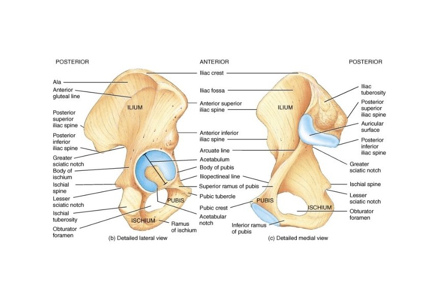
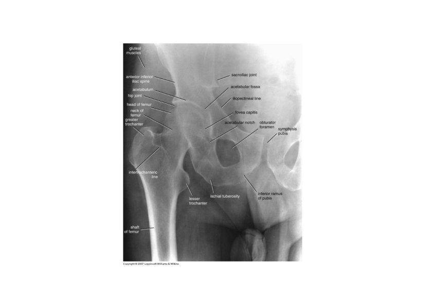
Ilium • Largest of the three hip bones • Ilium is the superior part of the hip bone • Consists of a superior ala and inferior body which forms the acetabulum (the socket for the head of the femur) • Superior border ‐ iliac crest • Hip pointer ‐ occurs at anterior superior iliac spine • iliac spines • Greater sciatic notch ‐ allows passage of sciatic nerve • Iliac fossa for muscle attachment • Gluteal lines indicating muscle attachment • Sacroiliac joint at auricular surface & iliac tuberosity

Ischium and Pubis • Ischium ‐ inferior and posterior part of the hip bone • Most prominent feature is the ischial tuberosity, it is the part that meets the chair when you are sitting • ischial spine • lesser sciatic notch • ramus • Pubis ‐ inferior and anterior part of the hip bone • Superior and inferior rami and body

False and True Pelves • Pelvic brim ‐ a line from the sacral promontory to the upper part of the pubic symphysis • False pelvis ‐ lies above this line • Contains no pelvic organs except urinary bladder (when full) and uterus during pregnancy • True pelvis ‐ the bony pelvis inferior to the pelvic brim, has an inlet, an outlet and a cavity • Pelvic axis ‐ path of baby during birth

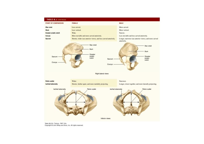
Comparing Male and Female Pelves • Males • Bone are larger and heavier • Pelvic inlet is smaller and heart shaped • Pubic arch is less the 90° • Female • Wider and shallower • Pubic arch is greater than 90° • More space in the true pelvis

Comparing Male and Female Pelves

Male pelvis

Female pelvis

Surface Anatomy of Pelvic Girdle • Iliac crest • ASIS • Pubic symphysis • Pubic tubercle • 2 cm from pubic symphysis • Ischial tuberosity

Lower Extremity • Each lower limb = 30 bones • • • femur and patella within the thigh tibia & fibula within the leg tarsal bones in the foot metatarsals within the forefoot phalanges in the toes • Joints • hip, knee, ankle • proximal & distal tibiofibular • metatarsophalangeal
 • longest & strongest bone in body • head articulates with acetabulum")
Femur (thighbone) • longest & strongest bone in body • head articulates with acetabulum (attached by ligament of head of femur) • Fovea capitis • neck is common fracture site • greater & lesser trochanters, linea aspera, & gluteal tuberosity-- muscle attachments • Adductor tubercle - - adductor magnus • medial & lateral condyles articulate with tibia • patellar surface anteriorly between condyles • intercondylar fossa - depression between the condyles • Angle of inclination • Normal range 115°- 140° • Male > Female

Patella • Largest sesamoid bone in the body • Forms the patellofemoral joint • Superior surface is the base • Inferior, narrower surface is the apex • Thick articular cartilage lines the posterior surface • Increases the leverage of the quadriceps femoris muscle

Surface Anatomy of Femur • Greater trochanter • Femoral epicondyles • Adductor tubercle • Patella

Tibia and Fibula • Tibia • medial & larger bone of leg • weight‐bearing bone • lateral & medial condyles • Intercondylar …… • tibial tuberosity for patellar lig. • proximal tibiofibular joint • medial malleolus at ankle • fibular notch • Fibula • not part of knee joint • muscle attachment only • Head, neck, shaft • lateral malleolus at ankle
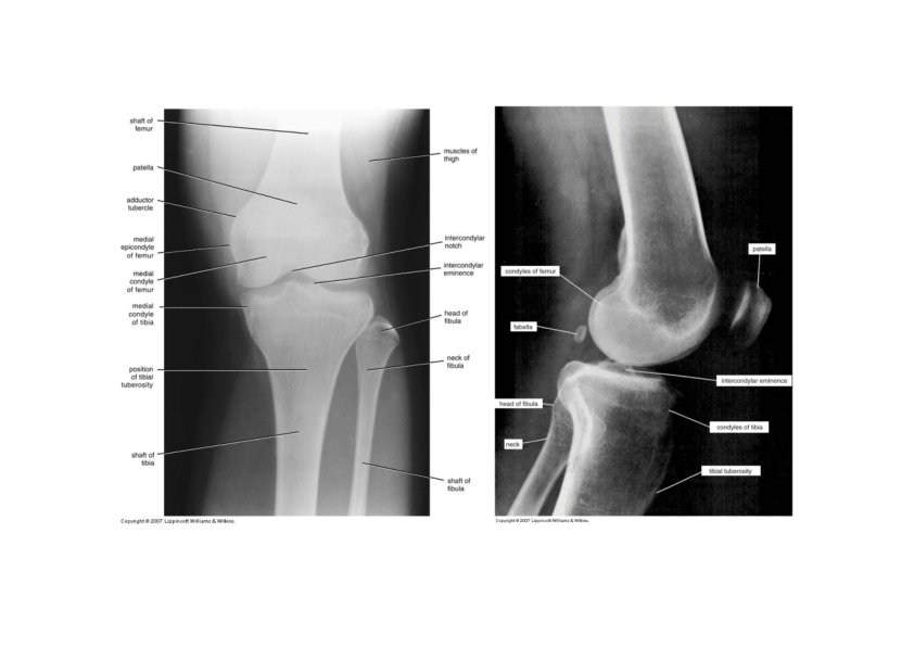
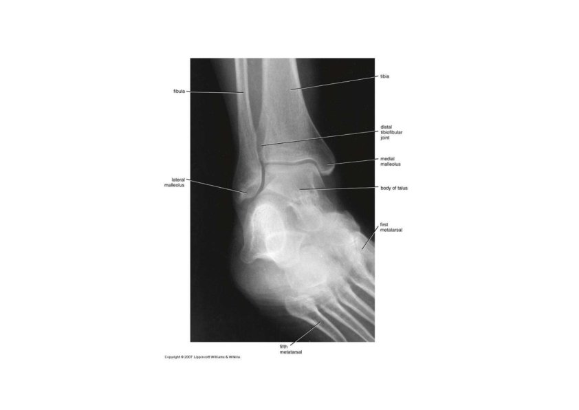

Surface Anatomy of Tibia & Fibula • Tibial tuberosity • Anteromedial surface of tibia • Head of fibula • Medial malleolus • Lateral malleolus
 • Talus = ankle")
Tarsus • Proximal region of foot (contains 7 tarsal bones) • Talus = ankle bone (articulates with tibia & fibula) • Calcaneus ‐ heel bone • Cuboid, navicular & 3 cuneiforms
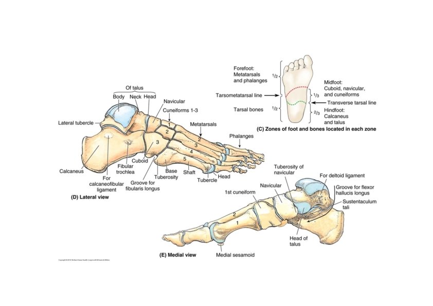

Tarsus • Calcaneum • • Tubercles Sustentaculum tali Sulcus calcanei Peroneal tubercle (fibular trochlea • Talus • Head, neck, and body • Sulcus tali • Sinus tarsi • Interosseous talocalcaneal ligament
 • each")
Metatarsus and Phalanges • Metatarsus • 5 metatarsals (1 is most medial) • each with base, shaft and head • Phalanges • distal portion of the foot • similar in number and arrangement to the hand • big toe is hallux
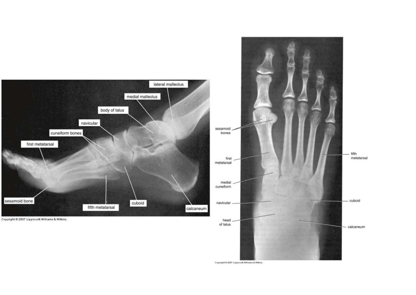

Surface Anatomy of Bones of Foot • Medial tubercle of calcaneus • Medial side • Head of talus • Sustentaculum tali • Lateral side • Fibular trochlea • Tuberosity of the 5 th metatarsal

Arches of the Foot • Function • distribute body weight over foot • yield & spring back when weight is lifted • Longitudinal arches along each side of foot • Medial longitudinal arch • Calcaneum, talus, navicular, cuniforms, and 1‐ 3 metatarsals • Lateral longitudinal arch • Calcaneum, cuboid, and 4‐ 5 metatarsals • Transverse arch across midfoot region • Cuboid, cuneiforms & bases of metatarsals • Flatfoot ‐ the arches decrease or “fall” • Clawfoot ‐ too much arch occurs due to various pathologies
- Slides: 30