
Lower GI Bleed Sameer Lapsia MD Anupama Chawla
Lower GI Bleed Sameer Lapsia, MD Anupama Chawla, MD Stony Brook Children’s Hospital 2013 Resident Education Series Reviewed by Christine Waasdorp Hurtado, MD of the Professional Education Committee
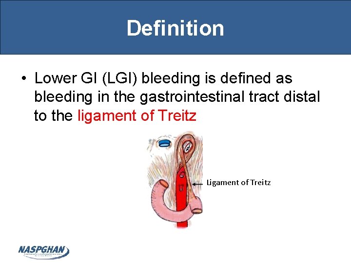
Definition • Lower GI (LGI) bleeding is defined as bleeding in the gastrointestinal tract distal to the ligament of Treitz Ligament of Treitz
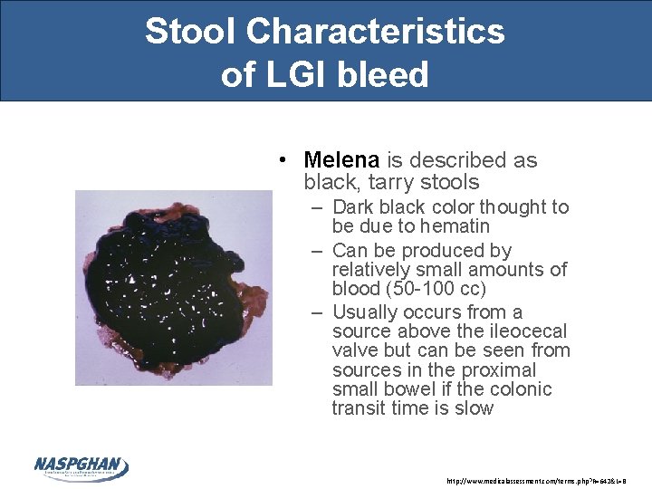
Stool Characteristics of LGI bleed • Melena is described as black, tarry stools – Dark black color thought to be due to hematin – Can be produced by relatively small amounts of blood (50 -100 cc) – Usually occurs from a source above the ileocecal valve but can be seen from sources in the proximal small bowel if the colonic transit time is slow http: //www. medicalassessment. com/terms. php? R=642&L=B
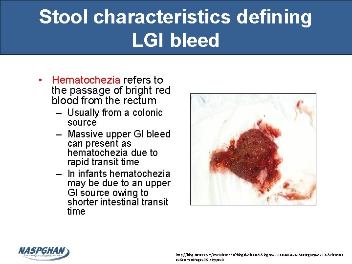
Stool characteristics defining LGI bleed • Hematochezia refers to the passage of bright red blood from the rectum – Usually from a colonic source – Massive upper GI bleed can present as hematochezia due to rapid transit time – In infants hematochezia may be due to an upper GI source owing to shorter intestinal transit time http: //blog. naver. com/Post. View. nhn? blog. Id=siasia 29&log. No=110084804249&category. No=135&view. Dat e=¤t. Page=1&listtype=0
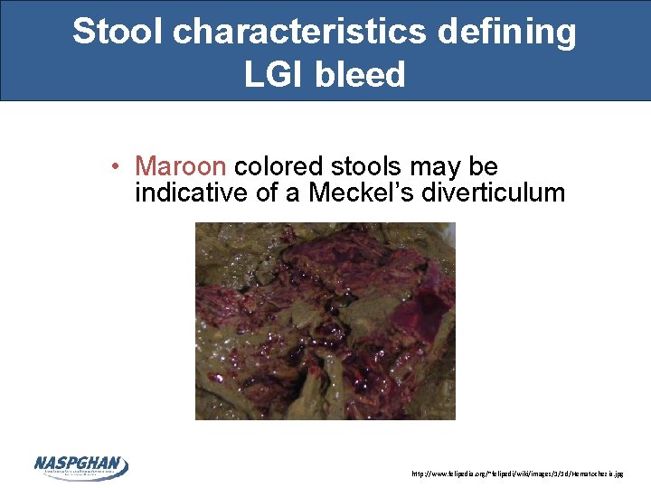
Stool characteristics defining LGI bleed • Maroon colored stools may be indicative of a Meckel’s diverticulum http: //www. felipedia. org/~felipedi/wiki/images/1/1 d/Hematochezia. jpg
Blood or not blood? : That is the question • Many items can appear as blood in the stools such as foods containing red food coloring (Jell-O, Kool Aid), syrup medications, beets, tomato and peach skins • Many items can appear as melena such as spinach, licorice, grape juice and certain medications such as Pepto-Bismol (due to bismuth) and Iron • Question is best answered by performing a guaiac test
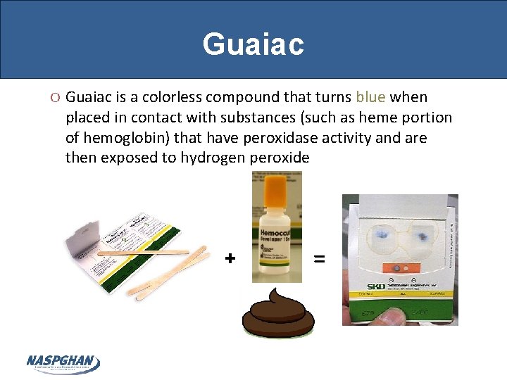
Guaiac O Guaiac is a colorless compound that turns blue when placed in contact with substances (such as heme portion of hemoglobin) that have peroxidase activity and are then exposed to hydrogen peroxide + =
Guaiac O “False-positives” can occur in foods containing peroxidase activity such as red meat, melons, grapes, radishes, turnips, cauliflower, and broccoli O “False-negative” can occur in patients taking Vitamin C due to anti-oxidant properties
Source of Blood : GI tract or not? That is another question • Melena may be seen in patients who have swallowed blood posttonsillectomy, or from epistaxis or traumatic nasopharayngeal lesions due to passage of a NG-tube • Swallowed maternal blood • Females who are menstruating may appear to have hematochezia and often their stool occult blood is positive • Hematuria may be mistaken for hematochezia • Munchausen syndrome by proxy should also be considered
Blood from the Lower or Upper GI tract? : • Upper GI bleed can mimic a lower GI bleed – Placement of a NG-tube and aspiration and guaiac of gastric contents can help • If negative guaiac may indicate bleeding from a distal source • However, a bleeding duodenal ulcer may not necessarily yield a positive guaiac. – If suspicion is high of an Upper GI source then an upper endoscopy is warranted
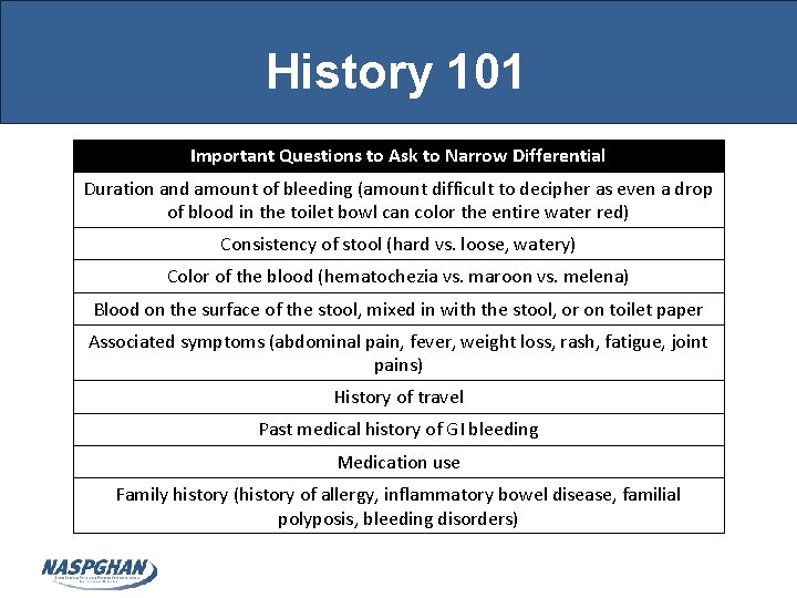
History 101 Important Questions to Ask to Narrow Differential Duration and amount of bleeding (amount difficult to decipher as even a drop of blood in the toilet bowl can color the entire water red) Consistency of stool (hard vs. loose, watery) Color of the blood (hematochezia vs. maroon vs. melena) Blood on the surface of the stool, mixed in with the stool, or on toilet paper Associated symptoms (abdominal pain, fever, weight loss, rash, fatigue, joint pains) History of travel Past medical history of GI bleeding Medication use Family history (history of allergy, inflammatory bowel disease, familial polyposis, bleeding disorders)
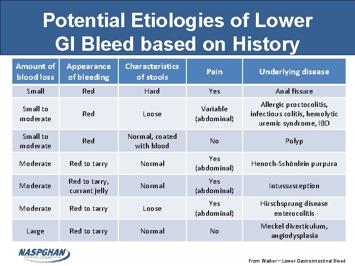
Potential Etiologies of Lower GI Bleed based on History Amount of blood loss Appearance of bleeding Characteristics of stools Pain Underlying disease Small Red Hard Yes Anal fissure Allergic proctocolitis, infectious colitis, hemolytic uremic syndrome, IBD Small to moderate Red Loose Variable (abdominal) Small to moderate Red Normal, coated with blood No Polyp Moderate Red to tarry Normal Yes (abdominal) Henoch-Schönlein purpura Moderate Red to tarry, currant jelly Normal Yes (abdominal) Intussusception Moderate Red to tarry Loose Yes (abdominal) Hirschsprung disease enterocolitis Large Red to tarry Normal No Meckel diverticulum, angiodysplasia From Walker – Lower Gastrointestinal Bleed
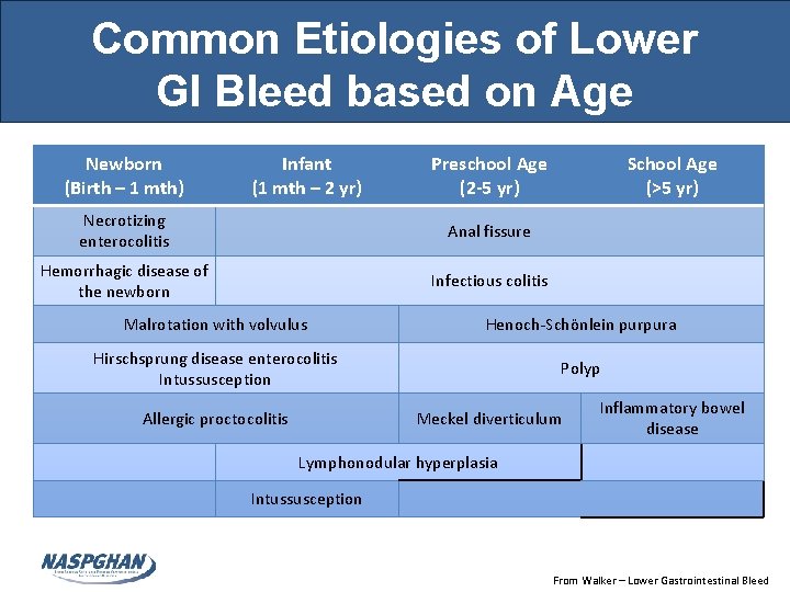
Common Etiologies of Lower GI Bleed based on Age Newborn (Birth – 1 mth) Infant (1 mth – 2 yr) Preschool Age (2 -5 yr) Necrotizing enterocolitis Anal fissure Hemorrhagic disease of the newborn Infectious colitis School Age (>5 yr) Malrotation with volvulus Henoch-Schönlein purpura Hirschsprung disease enterocolitis Intussusception Polyp Allergic proctocolitis Meckel diverticulum Inflammatory bowel disease Lymphonodular hyperplasia Intussusception From Walker – Lower Gastrointestinal Bleed
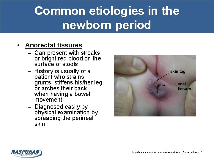
Common etiologies in the newborn period • Anorectal fissures – Can present with streaks or bright red blood on the surface of stools – History is usually of a patient who strains, grunts, stiffens his/her leg or arches their back when having a bowel movement – Diagnosed easily by physical examination by spreading the perineal skin http: //www. homeouniverse. com/category/diseases/stomach-diseases/
Common etiologies in the newborn period • Milk- or soy-induced colitis – Caused by ingestion of milk or soy protein which causes an inflammatory enteropathy in susceptible newborns – 25% - 50% of patients with cow’s milk protein intolerance will have a cross-reaction to soy protein – Treatment is with elimination of milk and soy protein from the mother’s diet if the infant is exclusively breastfed • Can challenge by adding milk or soy protein back in diet – Treatment is with elimination of milk and soy formula in formula fed infants – Casein-hydrolysate formula – Amino acid formula – Usually resolves by 1 year of age
Common etiologies in the newborn period • Necrotizing enterocolitis (NEC) – Intestinal pneumatosis of unclear etiology – Associated with prematurity, low birth weight, asphyxia, and sepsis (10% of NEC occurs in fullterm patients) • Correlation with PRBC transfusion – Symptoms include systemic instability, apnea, lethargy, poor feeding, abdominal distention, bilious emesis, and bloody stools – Medical and/or surgical management depends on the severity and course of the disease
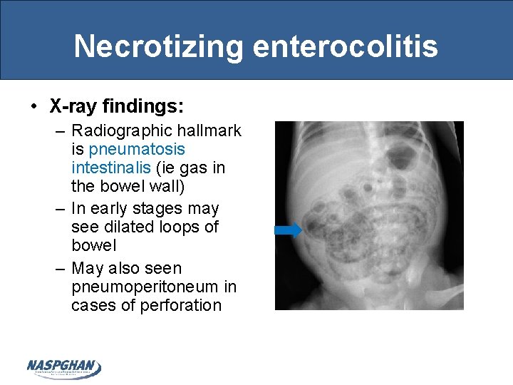
Necrotizing enterocolitis • X-ray findings: – Radiographic hallmark is pneumatosis intestinalis (ie gas in the bowel wall) – In early stages may see dilated loops of bowel – May also seen pneumoperitoneum in cases of perforation
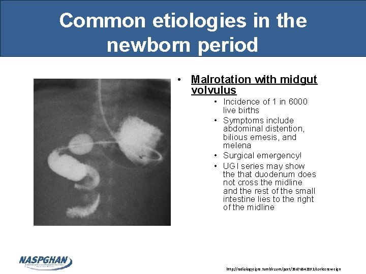
Common etiologies in the newborn period • Malrotation with midgut volvulus • Incidence of 1 in 6000 live births • Symptoms include abdominal distention, bilious emesis, and melena • Surgical emergency! • UGI series may show the that duodenum does not cross the midline and the rest of the small intestine lies to the right of the midline http: //radiologysigns. tumblr. com/post/35676542893/corkscrew-sign
Common etiologies in infancy • Hirschsprung Disease Enterocolitis – Typical presentation is that of severe constipation, abdominal distention, vomiting, and feeding intolerance. • 10 -30% of patients have Lower GI bleeding • Gold standard for diagnoses is via rectal biopsy demonstrating absence of ganglion cells in the Meissner and the Auerbach plexus • Treatment is via either a diverting colostomy followed by a second-stage intestinal pull-through of intestine containing nerves or a primary pull-through
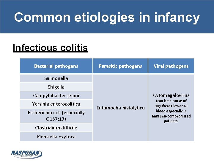
Common etiologies in infancy Infectious colitis Bacterial pathogens Parasitic pathogens Viral pathogens Salmonella Shigella Cytomegalovirus Campylobacter jejuni Yersinia enterocolitica Escherichia coli (especially O 157: 17) Clostridium difficile Klebsiella oxytoca Entamoeba histolytica (can be a cause of significant lower GI bleed especially in immuno-compromised patients)
Common etiologies in infancy • Meckel diverticulum – Rule of 2 s: – 2% of the population – 2: 1 male: female ratio – 2 feet from the ileocecal valve (usual location) – 2 inches in diameter – 2% of patients develop complications – 2 types of tissue are usual (gastric mucosa – most common; can also have pancreatic tissue) O Presentation: O The lower GI bleed is often brisk and painless O Stool often described as maroon colored O Diagnosis can be made by radionuclide 99 m pertechnetate scan showing the presence of heterotopic gastric mucosa; occasionally laparoscopy/laparotomy may be necessary O Rx via surgical excision
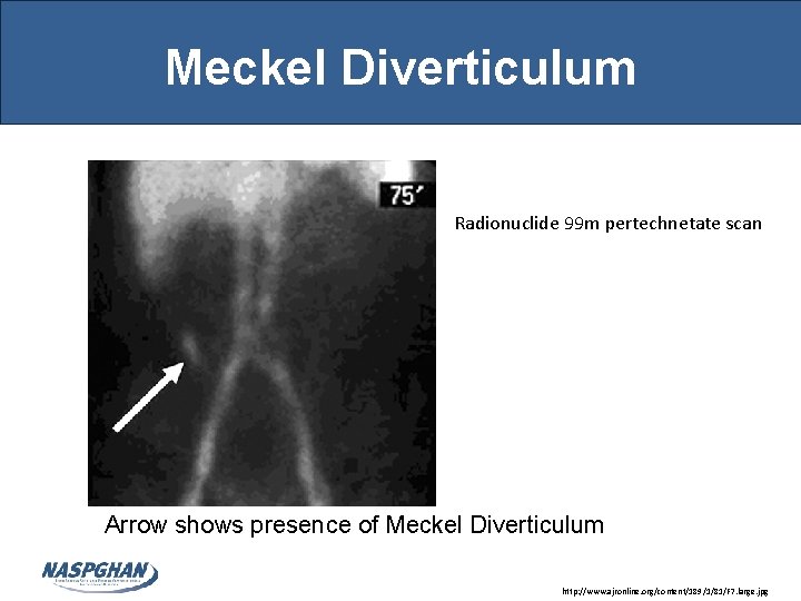
Meckel Diverticulum Radionuclide 99 m pertechnetate scan Arrow shows presence of Meckel Diverticulum http: //www. ajronline. org/content/189/1/81/F 7. large. jpg
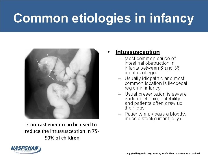
Common etiologies in infancy • Intussusception Contrast enema can be used to reduce the intussusception in 7590% of children – Most common cause of intestinal obstruction in infants between 6 and 36 months of age – Usually idiopathic and most common location is ileocecal region in infancy – Usual presentation is severe abdominal pain, irritability and patients often draw up their legs – Patients may pass a bloody, mucoid stool(currant jelly) http: //radiologyinthai. blogspot. com/2011/10/intussusception-reduction. html
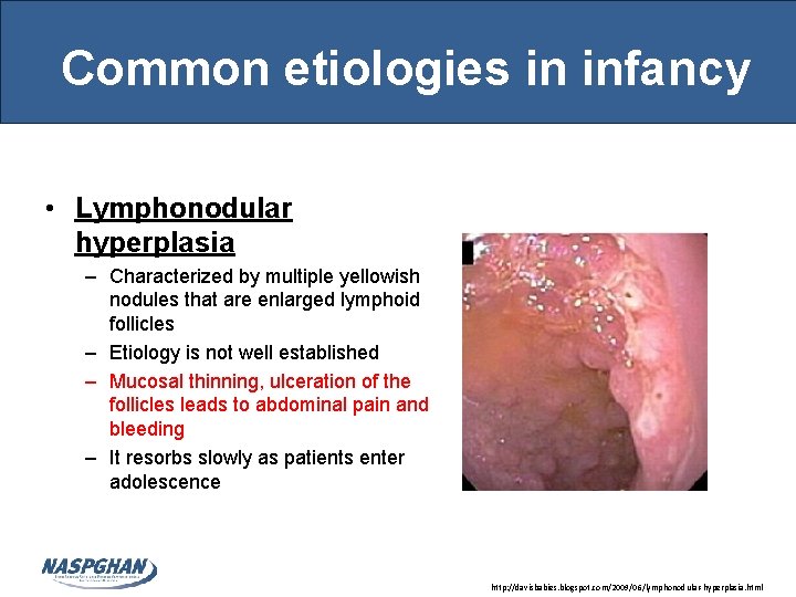
Common etiologies in infancy • Lymphonodular hyperplasia – Characterized by multiple yellowish nodules that are enlarged lymphoid follicles – Etiology is not well established – Mucosal thinning, ulceration of the follicles leads to abdominal pain and bleeding – It resorbs slowly as patients enter adolescence http: //davisbabies. blogspot. com/2009/06/lymphonodular-hyperplasia. html
Common etiologies during Preschool Age • Polyps • Intestinal polyps can cause isolated, recurrent, and painless hematochezia • Juvenile polyps account for more than 95% of all polyps found in children and have low malignancy potential – They are hamartomatous – Vast majority are solitary and occur mostly on the left side of the colon – Recurrent or multiple polyps can be seen in juvenile polyposis coli or generalized juvenile polyposis • Adenomatous polyps are more frequently found in adults and have high malignancy potential • Both are treated with excision via colonoscopy or if too numerous or malignant potential via surgery
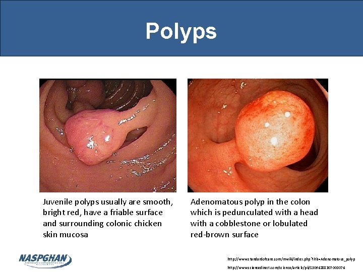
Polyps Juvenile polyps usually are smooth, bright red, have a friable surface and surrounding colonic chicken skin mucosa Adenomatous polyp in the colon which is pedunculated with a head with a cobblestone or lobulated red-brown surface http: //www. standardofcare. com/mwiki/index. php? title=Adenomatous_polyp http: //www. sciencedirect. com/science/article/pii/S 1096288307001076
Common etiologies during Preschool Age • Henoch-Schönlein Purpura • Systemic vasculitis involving the skin, GI tract, joints and kidneys • Typical presentation is an urticarial rash on the buttocks and lower extremities with peak onset from 3 -7 years followed by large joint arthralgias and papular purpuric lesions • GI manifestions occur in 45 -75% of patients » Include: vomiting, colicky abdominal pain, melena, and/or bloody stools due to diffuse mucosal hemorrhage » Intussusception associated with HSP can also cause melena or hematochezia
Common etiologies during School Age • Inflammatory bowel disease • 90 -95% of patients with Ulcerative Colitis and 25% of patients with Crohn’s disease present with rectal bleeding and/or bloody diarrhea • The peak incidence of IBD occurs in patients between the ages of 15 and 25 years. – Approximately 25 to 30 percent of patients with CD and 20 percent of patients with UC present before the age of 20 years
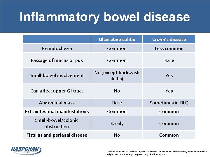
Inflammatory bowel disease Ulcerative colitis Crohn’s disease Hematochezia Common Less common Passage of mucus or pus Common Rare Small-bowel involvement No (except backwash ileitis) Yes Can affect upper GI tract No Yes Abdominal mass Rare Sometimes in RLQ Extraintestinal manifestations Common Small-bowel/colonic obstruction Rarely Common Fistulas and perianal disease No Common Modified from: Das KM. Relationship of extraintestinal involvements in inflammatory bowel disease: New insights into autoimmune pathogenesis. Dig Dis Sci 1999; 44: 1.
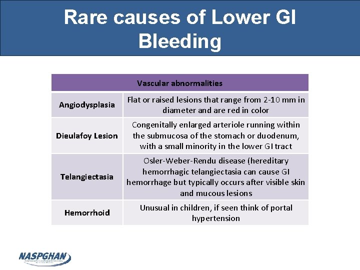
Rare causes of Lower GI Bleeding Vascular abnormalities Angiodysplasia Flat or raised lesions that range from 2 -10 mm in diameter and are red in color Dieulafoy Lesion Congenitally enlarged arteriole running within the submucosa of the stomach or duodenum, with a small minority in the lower GI tract Telangiectasia Osler-Weber-Rendu disease (hereditary hemorrhagic telangiectasia can cause GI hemorrhage but typically occurs after visible skin and mucous lesions Hemorrhoid Unusual in children, if seen think of portal hypertension
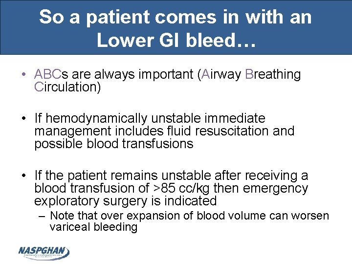
So a patient comes in with an Lower GI bleed… • ABCs are always important (Airway Breathing Circulation) • If hemodynamically unstable immediate management includes fluid resuscitation and possible blood transfusions • If the patient remains unstable after receiving a blood transfusion of >85 cc/kg then emergency exploratory surgery is indicated – Note that over expansion of blood volume can worsen variceal bleeding
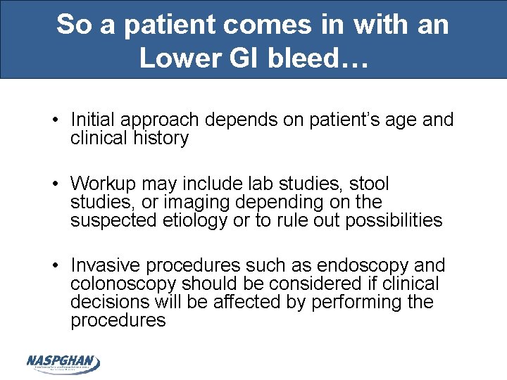
So a patient comes in with an Lower GI bleed… • Initial approach depends on patient’s age and clinical history • Workup may include lab studies, stool studies, or imaging depending on the suspected etiology or to rule out possibilities • Invasive procedures such as endoscopy and colonoscopy should be considered if clinical decisions will be affected by performing the procedures
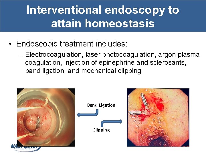
Interventional endoscopy to attain homeostasis • Endoscopic treatment includes: – Electrocoagulation, laser photocoagulation, argon plasma coagulation, injection of epinephrine and sclerosants, band ligation, and mechanical clipping Band Ligation Clipping
References • Turck D & Michaud L. Lower Gastrointestinal Bleeding. In Walker’s Pediatric Gastrointestinal Disease 5 Volume 2. Shelton, CT: People’s Medical Publishing House. • Abdullah BA, Gupta SK, & Croffie JM. The role of esophagogastroduodenoscopy in the initial evaluation of childhood inflammatory bowel disease: a 7 -year study. Journal of Pediatric Gastroenterology & Nutrition 2002; 35: 636.
- Slides: 34