LOWER GASTROINTESTINA L BLEEDING SUPERVISED BY DR EMAD

LOWER GASTROINTESTINA L BLEEDING SUPERVISED BY DR. EMAD ABU-RAJOOH DONE BY: SALWA AZZAWI SARA ZAIDEEN BALKEES NASER YALA JAWAZNEH

OBJECTIVES • Definition • Approach • Classification • Most common causes • Management

DEFINITION

DEFINITION • Acute lower gastrointestinal bleeding is hemorrhage arising distal to the ligament of Treitz. • The colon is the source of hemorrhage in more than 95% to 97% of cases, with the remaining 3 to 5%arising in small bowel sites.

DEFINITION • Massive LGIB is defined as follows: Passage of a large volume of red or maroon blood through the rectum with hemodynamic instability and shock.

DIFFERENTIATION Upper GI bleeding Lower GI bleeding More common 4: 1 Less common From nose to Treitz Distal to Teritz Hematamesis & Melena Hematocezia +/Melena Elevated BUN: Creatinine ratio >30

APPROACH EMERGENCY DEPARTMENT HISTORY PHYSICAL EXAMINATION INVESTIGATION

APPROACH • Most cases of acute colonic bleeding will stop spontaneously, thereby allowing non-urgent evaluation. However, for patients with severe hematochezia, defined as continued bleeding within the first 24 h of hospitalization with a drop in the hemoglobin of at least 4 -6 g/d. L and/or a transfusion requirement of at least 2 units of packed red blood cells, urgent diagnosis and intervention are required to control the bleeding. • Clinical factors predictive of severe colonic bleeding include aspirin use, at least two comorbid illnesses, pulse greater than 100/minute, and systolic blood pressure <115 mm. Hg. The overall mortality rate from colonic bleeding is 2. 4– 3. 9 %. Independent predictors of inhospital mortality are age over 70 years, intestinal ischemia, and two or more comorbidities.

APPROACH Emergency Department • patients with acute LGIB, who might have signs of hemodynamic instability should be resuscitated thoroughly according to the extent of blood loss. 1. Two wide bore cannulas are installed, 2. Blood is withdrawn for CBC, cross-match, coagulation profile and urea and electrolytes 3. Proper replacement with I. V fluids or blood products is done 4. Urinary catheter is important for input-output charts 5. Oxygen is administered as needed.

APPROACH History • Important historical points to assess include: 1. Abdominal pain and weight loss (non-specific, but may suggest inflammatory bowel disease, ischemia, and/or malignancy). 2. Medication use (NSAIDS and other medications that can cause ulcers or intestinal ischemia). 3. Recent colonoscopy with polypectomy (post-polypectomy bleed), prior abdominal/pelvic radiation (radiation proctitis/colitis). 4. Prior operations (possible anastomotic ulcers). 5. History of abdominal aortic aneurysm with or without surgical repair (possible aorto-enteric fistula). 6. History of alcoholism or chronic liver disease raises the suspicion for bleeding due to portal hypertension. 7. The manner in which the patient with bleeding presents can also suggest potential etiologies. Bright red blood is more often seen from ano-rectal and distal colonic sources, but brisk upper GI bleeding can also manifest this way. 8. Painless severe bleeding with clots is more common with diverticular hemorrhage. Bloody diarrhea often occurs with ischemic and inflammatory colitides.

APPROACH Physical examination should include measurement of orthostatic vital signs in patients without overt shock. Pertinent findings on physical examination may include: 1. Scars from previous abdominal incisions. 2. The presence of abdominal masses, or skin and oral lesions suggestive of polyposis syndromes. 3. Stigmata of cirrhosis suggestive of bleeding from hemorrhoids or varices secondary to portal hypertension should be considered. 4. The rectal examination is important to identify anorectal pathology, including tumors, ulcers, or polyps. 5. The color of the rectal contents and the presence of formed stool or blood clot should also be noted. 6. Anoscopic examination to exclude hemorrhage from hemorrhoids should be completed. 7. A nosogastric tube should be inserted to look for blood or coffee ground-like material to exclude an upper gastrointestinal source. 8. In patients with hematochezia and hemodynamic instability, emergency upper endoscopy is required.

APPROACH Investigations Patients with presumed LGIB may undergo: 1. Early sigmoidoscopy for the detection of obvious, low-lying lesions. However, the procedure is difficult with brisk bleeding, and it is usually not possible to identify the area of bleeding. Sigmoidoscopy is useful primarily in patients <40 years with minor bleeding. 2. Patients with hematochezia and hemodynamic instability (acute LGIB) should have upper endoscopy to rule out an upper Gl source before evaluation of the lower Gl tract, upper endoscopy is also indicated in patients with chronic LGIB suspected to have UGIB. 3. You can use NGT: if suction is blood this mean UGIB, if bile most likely no UGIB, if clear fluid can’t rule out. 4. Colonoscopy after an oral lavage solution is the procedure of choice in patients admitted with LGIB unless bleeding is too massive or unless sigmoidoscopy has disclosed an obvious actively bleeding lesion, colonoscopy provides both a diagnostic and therapeutic tool.

APPROACH Investigations 5. 99 MTc-labeled red cell scan allows repeated imaging for up to 24 h reveals bleeding even with a low rate of blood lose(01 ml/min). it does localize the site of the lesion but not identify the cause. 6. In active LGIB, angiography can detect the site of bleeding (extravasation of contrast into the gut) and permits treatment with intraarterial infusion of vasopressin or embolization. Even after bleeding has stopped, angiography may identify lesions with abnormal vasculature, such as vascular ectasias or tumours. 7. Occasionally, the LGIB source is in the small intestine. Push enteroscopy and capsule endoscopy have been used for the assessment of obscure bleeding.

CLASSIFICATION OF CAUSES
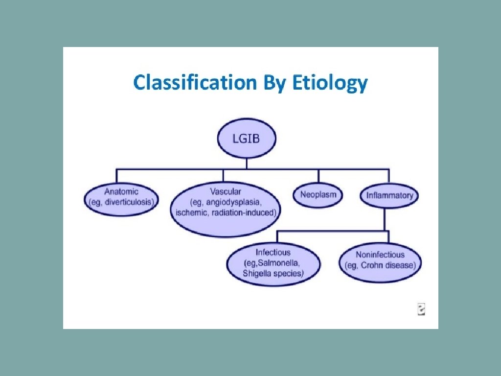

CLASSIFICATION BY SEVERITY Acute Subacute/Chronic Diverticular disease Anal disease Angiodysplasia Polyps Meckel’s diverticulum Carcinoma Ischemic colitis Solitary rectal ulcer Mesenteric ischemia Radiation enteritis

MOST COMMON CAUSES

DIVERTICULAR DISEASE In the United States, diverticula are the most common cause of significant lower GI bleeding. In the past diverticula were thought to be rare in patients younger than 40 years, but it is now an increasingly common diagnosis in this age group. Only 3% to 15% of individuals with diverticulosis experience any bleeding. Bleeding generally occurs at the neck of the diverticulum and is believed to be secondary to bleeding from the vasa recti as they penetrate through the submucosa.

DIVERTICULAR DISEASE • Of those that bleed, more than 75% stop spontaneously, although approximately 10% will rebleed within a year and almost 50% within 10 years. • Although diverticular disease is much more common on the left side, right-sided disease is responsible for more than half the bleeding.

ANGIODYSPLASIA • Angiodysplasias of the intestine, also referred to as arteriovenous malformations, are distinct from hemangiomas and true congenital arteriovenous malformations. • They are thought to be acquired degenerative lesions secondary to progressive dilation of normal blood vessels within the submucosa of the intestine. Angiodysplasias have an equal gender distribution and • are almost uniformly found in patients older than 50 years. These lesions are notably associated with aortic stenosis and renal failure especially in the elderly.

ANGIODYSPLASIA • The hemorrhage tends to arise from the right side of the colon, with the cecum being the most common location, although they can occur in the rest of the colon and small bowel. • Most patients present with chronic bleeding, but in up to 15%, hemorrhage may be massive. Bleeding stops spontaneously in most cases, but approximately 50% will rebleed within 5 years.

MANAGEMENT

MANAGEMENT In diverticular disease: • • The best method of diagnosis and treatment is colonoscopy, although success is sometimes limited by the large amount of bleeding. If the bleeding diverticulum can be identified, epinephrine injection may control the bleeding. Electrocautery can also be used, and most recently, endoscopic clips have been successfully applied to control the hemorrhage. If bleeding ceases with these maneuvers or spontaneously, expectant management may be appropriate; however, this requires clinical judgment based on the magnitude of the hemorrhage and the patient’s comorbidities, particularly cardiac disease.

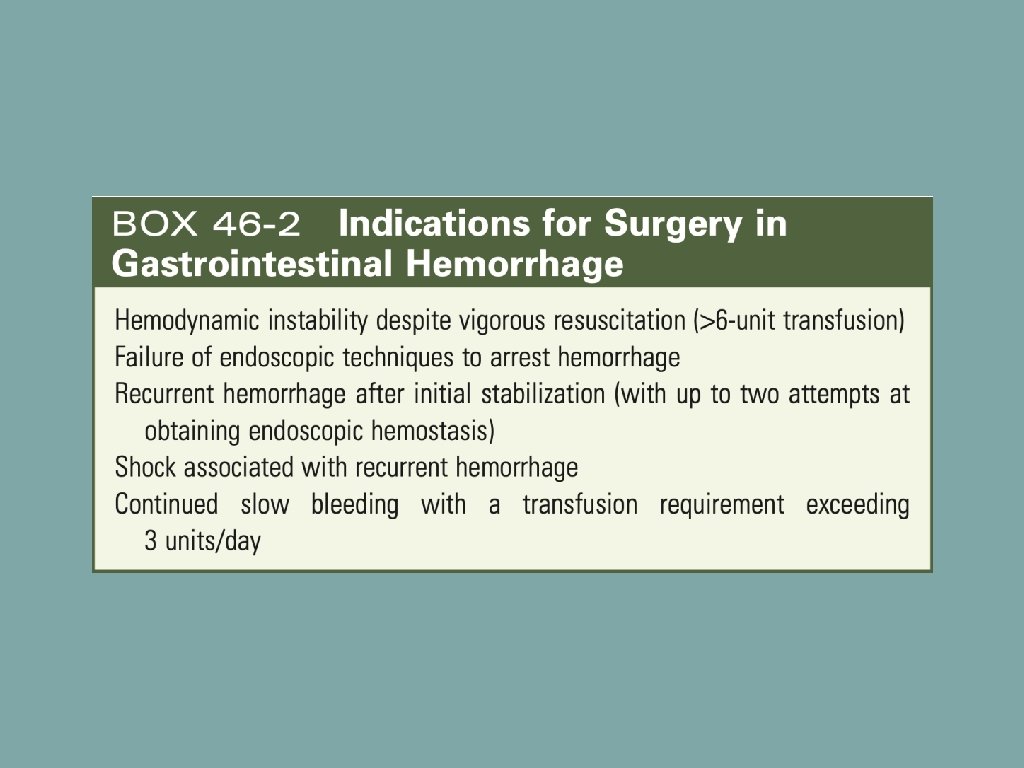
In diverticular disease: • • If none of these maneuvers is successful or if hemorrhage recurs, embolization can be considered. angiography with Superselective embolization of the bleeding colonic vessel has gained popularity with high success rates (>90%), although the risk of ischemic complications continues to be of concern. Under these circumstances, colonic resection is indicated. Certainty of the site of bleeding is critical. As surgery: Blind hemicolectomy is associated with rebleeding in more than 50% of patients, and operation based on RBC scan localization alone can result in recurrent hemorrhage in up to one third of patients. Subtotal colectomy does not eliminate the risk of recurrent hemorrhage and, compared with segmental resection, is accompanied by a significant increase in morbidity, particularly diarrhea in older patients, in whom the remaining rectum may never adapt.

MANAGEMENT In angiodysphagia : These lesions can be diagnosed by either colonoscopy or angiography. During colonoscopy, they appear as red stellate lesions with a surrounding rim of pale mucosa and can be treated with sclerotherapy or electrocautery. Angiography demonstrates dilated, slowly emptying veins and sometimes early venous filling. If these lesions are discovered incidentally, no furtherapy is indicated. In acutely bleeding patients, they have been successfully treated with: 1. Intra-arterial vasopressin (as a drug) 2. Selective gel foam embolization 3. Endoscopic electrocoagulation 4. Injection with sclerosing agents. As Surgery: If these measures fail or bleeding recurs and the lesion has been localized, most commonly right colectomy, is effective. segmental resection,
")
THANK YOU References: Sabiston Textbook of Surgery National Center for Biotechnology Information (NCBI)
- Slides: 27