Low voltage ECG The QRS is said to


- Slides: 4

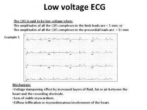
Low voltage ECG The QRS is said to be low voltage when: The amplitudes of all the QRS complexes in the limb leads are < 5 mm; or The amplitudes of all the QRS complexes in the precordial leads are < 10 mm Example 1 Mechanism: • Voltage dampening effect by increased layers of fluid, fat or air between the heart and the recording electrode. • Loss of viable myocardium. • Diffuse infiltration or myxoedematous involvement of the heart.

Low voltage ECG Causes • Fluid Pericardial effusion Pleural effusion • Fat Obesity • Air Emphysema Pneumothorax • Infiltrative / Connective Tissue Disorders Myxoedema Infiltrative diseases (restrictive cardiomyopathy) e. g. amyloidosis, sarcoidosis, haemochromatosis Constrictive pericarditis Scleroderma • Loss of viable myocardium Previous massive MI End-stage dilated cardiomyopathy Example 2 Most important cause: massive pericardial effusion (above) Produces a triad of: • Low voltage • Tachycardia • Electrical alternans ; consecutive, normally-conducted QRS complexes alternate in height. (heart swings backwards and forwards within a large fluid-filled pericardium. )

Example 3 Low voltage ECG Previous massive anterior MI: • Low QRS voltage in V 1 -6. • This ECG also demonstrates biphasic anterior T waves (Wellen’s syndrome) indicating new critical occlusion of the LAD artery.

Example 4 Low Voltage ECG Emphysema • Low voltages in the limb leads • Other features of emphysema include: rightward axis, peaked P waves (P pulmonale) and clockwise rotation (reduced R wave progression)


