Longlasting depressionlike behavior and epigenetic changes of BDNF

Long-lasting depression-like behavior and epigenetic changes of BDNF gene expression induced by perinatal exposure to methylmercury Onishchenko N, Karpova N, Sabri F, Castrén E, Ceccatelli S. Maneeshi Prasad 11/13/2009

Depression • 1 in 6 individual in US is affected from depression during their lifetime • Approximately 18. 8 million American adults, or about 9. 5 percent of the U. S. population age 18 and older in a given year, have a depressive disorder • Nearly twice as many women (12. 0 percent) as men (6. 6 percent) are affected by a depressive disorder each year • Depression affects all people regardless of age, geographic location, demographic or social position

Depression • Depression often exists with other diseases, including chronic pain, arthritis, diabetes and HIV • Depression is also known to weaken the immune system, making the body more susceptible to other medical illnesses • Anxiety disorders, such as post–traumatic stress disorder (PTSD), obsessive–compulsive disorder, panic disorder, social phobia and generalized anxiety disorder, often accompany depression

Causes of Depression • Biochemical. People with depression have physical changes in their brains with neurotransmitters and also could be a culprit may also play a role in depression • Genes. Depression is more common in people who are biologically related to a family member who also has the condition • Environmental factors such as the loss of a loved one, financial problems and high stress also leads to depression

Symptoms of depression Depression symptoms can vary greatly with age. Some common symptoms are: • Loss of interest in normal daily activities • Feeling sad or down • Feeling hopeless • Crying spells for no apparent reason • Problems sleeping • Trouble focusing or concentrating • Difficulty making decisions • Unintentional weight gain or loss • Irritability • Restlessness • Being easily annoyed • Feeling fatigued or weak • Feeling worthless • Loss of interest in sex • Thoughts of suicide or suicidal behavior • Unexplained physical problems, such as back pain or headaches

Neural circuitry of depression • Limbic regions have been implicated in depression and antidepressant action • Neural activity in amygdala and subgenual cingulate cortex is highly increased in individuals with depressive symptoms and goes back to normal on successful treatment

Neural circuitry of depression

Neurotrophins • Depressed patients show volumetric decrease in the hippocampus and forebrain regions • This relates to the decrease in neurotrophic factors • BDNF mediated signaling gets reduced due to stress • Chronic antidepressant treatment increases BDNF mediated signaling • Similar results have been seen in post-mortem hippocampus of patients with depression: low BDNF levels in hippocampus and serum

BDNF signaling • BDNF signaling has region and antidepressant specific effect • Antidepressant effects can be inhibited by blocking neurogenesis in hippocampus • Antidepressants increase the amount of growth factors (BDNF, VEGF) that influence neurogenesis in hippocampus

Neurotrophic hypothesis of depression • Reduction in brain BDNF levels leads to depression and increase in brain BDNF levels leads to antidepressant action • Antidepressant increase the Tr. KB activation and signaling – Activation of PLCg signaling • Phosphorylation of CREB – Increase in BDNF transcription
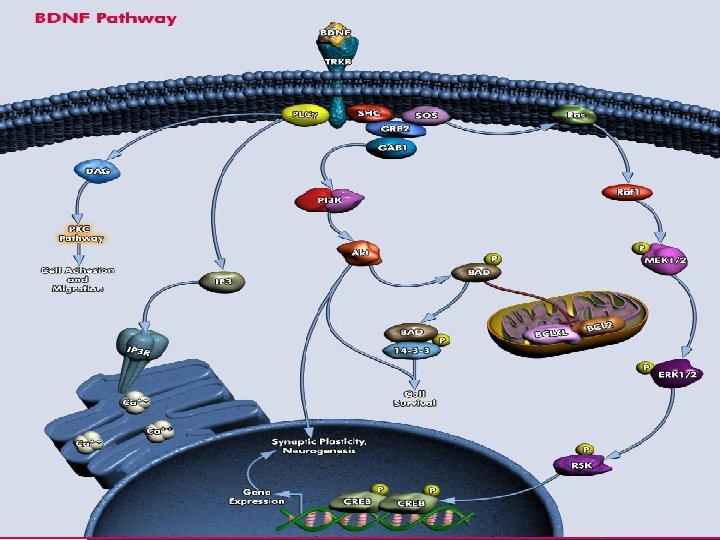

BDNF and depression

Neurodevelopmental disorders • Learning disabilities, sensory deficits, attention deficit, hyperactivity disorder • Autism and autism spectrum disorders, traumatic brain injury, communication, speech and language disorders • Genetic disorders such as fragile-X syndrome, Down syndrome and Rett syndrome, epilepsy, fetal alcohol syndrome, learning disorders, neurological and psychiatric

• Environmental conditions affect expression of genes, resulting in long lasting changes in structure and function of brain – Stress – Drug treatments – Maternal care – Toxins e. g. : Methyl mercury Environmental contaminant present in sea food

How People Are Exposed to Mercury • Eating fish or shellfish that is contaminated with methylmercury, which is the main source of general human exposures to mercury • Breathing air contaminated with elemental mercury vapors (e. g. , in workplaces such as dental offices and industries that use mercury or in locations where a mercury spill or release has occurred) • Having dental fillings that contain mercury; and • Practicing cultural or religious rituals that use mercury.

How Mercury Affects People’s Health • Short-term exposure to extremely high levels of elemental mercury vapors can result in lung damage, nausea, diarrhea, increases in blood pressure or heart rate, skin rashes, eye irritation, and injury to the nervous system • Prolonged exposure to lower levels of elemental mercury can permanently damage the brain and kidneys • The developing brain of a fetus can be injured if the mother is exposed to methylmercury

Levels of Mercury in U. S. Population • Survey period of 2005– 2006: 95 th percentile levels for total blood mercury in children aged 1 -5 years and females aged 16 -49 years were 1. 43μg/L and 4. 48 μg/L, respectively i. e. only 5 percent of the population will have values at this level or higher Mercury on neurodegeneration http: //www. epa. gov/mercury/effects. htm http: //www. epa. gov/mercury/advisories. htm

Long-lasting depression-like behavior and epigenetic changes of BDNF gene expression induced by perinatal exposure to methylmercury

Experimental Animals and methods • C 57 BL/6/Bkl mice – Pregnant dams exposed to Me. Hg @ 0. 5 mg/kg/day via drinking water from gestational day 7 till day 7 after delivery – No change in litter size, body weight gain – Results in brain mercury concentration similar to infants in human population who eat Me. Hg contaminated fish Ø In-situ hybridization, Ch. IP, Primer extension, Densitometry

Forced swim test • Animals starting at 9 weeks were tested • Individuals were tested for immobility for 6 min after a pretest of 15 min 24 hrs before the test • Immobility- passively floating in water for 2 sec or longer with only small movements enough to keep the head above water

Results: Depression-like behavior of Me. Hg-exposed mice

Results of FST • Immobility time was longer in perinatally Me. Hg-exposed mice than controls of 9 weeks, 12 weeks and 14 months • Confirming the long-lasting effect of Me. Hg • Treatment with fluoxetine (Prozac) reduced the immobility time in the Me. Hg treated animals

Hippocampal BDNF m. RNA level

Tr. KB m. RNA expression

Results of BDNF m. RNA expression levels • Me. Hg lead to significant decrease in BDNF m. RNA level in dentate gyrus but not in CA 1 and CA 3 regions of 12 week old mice and persisted in 14 -month old mice • No change was seen in Tr. KB m. RNA expression in the hippocampal formation • Fluoxetine treatment was able to restore the BDNF m. RNA expression in dentate gyrus

Epigenetic regulation in depression. H 3 K 27 -methylation : no transcription H 3 K 9 and H 3 K 14 - acetylating: transcription
")
Chromatin Immunoprecipitation (Ch. IP)

BDNF Gene locus with 8 promoters

Epigenetic state of BDNF gene

H 3 K 27 methylation and H 3 acetylation regulate BDNF gene expression in hippocampus • Increased H 3 K 27 tri-methylation and decrease in histone H 3 acetylation at the BDNF promoter IV of 12 week old mice that persisted through 14 -month old Me. Hg exposed mice leading to silencing of BDNF gene • Fluoxetine treatment had no effect on the H 3 k 27 trimethylation but it did increase the histone H 3 acetylation to overcome the repression of BDNF transcription

Methylation of Cp. G sites • Using MS-SNu. PE (Methylation Sensitive Single Nucleotide Primer Extension) DNA treated with bisulphite to convert unmethylated cytosine converted to uracil Primer extension with P 32 labeled TTP and CTP, extension will end just 5’ to the C or U Quantification of CTP/TTP ratio to estimate the amount of methylated C Methylated Cp. G result in repression of transcription

Epigenetic state of BDNF gene

Epigenetic state of BDNF gene • Significant increase in DNA methylation at BDNF promoter IV in hippocampus was seen in 14 month old mice exposed to Me. Hg prenatally

Discussion • Previous studies have shown that Me. Hg exposure during developmental stages have behavioral effects in rodent animal models similar to depression • Current data confirms the role of Me. Hg in incorporating long term epigenetic changes in BDNF gene locus leading to depression-like symptoms in mice model exposed to Me. Hg prenatally • Antidepressant treatment was able to overcome this Me. Hg induced repression of BDNF expression in the hippocampus

Discussion • Impairment in memory, attention, language and visuospatial perception has been seen in children exposed to Me. Hg prenatally • Depressive syndromes have been seen in adults with occupational exposure to mercury • These patients have reduced hippocampal and serum BDNF levels, which can be restored by antidepressant treatment
")
Prozac (selective 5 -HT reuptake inhibitor)

Discussion • During depression there is decrease in BDNF m. RNA in dentate gyrus • BDNF expression has been associated with increase in neurogenesis in the subgranular zone of dentate gyrus • Suggesting that Me. Hg may be involved in decrease in neurogenesis

Discussion • Promoter IV of BDNF gene is regulated by neuronal activity (Ca. RF and CREB) • Me. Hg was able to induce long lasting epigenetic changes in this promoter in the hippocampus by methylation of H 3 K 27 and decrease in acetylation of H 3 • Antidepressant was able to increase the acetylation of H 3 at this promoter to relieve the Me. Hg induced repression of BDNF gene expression • Antidepressants may act through downregulation of histone deacetylases resulting in increased acetylation of histones and thus gene transcription

- Slides: 39