Loffler 2008 Brodmanns areas Brodmanns areas Segier Vuilleumier


Loffler, 2008

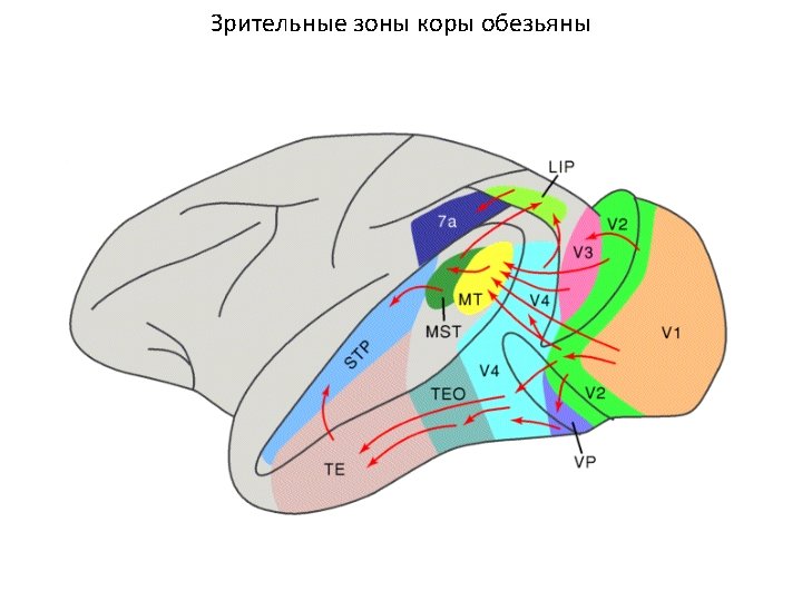

Brodmann’s areas

Brodmann’s areas

Segier, Vuilleumier, 2006
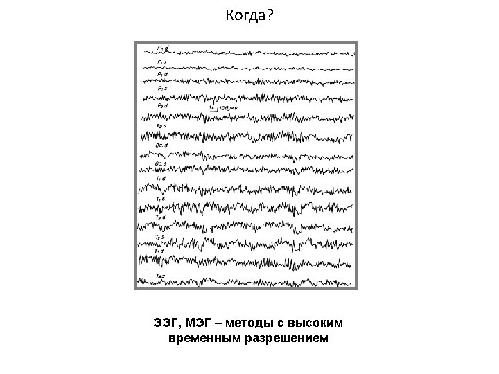

Kinsey et al. , 2009

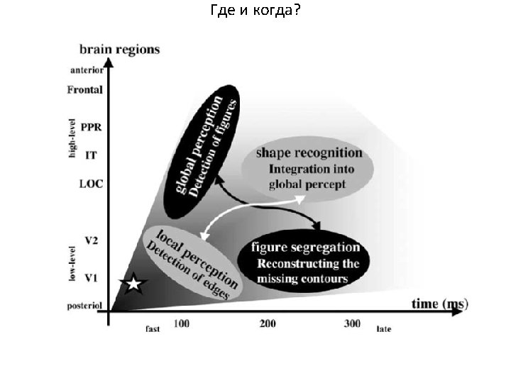
Fig. 2. Surface rendered group SAM images of the posterior brain depicting statistical estimates (pb 0. 05) of power changes within the 1– 4 Hz, 10– 30 Hz and 30– 95 Hz frequency bands between active and passive experimental states (500 ms) for viewing the illusory triangle (top), non-triangle pattern (middle) and real triangle pattern (bottom). The colour scale shows pseudo-t values for the oscillatory power changes: white-purple colours indicate a relative decrease in signal power during the active phase, while yellow–orange colours indicate a relative increase. Kinsey et al. , 2009

Fig. 3. Local maxima from group SAM images in the 10– 30 Hz frequency range for the illusory-(top row) non-(middle row) and realtriangles (bottom row). White dots represent local maxima for a post-stimulus time window of 0– 500 ms; black dots represent local maxima for a post-stimulus window of 300– 800 ms. Kinsey et al. , 2009


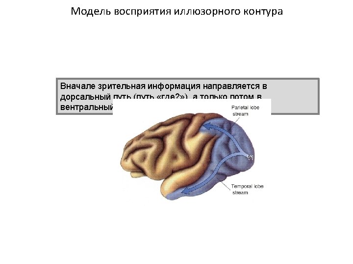
Kimberly et al. , 2010

Kimberly et al. , 2010

Kimberly et al. , 2010
- Slides: 26