
LOCALISING THE LESION WHERE IN THE CNS Kate


- Slides: 35
LOCALISING THE LESION: “WHERE IN THE CNS” Kate Hassan
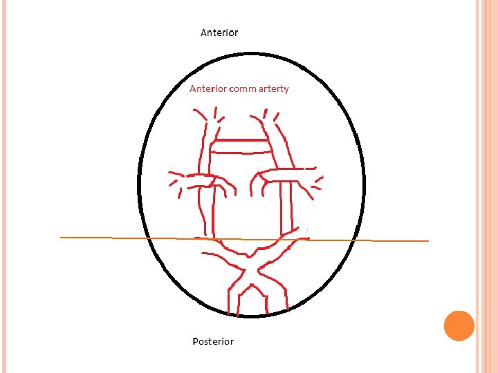
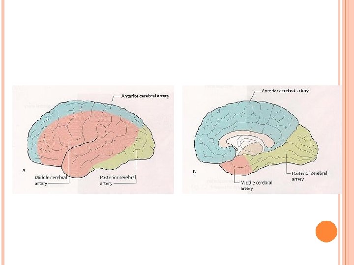
LEARNING OBJECTIVES Definition of CNS and PNS Definition of UMN and LMN Function of each of the cerebral lobes The homunculus Circle of willis and blood supply to the cerebral hemispheres Motor tracts – lateral corticospinal Sensory tracts – lateral spinothalamic and dorsal columns Stroke syndromes Clinical case scenarios
DEFINITIONS CNS = Brain and spinal cord PNS = anything outside brain and spinal cord � Also include autonomic nervous system and cranial nerves
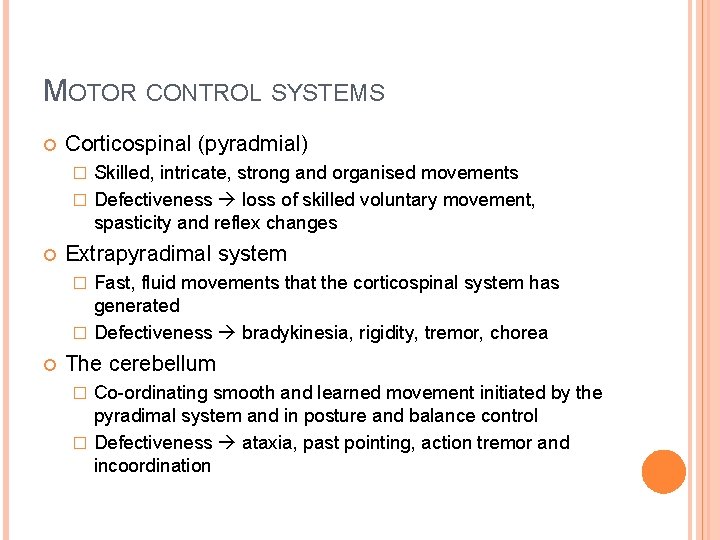
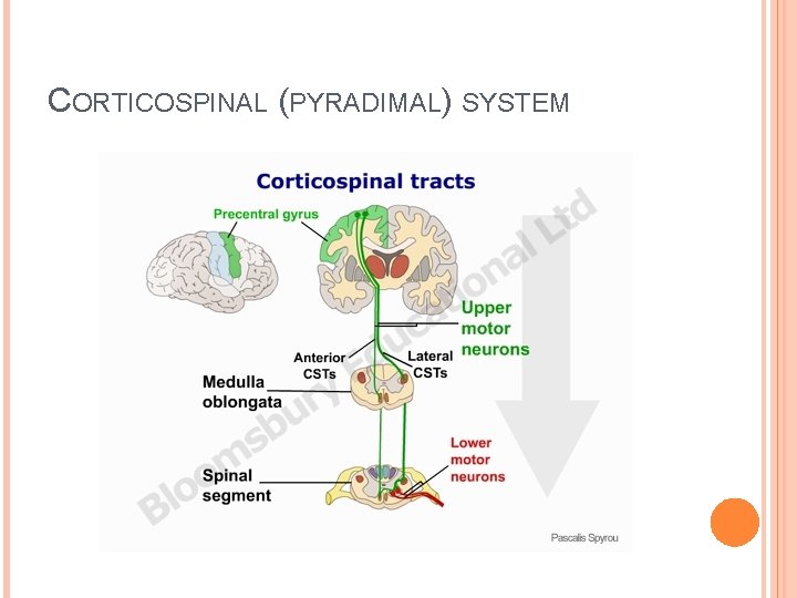
MOTOR CONTROL SYSTEMS Corticospinal (pyradmial) Skilled, intricate, strong and organised movements � Defectiveness loss of skilled voluntary movement, spasticity and reflex changes � Extrapyradimal system Fast, fluid movements that the corticospinal system has generated � Defectiveness bradykinesia, rigidity, tremor, chorea � The cerebellum Co-ordinating smooth and learned movement initiated by the pyradimal system and in posture and balance control � Defectiveness ataxia, past pointing, action tremor and incoordination �
CORTICOSPINAL (PYRADIMAL) SYSTEM
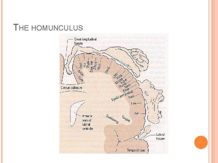
THE HOMUNCULUS
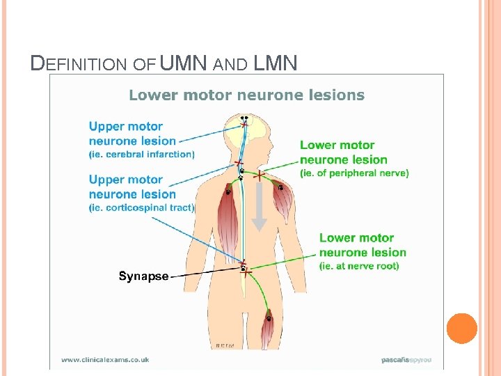
DEFINITION OF UMN AND LMN
UMN SIGNS VS LMN SIGNS UMN LMN wasting no yes fasciculation no yes tone increased decreased Power decreased increased reflexes increased decreased Plantars up going down going
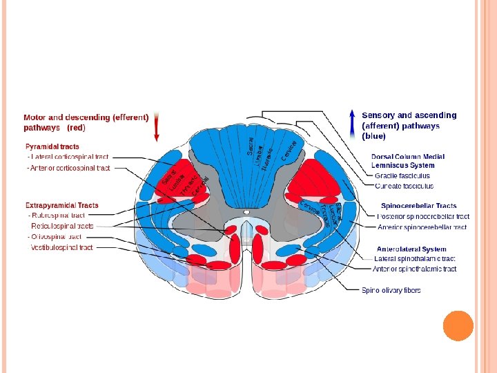
SENSORY PATHWAYS Peripheral nerves carry sensation from dorsal roots to the cord Posterior columns (dorsal columns) � Vibration, joint position, light touch and point discrimination � Cross in the brainstem passing to the thalamus Spinothalamic tracts � Pain and temperature � Cross within the cord and pass in the spinothalamic tracts to the thalamus and reticular formation Sensory cortex � Fibres from the thalamus pass to the parietal region sensory cortex and motor cortex
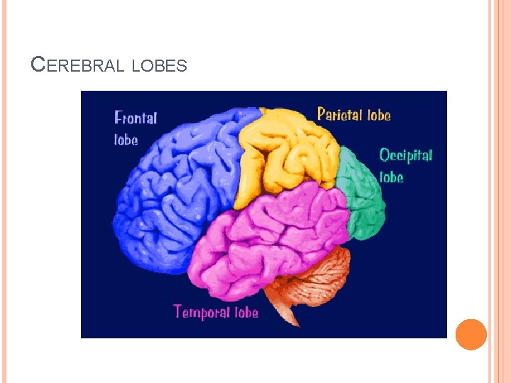
CEREBRAL LOBES
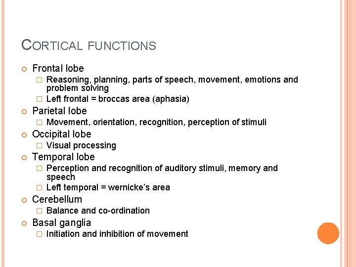
CORTICAL FUNCTIONS Frontal lobe Reasoning, planning, parts of speech, movement, emotions and problem solving � Left frontal = broccas area (aphasia) � Parietal lobe � Occipital lobe � Movement, orientation, recognition, perception of stimuli Visual processing Temporal lobe Perception and recognition of auditory stimuli, memory and speech � Left temporal = wernicke’s area � Cerebellum � Balance and co-ordination Basal ganglia � Initiation and inhibition of movement
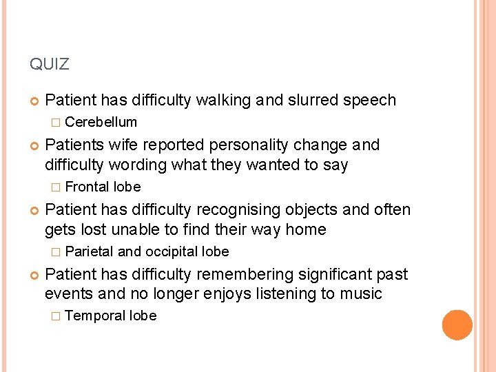
QUIZ Patient has difficulty walking and slurred speech � Cerebellum Patients wife reported personality change and difficulty wording what they wanted to say � Frontal Patient has difficulty recognising objects and often gets lost unable to find their way home � Parietal lobe and occipital lobe Patient has difficulty remembering significant past events and no longer enjoys listening to music � Temporal lobe
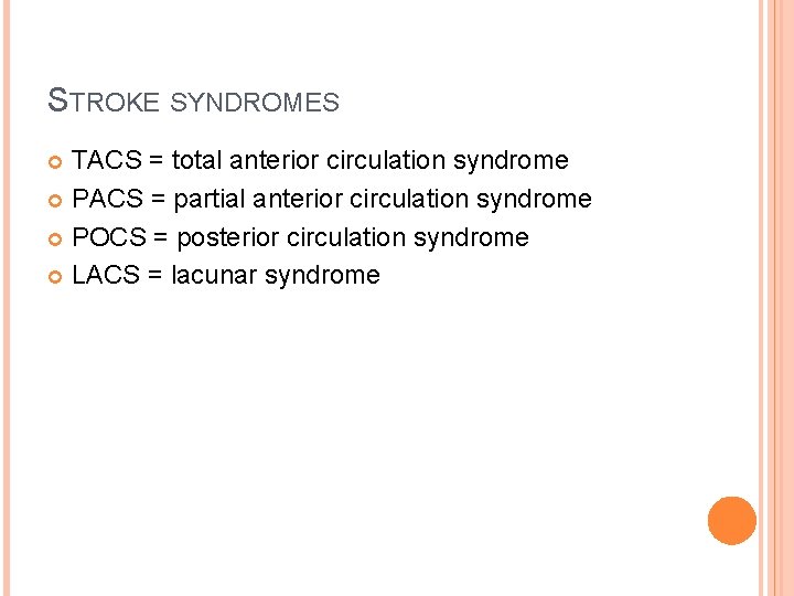
STROKE SYNDROMES TACS = total anterior circulation syndrome PACS = partial anterior circulation syndrome POCS = posterior circulation syndrome LACS = lacunar syndrome
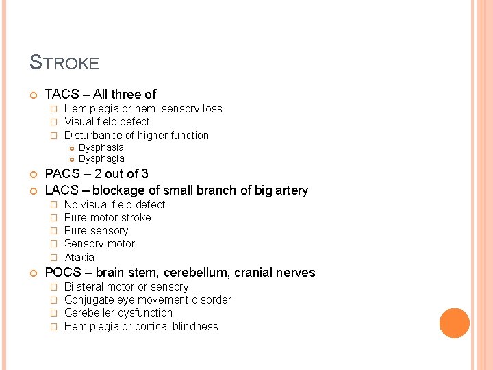
STROKE TACS – All three of � � � Hemiplegia or hemi sensory loss Visual field defect Disturbance of higher function PACS – 2 out of 3 LACS – blockage of small branch of big artery � � � Dysphasia Dysphagia No visual field defect Pure motor stroke Pure sensory Sensory motor Ataxia POCS – brain stem, cerebellum, cranial nerves � � Bilateral motor or sensory Conjugate eye movement disorder Cerebeller dysfunction Hemiplegia or cortical blindness
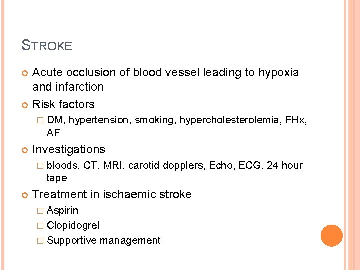
STROKE Acute occlusion of blood vessel leading to hypoxia and infarction Risk factors � DM, hypertension, smoking, hypercholesterolemia, FHx, AF Investigations � bloods, CT, MRI, carotid dopplers, Echo, ECG, 24 hour tape Treatment in ischaemic stroke � Aspirin � Clopidogrel � Supportive management
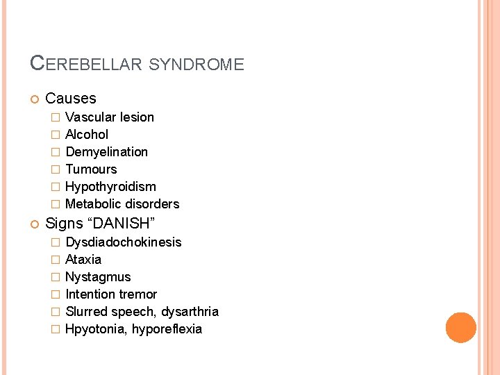
CEREBELLAR SYNDROME Causes � � � Vascular lesion Alcohol Demyelination Tumours Hypothyroidism Metabolic disorders Signs “DANISH” � � � Dysdiadochokinesis Ataxia Nystagmus Intention tremor Slurred speech, dysarthria Hpyotonia, hyporeflexia
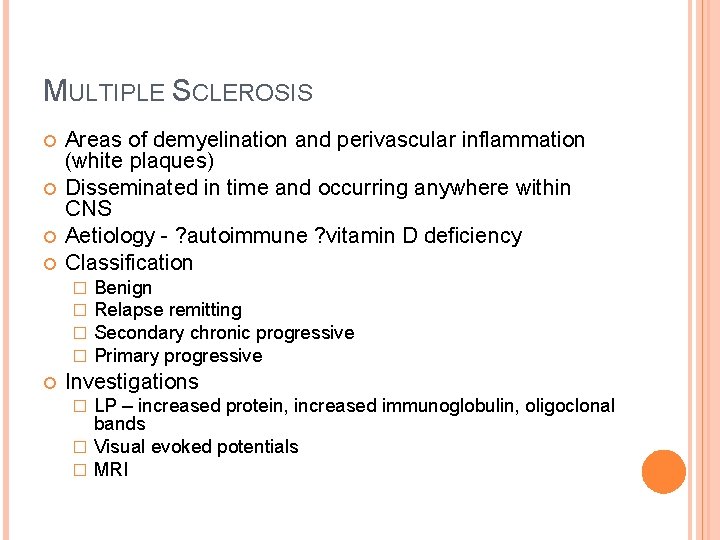
MULTIPLE SCLEROSIS Areas of demyelination and perivascular inflammation (white plaques) Disseminated in time and occurring anywhere within CNS Aetiology - ? autoimmune ? vitamin D deficiency Classification � � Benign Relapse remitting Secondary chronic progressive Primary progressive Investigations LP – increased protein, increased immunoglobulin, oligoclonal bands � Visual evoked potentials � MRI �
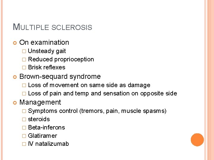
MULTIPLE SCLEROSIS On examination � Unsteady gait � Reduced proprioception � Brisk reflexes Brown-sequard syndrome � Loss of movement on same side as damage � Loss of pain and temp and sensation on opposite side Management � Symptoms control (tremors, pain, muscle spasms) � steroids � Beta-inferons � Glatiramer � IV natalizumab
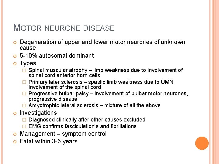
MOTOR NEURONE DISEASE Degeneration of upper and lower motor neurones of unknown cause 5 -10% autosomal dominant Types Spinal muscular atrophy – limb weakness due to involvement of spinal cord anterior horn cells � Primary later sclerosis – spastic limb weakness due to UMN involvement of the spinal cord � Progressive bulbar palsy – involvement of bulbar motor neurones, progressive disease � Amyotrophic lateral sclerosis – mixture of all the above � Investigations � � Diagnosed clinically after other causes excluded EMG confirms fasciculation's and fibrillations Management – symptom control Fatal within 3 -5 years
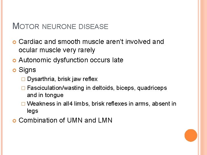
MOTOR NEURONE DISEASE Cardiac and smooth muscle aren’t involved and ocular muscle very rarely Autonomic dysfunction occurs late Signs � Dysarthria, brisk jaw reflex � Fasciculation/wasting in deltoids, biceps, quadriceps and in tongue � Weakness in all 4 limbs, brisk reflexes in arms, absent in legs Combination of UMN and LMN
CLINICAL CASE 1 23, female presents to her GP with a 2 week history of bilateral leg weakness having started with pins and needles and numbness in her hands and feet. She has had a few days of urinary incontinence which has resolved. 2 years ago she had an episode of blurred vision and pain in the right eye which lasted a month and fully resolved
CASE 1 What is the most likely diagnosis? What other signs or symptoms might you see in this condition? What is the pathological basis of this disorder? What further investigations would you do? How would you manage this patient?
CLINICAL CASE 2 61 female Becoming increasingly weak on her right side over a one week period. She is unable to walk and has slurred speech and right side of her face is drooping Past history of breast cancer o/e – right facial weakness, grade 4/5 weakness of the right arm and leg, right homonymous hemianopia and some difficulty naming objects and reflexes are brisk on the right side and her right plantar response is upgoing
CASE 2 What is the likely diagnosis?
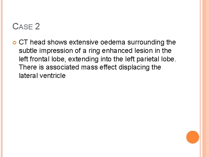
CASE 2 CT head shows extensive oedema surrounding the subtle impression of a ring enhanced lesion in the left frontal lobe, extending into the left parietal lobe. There is associated mass effect displacing the lateral ventricle
CASE 2 What is the likely cause? Other features that may be present? What management options are available?
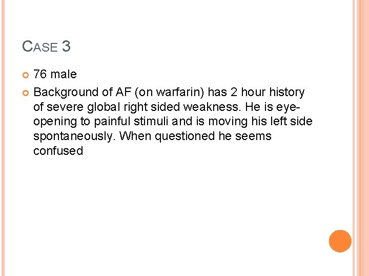
CASE 3 76 male Background of AF (on warfarin) has 2 hour history of severe global right sided weakness. He is eyeopening to painful stimuli and is moving his left side spontaneously. When questioned he seems confused
CASE 3 What is his GCS? What is the diagnosis? What investigations would you do? What are the important risk factors? How wound you manage this patient?
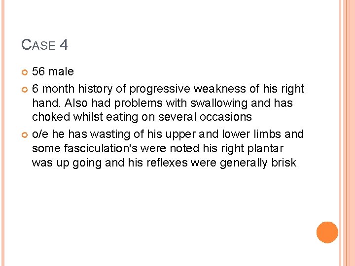
CASE 4 56 male 6 month history of progressive weakness of his right hand. Also had problems with swallowing and has choked whilst eating on several occasions o/e he has wasting of his upper and lower limbs and some fasciculation's were noted his right plantar was up going and his reflexes were generally brisk
CASE 4 Diagnosis? What investigations would you perform? How would you manage this patient?
THANK YOU FOR LISTENING Any questions?