
LIVER PANCREAS SPLEEN Objectives By the end of


LIVER, PANCREAS & SPLEEN Objectives: By the end of this lecture, the student should be able to describe: 1. The histological structure of liver with special emphasis on the classic hepatic lobule. 2. The histological structure of exocrine portion of pancreas. 3. The histological features of spleen.
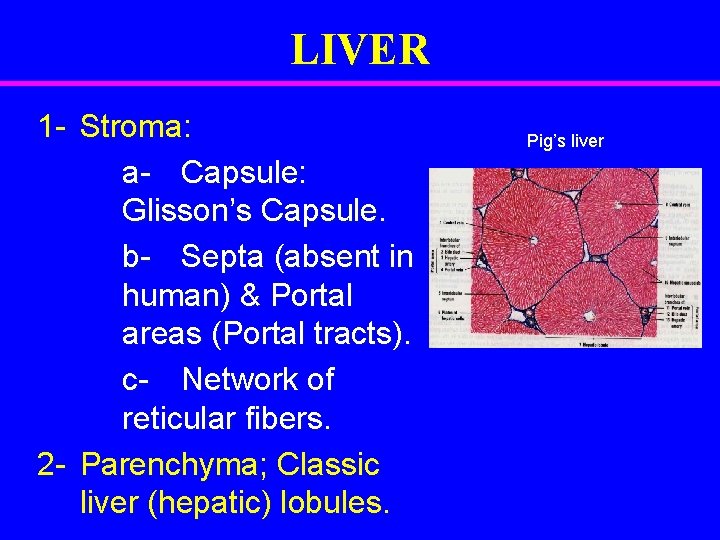
LIVER 1 - Stroma: a- Capsule: Glisson’s Capsule. b- Septa (absent in human) & Portal areas (Portal tracts). c- Network of reticular fibers. 2 - Parenchyma; Classic liver (hepatic) lobules. Pig’s liver
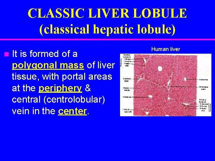
CLASSIC LIVER LOBULE (classical hepatic lobule) It is formed of a polygonal mass of liver tissue, with portal areas at the periphery & central (centrolobular) vein in the center. Human liver
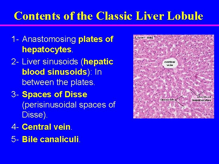
Contents of the Classic Liver Lobule 1 - Anastomosing plates of hepatocytes. 2 - Liver sinusoids (hepatic blood sinusoids): In between the plates. 3 - Spaces of Disse (perisinusoidal spaces of Disse). 4 - Central vein. 5 - Bile canaliculi.
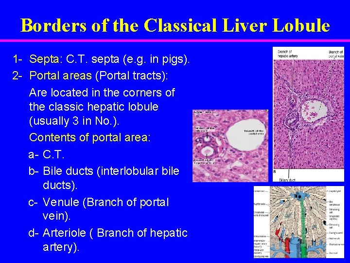
Borders of the Classical Liver Lobule 1 - Septa: C. T. septa (e. g. in pigs). 2 - Portal areas (Portal tracts): Are located in the corners of the classic hepatic lobule (usually 3 in No. ). Contents of portal area: a- C. T. b- Bile ducts (interlobular bile ducts). c- Venule (Branch of portal vein). d- Arteriole ( Branch of hepatic artery).
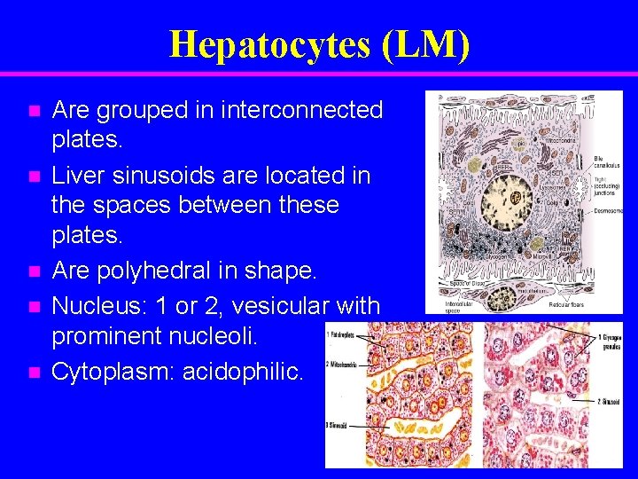
Hepatocytes (LM) Are grouped in interconnected plates. Liver sinusoids are located in the spaces between these plates. Are polyhedral in shape. Nucleus: 1 or 2, vesicular with prominent nucleoli. Cytoplasm: acidophilic.
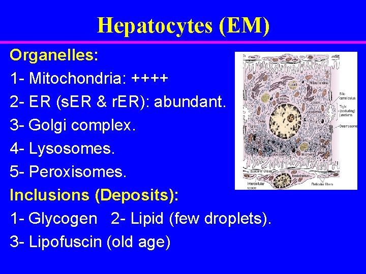
Hepatocytes (EM) Organelles: 1 - Mitochondria: ++++ 2 - ER (s. ER & r. ER): abundant. 3 - Golgi complex. 4 - Lysosomes. 5 - Peroxisomes. Inclusions (Deposits): 1 - Glycogen 2 - Lipid (few droplets). 3 - Lipofuscin (old age)

Liver Sinusoids (1) Endothelial Cells: – Fenestrated & discontinuous → free passage of plasma. – Basal lamina is absent. (2) Kupffer Cells: – Are macrophages. – Are found on the luminal surface of the endothelial cells. – Function: phagocytosis.

Space of Disse (Perisinusoidal Space) Contents: 1 - Fat-storing cells (Ito cells): – contain vitamin A-rich lipid. – form reticulin. 2 - Reticular fibers: (type III collagen). 3 - Plasma of blood. 4 - Microvilli of hepatocytes.
Pancreas
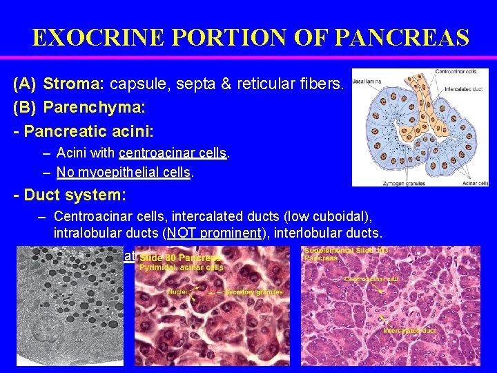
EXOCRINE PORTION OF PANCREAS (A) Stroma: capsule, septa & reticular fibers. (B) Parenchyma: - Pancreatic acini: – Acini with centroacinar cells. – No myoepithelial cells. - Duct system: – Centroacinar cells, intercalated ducts (low cuboidal), intralobular ducts (NOT prominent), interlobular ducts. – N. B. No striated ducts.
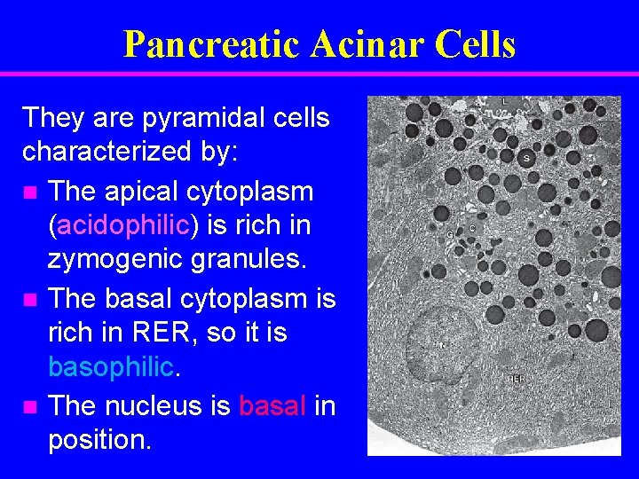
Pancreatic Acinar Cells They are pyramidal cells characterized by: The apical cytoplasm (acidophilic) is rich in zymogenic granules. The basal cytoplasm is rich in RER, so it is basophilic. The nucleus is basal in position.
SPLEEN
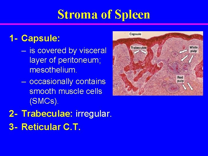
Stroma of Spleen 1 - Capsule: – is covered by visceral layer of peritoneum; mesothelium. – occasionally contains smooth muscle cells (SMCs). 2 - Trabeculae: irregular. 3 - Reticular C. T.
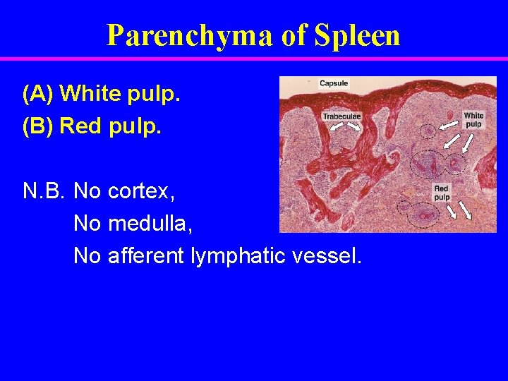
Parenchyma of Spleen (A) White pulp. (B) Red pulp. N. B. No cortex, No medulla, No afferent lymphatic vessel.
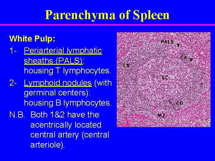
Parenchyma of Spleen White Pulp: 1 - Periarterial lymphatic sheaths (PALS): housing T lymphocytes. 2 - Lymphoid nodules (with germinal centers): housing B lymphocytes. N. B. Both 1&2 have the acentrically located central artery (central arteriole).
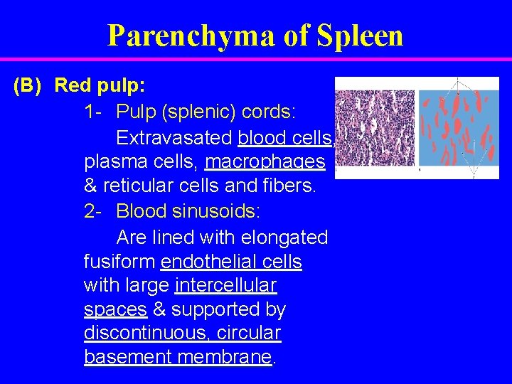
Parenchyma of Spleen (B) Red pulp: 1 - Pulp (splenic) cords: Extravasated blood cells, plasma cells, macrophages & reticular cells and fibers. 2 - Blood sinusoids: Are lined with elongated fusiform endothelial cells with large intercellular spaces & supported by discontinuous, circular basement membrane.
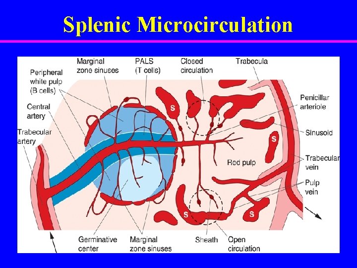
Splenic Microcirculation

BEST WISHES
- Slides: 20