liver failure The alterations that cause liver failure


 2 -Alcoholic")


 ↑ liver enz. Severe hepatic dysfunction is")


 is present")


 -Not necessarily correlated with other features")


 2 -Unpredictable (idiosyncratic) 56")


 is")


 is a benign, self-limited disease. •")


 - hepatocytes 2. HBe Ag(pre-core")


 • prevalence rate is 3% (0. 1% to 12%, depending")


")


 acute coinfection after exposure")
")


 -Necrosis of hepatocytes")


 those who harbor one of the viruses")

- Slides: 118
liver failure • The alterations that cause liver failure fall into 3 categories: • 1 - Acute liver failure with massive hepatic necrosis • 2 - Chronic liver disease • 3 - Hepatic dysfunction without overt necrosis. 1
1 -Acute liver failure. • This is most often caused by drugs or fulminant viral hepatitis. • Acute liver failure denotes clinical hepatic insufficiency that progresses from onset of symptoms to hepatic encephalopathy within 2 to 3 weeks. • A course extending as long as 3 months is called subacute failure. 2
• The histologic correlate of acute liver failure is massive hepatic necrosis. • It is an uncommon but life-threatening condition that often requires liver transplantation. 3
2 -Chronic liver disease • This is the most common route to hepatic failure and is the end point of relentless chronic liver damage ending in cirrhosis. 4
Hepatic dysfunction without overt necrosis. • Hepatocytes may be viable but unable to perform normal metabolic function: • 1 - acute fatty liver of pregnancy (which can lead to acute liver failure a few days after onset) • 2 - tetracycline toxicity • 3 - Reye syndrome 5
Clinical features 1 -Jaundice 2 -Hypoalbuminemia →edema 3 -Hyperammonemia 4 -Fetor hepaticus (musty or sweet & sour) 5 -Palmar erythema hyperestrogenemia 6 -Spider angiomas 7 -Hypogonadism & gynecomastia 6
Complications: 1 -Multiple organ failure e. g lung 2 -Coagulopathy → bleeding def. factors II, VII, IX, X 3 -Hepatic encephalopathy 4 -Hepatorenal Syndrome 7
Alcoholic liver disease -Alcohol is most widely abused agent -Excessive ethanol consumption causes more than 60% of chronic liver disease in most Western countries and accounts for 40% to 50% of deaths due to cirrhosis. -It is the 5 th leading cause of death in USA due to : 1. Accident 2. Cirrhosis 8
Pathogenesis • Short-term ingestion of as much as 80 gm of ethanol/d (8 beers or 7 ounces of 80 -proof liquor) generally produces mild, reversible hepatic changes. • Chronic intake of 50 to 60 gm/day is considered a borderline risk for severe injury. • women seem to be more susceptible to hepatic injury than are men because of low gastric metabolism of ethanol and differences in body composition. 9
-80 – 100 mg/dl is the legal definition for driving under the influence of alcohol -44 ml of ethanol is required to produce this level in 70 kg person -In occasional drinkers, bl. Level of 200 mg/dl produces coma & death & respiratory failure at 300 -400 mg/dl 10
- Habitual drinkers can tolerate levels up to 700 mg/dl without clinical effect due to metabolic tolerance explained by 5 -10 X induction of cytochrome P-450 system that includes enzyme CYP 2 E 1 which increases the metabolism of ethanol as well as other drugs as cocaine & acetominophen 11
Forms of alcoholic liver disease 1 -Hepatic steatosis (90 -100% of drinkers) 2 -Alcoholic hepatitis ( 1 - 35% of drinkers) 3 -Cirrhosis ( 14% of drinkers) - Steatosis & hepatitis may develop independently 12
Hepatic steatosis -Can occur following even moderate intake of alcohol in form of microvesicular steatosis - initially centrilobular but in severe cases it may involve the entire lobule. -Chronic intake → diffuse steatosis -Liver is large ( 4 – 6 kg) soft yellow & greasy -Continued intake →fibrosis -Fatty change is reversible with complete absention from further intake of alcohol 13
Alcoholic hepatitis Characteristic findings : 1 -Hepatocyte swelling & necrosis -Accumulation of fat & water & proteins -Cholestasis -Hemosiderin deposition in hepatocytes & kupffer cells 2 -Mallory-hayline bodies -eosinoplilic cytoplasmic inclusions in degenerating hepatocytes formed of cytokeratin infermediate filaments & other proteins 14
-Mallory-hayline inclusions are characteristic but not pathognomonic of alcoholic liver disease, they are also seen in : 1 -Primary biliary cirrhosis 2 -Wilson disease 3 -Chronic cholestatic syndromes 4 -Hepatocellular carcinoma 15
3 -Neutrophilic reaction 4 -Fibrosis -Sinusoidal & perivenular fibrosis -Periportal fibrosis 5 -Cholestasis 6 -Mild deposition of hemosiderin in hepatocytes & kupffer cells 16
Alcoholic cirrhosis -Usually it develops slowly -Initially the liver is enlarged yellow but over years it becomes brown shrunken non-fatty organ s. t < l kg in wt. -Micronodular → mixed micro & macronodular -Laennec cirrhosis = scar tissue -Bile stasis -Mallory bodies are only rarely evident at this stage -Irreversible -It can develop rapidly in the presence of alcoholic hepatitis (within 1 -2 yrs). 17
Ethanol metabolism Ethanol → acetaldehyde CH 3 CH 2 OH CH 3 C=O H ↑ -Alcohol dehydrogenase (stomach + liver) -Cytochrome P-450 -Catalase ( liver) 18
Acetaldehyde → Acetic acid ↑ Aldehyde dehydrogenase 19
- After absorption ethanol is distributed as Acetic acid in all tissues & fluid in direct proportion to blood level - Women have lower levels of gastric alcohol dehydrogenase activity than men & they may develop higher blood Levels than men after drinking the same quantity of ethanol. 20
- Less than 10% of absorbed ethanol is excreted unchanged in urine , sweat & breathe -There is genetic polymorphism in aldehyde dehydrogenase that affect ethanol metabolism e. g 50% of chinese , vietnamase & Japanese have lowered enzyme activity due to point mutation of the enzyme. → accumulation of acetaldehyde → facial flushing, tachycardia & hyperventilation. 21
Clinical features -Hepatic steatosis ( reversible ) ↑ liver enz. Severe hepatic dysfunction is unusual -Alcoholic hepatitis • 15 -20 yr. of excessive drinking • Non-specific symptoms, malaise, anorexia, wt. loss • Hepatosplenomegaly • ↑ LFT Each bout of hepatitis → 10 -20% risk of death → cirrhosis in 1/3 in few yrs. -Cirrhosis Portal hypertension 22
• Causes of death in alcoholic liver disease 1 -hepatic failure 2 -Massive GI bleeding 3 -Infections 4 -Hepatorenal syndrome 5 -HCC in 3 -6% of cases 23
Cirrhosis • It is a diffuse process characterized by fibrosis & the conversion of liver parenchyma into nodules 24
• Main characteristics 1. Bridging fibrous septae 2. Parenchymal nodules encircled by fibrotic bands 3. Diffuse architecture disruption 25
• Types : Micronodules < 3 mm in diameter Macronodules > 3 mm in diameter 26
Causes of cirrhosis 1. Chronic alcoholism 2. Chronic viral infection HBV & HCV 3. Biliary disease 4. Hemochromatosis 5. Autoimmune hepatitis 6. Wilson disease 7. α-1 - antitrypsin deficiency 27
8. Rare causes Galactosemia Tyrosinosis Glycogen storage disease III &IV Lipid storage disease Hereditary fructose intolerance Drug induced e. g methyldopa 9. Cryptogenic cirrhosis 10% 28
Pathogenesis of cirrhosis -The mechanism of cirrhosis involves: 1 -Hepatocellular death 2 -Regeneration 3 -Progressive fibrosis 4 -Vascular changes 29
• The development of cirrhosis requires that cell death occur over long periods of time and be accompanied by fibrosis. • Fibrosis progresses to scar formation when the injury involves not only the parenchyma but also the supporting connective tissue. 30
-In normal liver the ECM collagen (types I, III, V & XI) is present only in : Liver capsule Portal tracts Around central vein 31
-delicate framework of type IV collagen & other proteins lies in space of Disse -In cirrhosis types I & III collagen & others are deposited in the space of Disse 32
• Vascular changes consisting of the loss of sinusoidal endothelial cell fenestrations and the development of portal vein-hepatic vein and hepatic artery-portal vein vascular shunts contribute to defects in liver function. 33
• Collagen deposition converts sinusoids with fenestrated endothelial channels that allow free exchange of solutes between plasma and hepatocytes to higher pressure fast-flowing vascular channels without such solute exchange. 34
• The movement of proteins (e. g. , albumin, clotting factors, lipoproteins between hepatocytes and the plasma is markedly impaired. • These functional changes are aggravated by the loss of microvilli from the hepatocyte surface which diminishes the transport capacity of the cell. 35
- The major source of collagen in cirrhosis is the perisinusoidal stellate cells (Ito cells) which lie in space of Disse - Perisinusoidal stellate cells act normally as storage cells for vit A & fat 36
• Activated stellate cells produce growth factors, cytokines, and chemokines that cause their further proliferation and collagen synthesis. • TGF-β is the main fibrogenic agent for stellate cells. 37
• Fibrosis is a dynamic process that involves the synthesis and deposition of ECM components activation of inhibitors of metalloproteinases 38
-The stimuli for the activation of stellate cells & production of collagen are : 1 -reactive oxygen species 2 -Growth factors 3 -cytokines TNF, IL-I, lymphotoxins 39
-Clinical features of cirrhosis : -Silent -Anorexia, wt loss, weakness -Complications : 1 -Progressive hepatic failure 2 -Portal hypertension 3 -Hepatocellular carcinoma 40
Portal hypertension - ↑ resistance to portal blood flow at the level of sinusoids & compression of central veins by perivenular fibrosis & parenchymal nodules - Arterial – portal anastomosis develops in the fibrous bands →increase the blood pressure in portal venous system 41
Causes of portal hypertension I. Prehepatic 1 -Portal vein thrombosis 2 -Massive splenomegaly II. Post hepatic 1 -Severe Rt. - sided heart failure 2 -Constrictive pericarditis 3 -Hepatic vein out flow obstruction III. Hepatic 1 -Cirrhosis 2 -Schistosomiasis 3 -Massive fatty change 4 -Diffuse granulomatosis as sarcoidosis, TB 5 -Disease of portal microcirculation as nodular regenerative hyperplasia 42
Clinical consequence of portal hypertension 1 -Ascitis 2 -Portosystemic shunts 3 -Hepatic encephalopathy 4 -Splenomegaly 43
Ascitis -Collection of excess fluid in peritoneal cavity -It becomes clinically detectable when at least 500 ml have accumulated -Features 1 -Serous fluid 2 -Contains as much as 3 g/ml of protein (albumin) 3 -It has the same concentration as blood of glucose, Na+, & K+ 4 -Mesothelial cells & lymphocytes 5 -Neutrophils = infection 6 -RBCs = DISSEMINATED CANCR 44
-Pathogenesis 1 -Sinusoidal ↑ Bp 2 -Hypoalbuminemia 3 -Leakage of hepatic lymph into the peritoneal cavity N- thoracic duct lymph flow is 800 -1000 ml/d in cirrhosis it may approach 20 L /day 4 -Renal retention of Na+ & water due to 2 ry hyperaldosteronism 45
Portosystemic shunt -Because of ↑portal venous pressure bypasses develop wherever the systemic & portal circulation share capillary beds -Sites: 1 -Around & within the rectum (Hemorrhoids) 2 -Gastroesophageal junction (varicies ) 3 -Retroperitoneum 4 -Falciform ligament of the liver (periumbilical & abdominal wall collaterals ) → caput medusae 46
- Gastroesophageal varicies appear in 65% of pts. with advanced cirrhosis & cause death in 50% of then due to UGI bleeding
Splenomegaly -Usu. 500 -1000 gms (N <300 gms) -Not necessarily correlated with other features of portal ↑Bp -May result in hypersplenism 48
Hepatic Encephalopthy -It is a complication of acute & chronic hepatic failure -Disturbance in brain function ranging from behavioural changes to marked confusion & sutpor to deep coma & death -The changes may progress over hrs. or days 49
-Neurological signs: Rigidity Hyper-reflexia Non – specific EEG Seizures Asterixis ( non-rhythmic rapid extension flexision movements of head & extremities). -Brain shows edema & astrocytic reaction
Pathogenesis -Physiologic factors important in development of hepatic encephalopathy : 1 -Severe loss of hepatocellular function 2 -Shunting of blood around damaged liver ↓↓ -Exposure of Brain to toxic metabolic products -Acute insult : -Chronic insult: 51 ↑ NH 3 level in blood → generalized brain edema impaired neuronal function alteration in central nervous system AA metabolism
Hepatorenal Syndrome • appears in individuals with severe liver disease. • consists of the development of renal failure without primary abnormalities of the kidneys themselves. 52
• Excluded by this definition are concomitant damage to both liver and kidney, as may occur with exposure to CCL 4 and certain mycotoxins and the copper toxicity of Wilson disease. • Also excluded are instances of advanced hepatic failure in which circulatory collapse leads to acute tubular necrosis & acute renal failure. 53
• Kidney function promptly improves if hepatic failure is reversed. • the exact cause is unknown. • systemic vasoconstriction leading to severe reduction of renal blood flow particularly to the cortex. 54
• Onset of this syndrome is typically by a drop in urine output associated with rising BUN and creatinine values. • The renal failure may increase the risk of death in the patient with acute fulminant or advanced chronic hepatic disease. 55
Drug – lnduced liver disease -Drug reactions: 1 -Predictable (intrinsic) 2 -Unpredictable (idiosyncratic) 56
• Predictable drug reactions may occur in anyone who accumulates a sufficient dose (dose-dependent). • Unpredictable reactions depend on idiosyncrasies of the host: • 1 -the host's propensity to mount an immune response to the antigenic stimulus. • 2 -the rate at which the host metabolizes the agent.
• The injury may be immediate or take weeks to months to develop. • drug-induced chronic hepatitis is clinically and histologically indistinguishable from chronic viral hepatitis or autoimmune hepatitis and hence serologic markers of viral infection are critical for making the distinction.
Predictable drugs: Acetaminophen Tetracycline Antineoplastic agents CCL 4 Alcohol Unpredictable drugs Chlorpromazine Halothane Sulfonamides Methyldopa Allopurinol
-Mechanism of drug injury : 1 -Direct toxic damage e. g acetaminophen CCl 4 mushroom toxins 2 -Immune-mediated damage 60
-Patterns of injury 1 -Hepatocellular necrosis 2 -Cholestasis 3 -Steatosis 4 -Steatohepatitis 5 -Fibrosis 6 -Vascular lesions 7 -Granuloma 8 -Neoplasms benign & malignant 61
• Pattern of Injury Morphology Examples • Cholestatic Bland hepatocellular cholestasis, without inflammation Contraceptive and anabolic steroids • Cholestatic hepatitis Cholestasis with lobular necroinflammatory activity antibiotics; phenothiazines • Hepatocellular Spotty hepatocyte necrosis Methyldoya, phenytoin necrosis Submassive necrosis, zone 3 Acetaminophen, halothane • Massive necrosis Isoniazid, phenytoin • Steatosis Macrovesicular Ethanol, methotrexate, corticosteroids, total parenteralnutrition
• Steatohepatitis Microvesicular Mallory bodies Amiodarone, ethanol • Fibrosis and Periportal and Methotrexate, isoniazid cirrhosis pericellular fibrosis enalapril • Granulomas non-caseating Sulfonamides • Vascular lesions Sinusoidal obstruction High-dose chemotherapy syndrome (veno- bush teas occlusivedisease) Budd-Chiari Oral contraceptives(OCP) syndrome Sinusoidal dilatation Oral contraceptives (OCP) Peliosis hepatis Anabolic steroids (blood-filled cavities) tamoxifen
• Neoplasms Hepatic adenoma OCP anabolic steroids HCC Thorotrast Cholangiocarcinoma Thorotrast Angiosarcoma Thorotrast, vinyl chloride
Drugs that may cause acute liver failure 1 -Acetaminophen 2 -Halothane 3 -antituberculosis drugs (rifampin, isoniazid) 4 -antidepressant monoamine oxidase inhibitors 5 -toxins as CCL 4 & mushroom poisoning 65
• The most common cause (46% of cases of acute liver failure) is acetaminophen intoxication. • about 60% of these are a consequence of accidental overdosage.
Morphology: Massive necrosis → 500 – 700 gm liver Submassive necrosis Patchy necrosis 67
• Patient survival for more than a week permits regeneration of surviving hepatocytes. • Regeneration is initially in the form of strings of ductular structures which mature into hepatocytes. • If the parenchymal framework is preserved liver architecture is restored. • With massive destruction of lobules leads to formation of nodular masses of liver cells. • Scarring may occur in patients with a protracted course of submassive or patchy necrosis representing a route for developing so-called macronodular cirrhosis
Infections of Liver 1 -Viral infections a-I. M EBV b-CMV c-Yellow fever d-Rubella , herpesvirus e-Adenoviruses enterovirus f-Hepatitis viruses A B C D E G 2 -Miliary tuberculosis 3 -Malaria 4 -Staphylococcal bacteremia 5 -Salmonelloses 6 -Candida 7 -Amebiasis 69
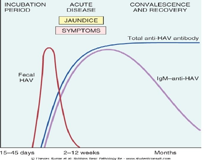
Hepatitis A virus • Hepatitis A ("infectious hepatitis") is a benign, self-limited disease. • incubation period of 15 to 50 days (average 28 days). • HAV does not cause chronic hepatitis or a carrier state and only rarely causes fulminant hepatitis. • Fatality rate is 0. 1%
-Transmission : Feco-oral rout -Endemic in developing countries with low hygiene & sanitation → anti-HAV Abs by the age of 10 yrs. → 50% by the age of 50 yrs.
-Clinically the disease is mild to asymptomatic affecting children of school age & rare thereafter -The virus is shed in bile & feces -The virus is shed is the stool 2 -3 wks before & 1 wk after the onset of jaundice -HAV is not shed in saliva, urine, or semen -HAV viremia is transient & b. I. Donors are not screened for the virus 72
• Waterborne epidemics may occur in developing countries where people live in overcrowded, unsanitary conditions. • Among developed countries, sporadic infections may be contracted by the consumption of raw or steamed shellfish (oysters, mussels, clams), which concentrate the virus from seawater contaminated with human sewage. • Ingestion of raw green onions contaminated with HAV caused outbreaks of the disease in the United States in 2003
Serelogic dx Anti HAV Ig. M: at the onset of symptoms → ↓ in few months Anti HAV Ig. G: appears later & persists for life -HAV vaccine is effective
Hepatitis B Virus • carrier rate of approximately 400 million. • About 80% of all chronic carriers live in Asia and the Western Pacific rim, where prevalence of chronic hepatitis B is more than 10%. • In the United States there approximately 185, 000 new infections per year.
-HBV is a hardy virus can withstand extremes of temperature & humidity -Prolonged IP 4 -26 wks -Prolonged viremia HBV remains in blood during the last stages of incubation period and during active episodes of acute and chronic hepatitis -Present in all body fluids as tears, saliva, sweat, breast milk, vaginal sec. , semen & pathological body fluids except stool 77
• vertical transmission from mother to child during birth constitutes the main mode of transmission. • horizontal transmission via: • 1 - transfusion • 2 - blood products • 3 - dialysis • 4 - needle-stick accidents among health care workers • 5 -IV drug abuse • 6 -sexual transmission (homosexual or heterosexual) • 7 -In 1/3 of patients the source of infection is unknown.
• HBV infection in adults is mostly cleared, but vertical transmission produces a high rate of chronic infection.
-Phases of infection : 1. Proliferative phase 2. Integrative phase 80
HBV antigens : 1. HBc Ag(hepatitis B core antigen) - hepatocytes 2. HBe Ag(pre-core protein) -blood 3. HBs Ag -blood -hepatocytes 4. DNA polymerase (HBV-DNA) (reverse transcriptase activity) 5. HBx protein ( transcriptional transactivator ) required for viral infectivity and may have a role in the causation of hepatocellular carcinoma by regulating p 53 degradation and expression 81
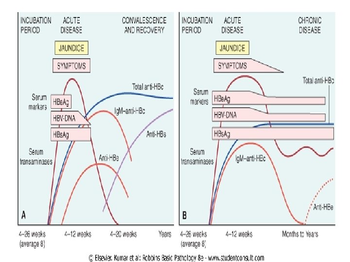
• HBs. Ag appears before the onset of symptoms, peaks during overt disease, and then declines to undetectable levels in 3 to 6 months. • Anti-HBs antibody does not rise until the acute disease is over and is usually not detectable for a few weeks to several months after the disappearance of HBs. Ag. • Anti-HBs may persist for life conferring protection • HBV-DNA, and DNA polymerase appear in serum soon after HBs. Ag, and all signify active viral replication
• Persistence of HBe. Ag is an important indicator of continued viral replication, infectivity, and probable progression to chronic hepatitis. • The appearance of anti-HBe Abs shortly after the disappearance of HBe. Ag indicates the end of the infection. • Ig. M anti-HBc becomes detectable in serum shortly before the onset of symptoms • Over a period of months the Ig. M anti-HBc antibody is replaced by Ig. G anti-HBc.
• Anti – HBs Ig. G: rise after the acute phase is over & remains detectable after wks or months after disappearance of HBs. Ag • Hepatitis B can be prevented by vaccination and by the screening of donor blood, organs, and tissues 84
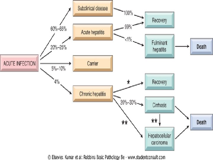
Clinical syndromes associated with HBV infection 1 -Acute hepatitis with recovery 2 -Nonprogressive chronic hepatitis 3 -Progressive chronic hepatitis ending in cirrhosis 4 -Fulminant hepatitis with massive liver necrosis 5 -Asymptomatic carrier state 86
Hepatitis C Virus (HCV) • prevalence rate is 3% (0. 1% to 12%, depending on the country). • Persistent chronic infection exists in 3 to 4 million persons in the United States, where the number of newly acquired HCV infections per year dropped from 180, 000 in the mid-1980 s to about 28, 000 in the mid-1990 s due to the marked reduction in transfusion-associated HCV as a result of screening procedures and a decline of infections in intravenous drug abusers.
• The major route of transmission is: • 1 - through blood inoculation • 2 - with intravenous drug use accounting for over 40% of cases in the United States. • 3 -via blood products is now fortunately rare, accounting for only 4% of all acute HCV infections. • 4 -Occupational exposure among health care workers accounts for 4% of cases. • 5 -The rates of sexual transmission and vertical transmission are low. • 6 - Sporadic hepatitis of unknown source accounts for 40% of cases.
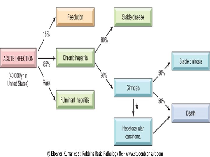
• HCV infection has a much higher rate than HBV of progression to chronic disease and eventual cirrhosis.
epidemiology -40000 new cases/yr in USA -1. 8% of the population ( 4 millions) are seropositive 70% of which have chronic liver disease -Anti HCV Ig. G occuring after active infection do not confer effective immunity due to genomic instability of the virus & antigenic variability -Anti HCV vaccine is not effective -Repeatd bouts of HCV infection are common causing hepatic damage -is characteristic due to reactivation of a pre existing infection or emergence of newly mutated strains 92
• The IP 2 to 26 weeks ( mean of 6 to 12 weeks). • The clinical course of acute hepatitis C is asymptomatic in 75% of individuals and is easily missed. • HCV RNA is detectable in blood for 1 to 3 weeks and is accompanied by elevations in serum aminotransferase.
• Clinical syndromes associated with HCV: • 1. Persistent infection with subclinical or asymptomatic acute infection • 2. Chronic hepatitis • 3. Fulminant hepatitis rare • 4. Cirrhosis 20% • 5. Hepatocellular carcinoma 94
Serological diagnosis • HCV RNA is detectable in bl. For 1 – 3 wks peak coincides with ↑ in serum transaminases • Anti HCV Abs detected in 50 – 70% of patients during symptomatic acute infection • In 30 – 50% of patients the anti HCV Abs emerge after 3 – 6 wks • In chronic HCV infection circulating HCV-RNA persists despite the presence of Abs in many patients ( > 90%) 95
Hepatitis D Virus -Hepatitis delta virus -Replication defective virus -Causes infection only when it is encapsulated by HBs. Ag -I. P 4 – 7 wks in superinfection 97
• 8% among HBs. Ag carriers in southern Italy to as high as 40% in Africa and the Middle East. • HDV infection is uncommon in Southeast Asia and China, areas in which HBV infection is endemic. • In the United States HDV infection is largely restricted to drug addicts and individuals receiving multiple transfusions (e. g. hemophiliacs who have prevalence rates of 1% to 10%).
• Delta hepatitis arises in two settings: • (1) acute coinfection after exposure to serum containing both HDV and HBV • (2) superinfection of a chronic carrier of HBV with a new inoculum of HDV. • Most coinfected individuals can clear the viruses and recover completely. • in superinfected individuals there is an acceleration of hepatitis, progressing to more severe chronic hepatitis 4 to 7 weeks later.
• Routes of transmission: • Parenteral (close personal contact)
• HDV Ag are detectable in the blood and liver just before and in the early days of acute symptomatic disease. • Ig. M anti-HDV antibody is the most reliable indicator of recent HDV exposure, but its appearance is transient. • acute coinfection by HDV and HBV is best indicated by detection of Ig. M against both HDV Ag and HBc. Ag • With HDV superinfection, HBs. Ag is present in serum; and anti-HDV antibodies (Ig. M and Ig. G) persist in low titer for months or longer.
Serologic diagnosis. HDV-RNA is detectable in blood & liver just prior to & in early days of acute symptomatic disease . Anti HDV Ig. M = recent HDV infection. Anti HDV Ig. M appears late & freq. short-lived. Coinfection : Ig. M against HDV Ag & HBV Ag. Superimposed infection: anti HDV Ig. M & HBs. Ag 102
Hepatitis E virus • HEV hepatitis is an enterically transmitted, waterborne infection occurring primarily beyond the years of infancy. • HEV is endemic in India • Prevalence rates of anti-HEV Ig. G antibodies approach 40% in the Indian population. • Sporadic infection seems to be uncommon & occurs mainly in travelers and accounts for more than 50% of cases of sporadic acute viral hepatitis in India.
- Water-borne infection Young – middle – aged adults Rare in children Endemic infection in India, Africa, Mexico…… Sporadic infection is uncommon & occurs mainly in travelers - Self-limiting mild disease except in pregnant women with high mortality rate (20%) - I. P: 6 wks ( range 2 -8 wks) - No chronic liver disease or carrier state 104
Serelogy -HEV-RNA can be detected in stool & liver before the onset of clinical symptoms -Anti HEV-Ig. M appears during acute illness & replaced by Ig. G when symptoms resolve (ie in 2 – 4 wks) 105
Clinicopathologic Syndromes 1 -Acute asymptomatic : serologic evidence only A B C D E 2 -Acute symptomatic hepatitis icteric or anicteric A B C D E 3 -Chronic hepatitis with or without progression to cirrhosis B &C 4 -Fulminant hepatitis with massive or submassive hepatic necrosis B, D A & C very rare 5 -Chronic carrier state B, C 106
Acute asymptomatic infection with recovery -Minimally ↑ serum tranaminases -HAV & HBV infections are freq. subclinical in childhood period -HCV infection is subclinical in 75% of the cases 107
Acute symptomatic infection with recovery -Can be caused by any hepatotropic viruses although it is uncommon in HCV infection -Phases: 1 -Incubation period 2 -Symptomatic preicteric phase . Malaise. General fatigability. Nausea. Loss of appetite. Fever, headaches, muscle pain, diarrhea. 10% of pts. Develop serum sickness-like synd. esp. with HBV infection (fever, rash, arthralgia ) due to circulating immune complexes 108
3 -Symptomatic icteric phase. Usual in adults but not children with HAV. Absent in 50% of cases of HBV & the majority of HCV. Conj. hyperbilirubinemia, dark colored urine , dark stool, pruritus . Prolonged PT, hyperglobulinemia, ↑ serum alkaline phosphatase 109
• 1 - diffuse swelling (ballooning degeneration • 2 - cholestasis, with bile plugs in canaliculi and brown pigmentation of hepatocytes. • 3 -Fatty change is mild and is unusual except with HCV infection. • 4 - Whether acute or chronic, HBV infection may generate "ground-glass" hepatocytes • a finely granular, eosinophilic cytoplasm shown by electron microscopy to contain massive quantities of HBs. Ag in the form of spheres and tubules. • Other HBV-infected hepatocytes may have "sanded" nuclei, resulting from abundant intranuclear HBc. Ag. Body_ • 5 - patterns of hepatocyte death are seen.
• 6 -confluent necrosis of hepatocytes may lead to bridging necrosis • 7 -lobular disarray • 8 -Inflammation. • 9 - Kupffer cells undergo hypertrophy and hyperplasia, and are often laden with lipofuscin pigment caused by phagocytosis of hepatocellular debris. • 10 The portal tracts are usually infiltrated with a mixture of inflammatory cells. • 11 -interface hepatitis). • 12 -bile duct proliferation
Fulminant hepatitis • Hepatic insufficiency that progresses from onset of symptoms to hepatic escepholopathy in 2 -3 wks • Subfulminant ( up to 3 mon) 112
Causes : 1 -Viral hepatitis 50 – 65% B, C, E HBV 2 x > HCV 2 -Drugs & chemical 25 - 50% e. g Isoniazid , halothane , methyldopa & acetominophen 3 -Obstruction of hepatic vein 4 -Wilson’s disease 5 -Acute fatty change of pregnancy. 6 -Massive tumor infiltration 7 -Reactivation of chronic hepatitis B 8 -Acute immune hepatitis
• Morphology -↓ liver size ( 500 – 700 gm) -Necrosis of hepatocytes -Collapsed reticulin tissue -Inflammatory infillrate -Regenerative activity of hepatocytes -Fibrosis 114
Chronic Hepatitis • Symptomatic, biochemical or serelogic evidence of continuing or relapsing hepatic disease for more than 6 months with histologically documented inflammation and necrosis • Progressive or non progressive • HBV , HCV, HBV-HDV 115
Morphology of chronic hepatitis • Mild to severe 1. Protal inflammation 2. Lymphoid aggregate 3. Necrosis of hepatocytes-councilman bodies 4. Bile duct damage 5. Steatosis 6. Interface hepatitis 7. Bridging necrosis & fibrosis 8. Fibrosis 9. Ground-glass appearance 10. Sanded nuclei 11. Lobular disarray 116
Carrier state • carriers are • (1) those who harbor one of the viruses but are suffering little or no adverse effects • (2) those who have nonprogressive liver damage but are essentially free of symptoms or disability • Both constitute reservoirs of infection. 117
Predisposing factors • 1 -HBV infection early in life, particularly through vertical transmission during childbirth, produces a carrier state 90% to 95% of the time. • only 1% to 10% of HBV infections acquired in adulthood yield a carrier state. • 2 -impaired immunity • 3 -HBV, HCV, ? HDV