
LIVER CIRRHOSIS PORTAL HTN By Abdel Rahman Salman
LIVER CIRRHOSIS & PORTAL HTN By: Abdel Rahman Salman
LIVER CIRRHOSIS • Definition: Cirrhosis is defined as fibrosis & nodular regeneration resulting from chronic hepatic injury, ending in loss of normal liver architecture. • Etiology: any chronic liver disease can lead to cirrhosis such as: 1 - Alcoholic liver disease 2 - Chronic viral hepatitis 3 - Biliary cirrhosis 4 - Cardiac cirrhosis 5 - Metabolic diseases: Hemochromatosis, Wilson disease 6 - Schistosomiasis 7 - Sever malnutrition 8 - Alfa 1 antitrypsin deficiency 9 - Cryptogenic 10 - Drugs: Methotrexate, Isoniazid, Methyldopa, Amiodarone
CLINICAL PICTURE • 1 - Compensated cirrhosis: Many patients with liver cirrhosis are asymptomatic for years • 2 - Decompensated cirrhosis: Clinical features: A) Liver: Firm & sharp border, Mostly it is shrunken but may be enlarged in early cases B) Clinical picture of the cause C) Clinical picture of the complications: • Liver cell failure • Portal HTN • Hepatocellular carcinoma
INVESTIGATIONS • Investigations for the liver: - Liver imaging: US to detect liver cirrhosis, splenomegaly, ascites/ CT - Fibroscan • Investigations for the cause • Investigations for the complications: - Liver cell failure: LFT - Portal HTN - Hepatocellular carcinoma: Tumor markers ( AFP, Carboxy prothrombin) • Child- Turcotte Pugh score : to assess the severity of liver cirrhosis

CHILD- TURCOTTE PUGH SCORE
TREATMENT • Liver damage from cirrhosis can't be reversed, but treatment can stop or delay further progression and reduce complications. 1 - Treatment of the cause 2 - Treatment of the complications (Portal HTN & Ascites) 3 - Drugs to decrease liver fibrosis: Colchicine, Penicillamine 4 - The definitive treatment is Liver transplant
PRIMARY BILIARY CHOLANGITIS • Autoimmune disease that leads to destruction of intra-hepatic bile duct • Clinical picture: 1 - may be asymptomatic 2 - Fatigue 3 - Obstructive jaundice & Itching 4 - Clubbing 5 - Hepatomegaly 6 - Malabsorption of fat-soluble vitamins (ADKE) 7 - Associated autoimmune diseases such as SLE 8 - Xanthelasma 9 - Features of Portal HTN & Liver cell failure
INVESTIGATIONS & TREATMENT • Anti-mitochondrial antibody ( AMA ) • Ig. M • LFT: high Alkaline phosphatase & direct bilirubin • Liver imaging: US/CT Enlarged liver with no dilated intra-hepatic biliary ducts • Liver biopsy is diagnostic • Tx: Ursodeoxycholic acid
SECONDARY BILIARY CIRRHOSIS • Long standing extrahepatic biliary obstruction due to stones or cancer of the head of pancreas • Tx: Treat the cause
CARDIAC CIRRHOSIS • Etiology: Long standing liver congestion caused by: 1 - RSHF 2 - Tricuspid valve disease 3 - Constrictive pericarditis & pericardial effusion 4 - Inferior vena cava obstruction 5 - Budd - Chiari syndrome • Clinical picture: Liver is enlarged, tender , soft then firm with sharp border • Investigations: As liver cirrhosis + Echo • Tx: Treat the cause such as Heart Failure
BUDD - CHIARI SYNDROME • It's obstruction of large hepatic veins due to : -Thrombosis e. g. Polycythemia, OCP, Behcet syndrome, Atrial myxoma, Antiphospholipid syndrome. -Obstruction (tumor). - Idiopathic. • Clinical picture: 1 - Acute stage: Triad of Abdominal Pain, Ascites , Rapidly enlarged tender liver 2 - Chronic stage: features of cardiac cirrhosis
INVESTIGATIONS • LFT: Elevated liver enzymes & bilirubin • Assessment of hypercoagulability state • Duplex ultrasound. • CT & MRI of the abdomen. • Hepatic venography • Liver biopsy
TREATMENT • Long term anticoagulant in a chronic cases • Thrombolytic therapy in acute cases • Surgical shunt or TIPS to divert blood flow around the obstruction • Control of ascites & hepatocellular failure • Liver transplant
HEMOCHROMATOSIS • Autosomal recessive disorder, The responsible gene is : HFE gene on chromosome 6 • It's a hereditary disease characterized by excessive absorption of iron resulting in increase of total body iron with its deposition in several organs • Etiology: Excessive iron absorption due to absence of mucosal block • Clinical picture: 1 - Liver : liver cirrhosis ( with an increased risk of hepatoma 20% ) 2 - Heart : Cardiomyopathy , arrhythmias. 3 - Skin : Bronzed color due to stimulation of melanocyte. 4 - Pancreas : Diabetes mellitus. 5 - Mental disturbance. 6 - Testicular atrophy. 7 - Adrenal gland : adrenal failure. 8 - Arthritis.
INVESTIGATIONS & TREATMENT • • Increase Serum ferritin & transferrin Decrease TIBC Liver imaging : abdominal CT & MRI Liver biopsy • Tx: - Repeated venesection: once/week (every bag of blood ( 500 ml ) contains 250 mg iron) - Iron chelator: Desferal - Symptomatic treatment : DM , Cardiomyopathy, …. . - Liver transplant

WILSON DISEASE • Autosomal recessive disorder, The responsible defective gene is: ATP 7 B on chromosome 13. • In Wilson’s disease copper absorption is normal but there is a deficiency in hepatic formation of "Ceruloplasmin" which is the principal transport protein for copper & necessary for biliary excretion so free copper will be released to the blood Abnormal deposition of Cu in several organs ( liver , brain , eye ) & urinary excretion of copper is increased • Clinical picture: - Liver : hepatitis (self limited) , may progress to cirrhosis. - Lentiform nucleus (Basal ganglia): extrapyramidal manifestation e. g. tremors. - Kidney: renal tubule damage which lead to albuminuria & aminoaciduria. - Eye: Kayser – Fleischer ring
INVESTIGATION & TREATMENT • Decreased ceruloplasmin level • Decreased blood copper level • Liver biopsy • Tx: - Penicillamine: increase urinary excretion of Cu - Zink acetate: blocks the absorption of copper. - Restriction of copper in diet : liver , mushrooms - Liver transplant

LIVER CELL FAILURE • Clinical Picture: 1. Fatigue, anorexia, loss of weight 2. Fever: release of pyrogens & Bacteremia 3. Fetor hepaticus: failure of hepatic detoxification of mercaptans 4. Skin manifestations: Palmar erythema, Spider navi, leukonychia, dupuytren’s 5. Jaundice 6. Ascites 7. Encephalopathy 8. Endocrinal manifestations 9. Cardiovascular manifestations 10. Hematological manifestations 11. Hepatorenal syndrome 12. pulmonary syndromes
INVESTIGATIONS • Liver function tests : - Serum enzymes ( AST , ALT ) : increased - Serum bilirubin : increased ( both direct & indirect ) - Plasma proteins : decreased albumin , increased globulin - Prothrombin time : prolonged , not corrected by parenteral vitamin K. • Investigations for the cause : - Abdominal US , CT : for cirrhosis. - Hepatitis markers : for hepatitis. • Investigations of ascites. • Investigations of hepatic encephalopathy. *Biopsy is very difficult due to bleeding & Ascites.
TREATMENT • Treatment of the cause : if possible. • Treatment of ascites. • Treatment of encephalopathy. • Liver transplantation.
ASCITES • Pathogenesis: 1 - Hypoalbuminemia (most important): Due to diminished synthesis of albumin by the liver 2 - Portal hypertension: it acts as just a localizing factor 3 - Increase Aldosterone Salt & water retention: In liver failure decrease destruction of aldosterone 4 - Peripheral arterial & splanchnic vasodilatation theory: Accumulation of vasodilators (mainly nitric oxide) pooling of blood into peripheral & splanchnic arteries vascular underfilling is sensed by the kidney This leads to activation of the reninangiotensin system Salt retention Ascites 5 - Associated factors: SBP, malignancy, T. B. peritonitis
INVESTIGATIONS • Abdominal ultrasound & CT : detect ascites even in very small amount • LFT • Ascetic fluid analysis: - Cell count : An ascetic fluid with high RBCs suggests : Malignancy, TB, Pancreatitis. An ascetic fluid with high WBCs: PMN > 250 suggests SBP. TB peritonitis : > 70% lymphocytes - SAAG
SAAG Serum Albumin - Ascitic fluid Albumin concentration - Clinical value : • SAAG > 1. 1 indicates that the ascites is due to portal hypertension regardless of its cause e. g. Liver cirrhosis, Congestive heart failure, Budd-Chiari syndrome. • SAAG < 1. 1 Exudative ascites: -Infection : TB , SBP. -Cancer : 1 ry or metastasis. -Pancreatitis. -Vasculitis -Nephrotic syndrome.
TREATMENT 1 - Salt restriction. < 2 g /d 2 - Diuretics : - Spironolactone in full dose 100 -400 mg/d… drug of choice. - Lasix ( 40 -160 mg/d ): Don’t give more due to risk of dehydration 3 - paracentesis: indicated in: - Sampling - To relieve respiratory distress - To relieve compression on the kidney 4 - Albumin 5 - Treatment of the cause : Antibiotics for SBP , liver transplantation
SPONTANEOUS BACTERIAL PERITONITIS (SBP) • Infection of the peritoneal cavity in absence of known etiology. • Occurs in 10% of cirrhotic patients. • Due to loss of detoxification function of the liver. • The prognosis is very bad. • Clinical picture : - deterioration of liver function, e. g. encephalopathy, ascites or jaundice. - resistance to diuretics may be a sign of SBP. - High fever, sever abdominal pain, tenderness.
DIAGNOSIS & TREATMENT • The key of diagnosis is examination of the ascetic fluid. - Neutrophils count > 250 cells / mm 3 with symptoms or >500 even without symptoms - Culture: E. coli. • Treatment : -Cefotaxime: 2 gm/ 3 times per day IV or Ciprofloxacin 400 mg/12 h for 5 - 10 days.
ENDOCRINE MANIFESTATIONS • In males: - Gynecomastia - Female distribution of suprapubic hair. - Impotence, ↓ libido & testicular atrophy. • In females: - Amenorrhea & sterility. - Atrophy of breast & ↓ libido. • In both: increase Aldosterone & ADH
CARDIOVASCULAR MANIFESTATIONS 1 - Hyperdynamic circulation: due to : Anemia & vasodilators 2 - Cyanosis & clubbing 3 - Cardiac dysfunction : due to • RAAS hyperactivity: leading to myocardium growth and fibrosis with disturbed relaxation. • An inhibitory effect of circulating cytokines ( e. g. nitric oxide ) on ventricular function. 4 - Decreased blood pressure due to vasodilators * This cardiac dysfunction remains clinically silent masked by the afterload reduction that is observed in cirrhosis. So, cardiac reserve may be diminished and acute heart failure may manifest after TIPS or liver transplantation.
HEMATOLOGICAL MANIFESTATIONS 1 - Anemia: • Normocytic normochromic : BM depression by toxins, Hypersplenism. • Microcytic hypochromic: Iron deficiency anemia due to chronic Blood loss. • Macrocytic : Vit B 12 & folic acid deficiency. 2 - Bleeding tendency: due to • Hypoprothrombinemia, Hypofibrinogenemia. • Decrease factors II, VII, IX, X ( 1972 ) • Decrease vit. K • Hypersplenism thrombocytopenia.
HEPATO-RENAL SYNDROME • Functional renal failure with normal renal histology in patients with advanced liver cirrhosis or fulminant hepatic failure • Pathophysiology: The hallmark of HRS is renal vasoconstriction due to: 1 - Peripheral splanchnic arterial vasodilation theory 2 - Hypovolemia due to hypoalbuminemia, ascites, diuretics. 3 - Cardiac dysfunction associated with hepatic disease • Types : - Type 1 HRS : Rapidly progressive type with high mortality rate. There is a doubling of serum creatinine to a level greater than 2. 5 mg/d. L in <2 weeks. - Type 2 HRS : Slower progressive course with a serum creatinine of >1. 5 mg/dl with better prognosis.
• Precipitating factors : 1. Large volume paracentesis of ascites without volume expansion. 2. Systemic infections e. g. SBP. 3. Excessive diuretic therapy. 4. Diminished intravascular volume e. g. diarrhea. • Prognosis : is very bad. • Treatment : - IV albumin ( plasma expansion ) - Systemic vasoconstrictors e. g. somatostatin analogs (octreotide). - Liver transplantation is the best choice.
HEPATO-PULMONARY SYNDROME • Triad of: Liver disease, Hypoxia, Pulmonary A-V shunting (Rt to Lt shunt) • Characterized by platypnea-orthodeoxia (dyspnea & deoxygenation in an upright position) • Evidence of intrapulmonary vascular abnormalities • Tx: Liver transplant
PORTO-PULMONARY SYNDROME • Pulmonary HTN in patients with Portal HTN • Characterized by fatigue, dyspnea, peripheral edema, chest pain & syncope • Dx is suggested by echo and confirmed by right heart catheterization
HEPATIC HYDROTHORAX • Pleural effusion in a patient with cirrhosis and no evidence of underlying cardiopulmonary disease. • Usually right-sided • Treatment with diuretics & Na restriction • No chest tubes • Thoracocentesis can be done in refractory cases • TIPS
HEPATIC ENCEPHALOPATHY • It's a neuropsychiatric syndrome that is seen in patients with acute or chronic liver cell failure or porto-systemic shunting. • Pathophysiology: 1 - Production of toxic substances : by the action of bacteria on intestinal proteins: Ammonia & inhibitory neurotransmitters e. g aminobutyric acid (GABA), Mercaptan, benzodiazepine like compounds. 2 - Disturbance of amino acids : Decreased branched-chain & increased aromatic amino acids leading to : - Decreased production of normal neurotransmitters ( as noradrenaline ) - Formation of false neurotransmitters ( e. g. octopamine ). 3 - Alkalosis & hypokalemia : - Decrease glucose entry to cerebral cells coma. - increase renal production of ammonia. - Increase the permeability of BBB to some of toxic substances.
PRECIPITATING FACTORS 1 - GIT bleeding. 2 - High protein intake 3 - Old Blood transfusion. 4 - Aspiration of ascites. 5 - Diuretics : Frusemide 6 - Drugs : Morphine , Benzodiazepines. 7 - SBP ( spontaneous bacterial peritonitis ). 8 - Surgery e. g. porto-systemic shunts.
CLINICAL PICTURE • Pre-Coma: - Sleep: Inverted sleep rhythm ( day/night reversal ) - Speech: slow, slurred or monotonous. - Apathy: slow response to questions. - Asterixis: (Flapping tremors). - Disorientation: for time, place, persons. -Disturbed behavior : childish attitude, phasic excitation & depression. • Coma: Irritable coma.
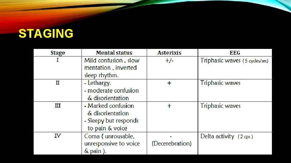
STAGING
DIAGNOSIS • The diagnosis can usually be made clinically, but when doubt exists : -Electroencephalogram (EEG) : shows diffuse slow triphasic waves. -Ammonia level : increased , It is not a sensitive or specific test for hepatic encephalopathy
TREATMENT • Correcting underlying cause • Lactulose: non absorbable disaccharide (acidifies colon, catharsis to get rid of nitrogenous products • Rifaximin • Zinc supplement. • No role for dietary protein restriction
PORTAL HYPERTENSION • Elevation of portal venous pressure above 12 mm Hg ( N: 7 -10 mm Hg ) • Etiology: The most common cause at all is Liver cirrhosis 1 - Supra hepatic ( Cardiac cirrhosis ) e. g. RSHF, Pericarditis, IVC obstruction & Budd Chiari syndrome. 2 - Hepatic (most common) : ---sinusoidal & post sinusoidal : -Liver cirrhosis "the most common cause". -Veno-occlusive disease. ---pre sinusoidal : -Schistosomiasis -Congenital fibrosis of portal tract. -Hodgkin's lymphoma, leukemia.
3 - Infra-hepatic : rare -Portal vein thrombosis ( Hepatoma ). -Congenital stenosis of portal vein. -Extrinsic compression ( tumor )
CLINICAL PICTURE 1 - Splenomegaly: - Dragging pain in the left hypochondrium. - May be the only clinical evidence of portal hypertension. - May be associated with hypersplenism, which by turn lead to pancytopenia & bleeding tendency. 2 - Intestinal congestion: Distension & Dyspepsia. 3 - Liver: firm & sharp border (cirrhotic liver) 4 - Ascites : SAAG > 1. 1 5 - Encephalopathy
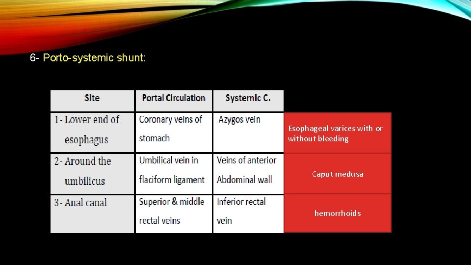
6 - Porto-systemic shunt: Esophageal varices with or without bleeding Caput medusa hemorrhoids
7 - ESOPHAGEAL VARICES • Dilated, elongated, tortuous veins located at lower end of the esophagus • Clinical picture: Approximately 90% of cirrhotic patients will develop esophageal varices, over 10 years, but only on third of these will bleed. • Bleeding is likely to occur with large varices, red signs on varices (diagnosed by endoscope) and in severe liver disease 1 - before rupture: - Asymptomatic (silent) - Dysphagia ( rare ) 2 - At rupture: Upper GIT bleeding - Painless massive hematemesis. - Melena.
INVESTIGATIONS • 1 - Endoscopy: - Detects early varices. (Esophageal varices confirm the diagnosis of portal hypertension) - Detects signs of impending rupture (red signs). - Detects active bleeding & its site. - Can be used for sclerotherapy or banding of varices. • 2 - Imaging of the portal circulation : a) Doppler or Duplex ultrasound : Detect pressure , blood Velocity & patency. b) CT , MRI.
3 - Measurement of the hepatic venous pressure gradient: portal HTN is present if > 6 mm. Hg, when >12 mm. Hg Risk of varices 4 - Spleno-portography: needle introduced into the spleen inject dye for patency of portal vein 5 - Evaluation of the liver: liver function tests , liver biopsy
TREATMENT • During attack: ( Control of acute bleeding ) 1 - Initial resuscitation with replacement of blood volume. 2 - Endoscopy to detect the site of bleeding. 3 - Pharmacological therapy : - Vasopressin: generalized V. C may result in peripheral vascular ischemia, Ischemic heart disease & hypertension so, we may use nitrate with vasopressin - Somatostatin. - Octreotide. 4 - Injection sclerotherapy and / or band ligation 5 - Sengstaken- Blakemore tube (balloon) 6 - When all the above measures fail, Trans jugular intrahepatic portosystemic shunt (TIPSS) or portosystemic shunt operations are considered
PREVENTION • Primary prevention: 1 - Small varices: no treatment is required. 2 - Large varices: β blockers ( propranolol ), Nitrates 3 - Impending rupture: band ligation • Secondary prevention: 1 - blockers (propranolol) 2 - Repeated injection sclerotherapy or band ligation 3 - TIPPS & liver transplant
SURGICAL OPTIONS • TIPSS: Inserting of short metal tube through Jugular vein to the liver & connect portal vein with hepatic vein (porto-systemic). - It requires only light sedation & local anesthesia. • Porto-systemic shunts: - Portocaval. ( SE : encephalopathy ) - mesenterico-caval. - lienorenal: minimal encephalopathy. • Splenectomy
HEPATOCELLULAR CARCINOMA • Risk is increased in liver cirrhosis & chronic HBV (lowest in Wilson, autoimmune) • Could be asymptomatic in early stages • Symptoms due to mass effect: abdominal pain, jaundice, mass, early satiety, liver decompensation • Can rupture causing hemoperitoneum • High AFP • Median survival 6 -20 months • 6 -monthly U/S surveillance
TREATMENT • Surgical resection • Transarterial chemoembolization (TACE) • Radiofrequency ablation, microwave ablation, and cryoablation • Percutaneous ethanol or acetic acid ablation • Systemic chemotherapy, with cytotoxic agents • Liver transplant

THANK YOU
- Slides: 55