Liver and Spleen By Dr Muslim Kandel 2018

Liver and Spleen By Dr. Muslim Kandel 2018 -19

The liver
 --The most")
The liver is central organ of metabolism (digestion , anabolisim and catabolism) --The most chemical levels in the blood in hepatic artery regulate and excretes as bile. ---All of the blood leaving (GIT)passes through the liver via portal vein back to circulation via hepatic vv to IVC

Lobes of liver Anatomical lobes Rt, & Lt lobes by (falciform lig, anteriorly &lig. Venosum posteiorly ) Rt. Lobe caudate lobe , quadrate lobe ) Lt. lobe small Borders ill-defined except sharp inf border Surgical (structural ) lobes 2 lobes equal Depend on bases of intrahepatic disterbution of hepatic a, portal v &biliary ducts 8 segments 4 in each lobe Each segment regard small liver

Anatomical lobes
 lobes Couinaud (or \"French\") system")
Surgical (structural ) lobes Couinaud (or "French") system
")
Surfaces of liver Smooth Diaphragmatic surfaces There is space between liver and diaphragm(subphrinc recess) Sup. surface related to diaphragm (lung & Ht) Rt. lat. Surface related to diaphragm (costodiaphragmatic recess of Rt plura, base of lung , ribs (7 -11) Ant. Surface attached to falciform lig, related to costal margin.
")
Post. Surface Adherent to diaphragm 1 -oesophageal groove 2 -fissure for lig. Venosum(ductus venosum) 3 -caudate lobe 4 -groove for IVC 5 -bare area
 Irregular surface due to visceral impressions A- inf surface")
Inf. Surface ( visceral ) Irregular surface due to visceral impressions A- inf surface of Lt lobe 1 -stomach 2 -lesser omentum B-Fissuer of lig. Teres C-quadrate lobe D-GB fossa E-abd. Organs 1 -colon 2 -renal 3 -duodenal 4 -suprarenal

Porta hepatis Its deep transverse slit on the post. part of inf. Surface of liver Bounderies Post—caudate process of caudate lobe Ant. --quadrate lobe Lt –lig. Teres Rt --- GB fossa Contenets 1 - portal v & its 2 br (Rt, &Lt) post 2 -hepatic a & its 2 br ant. To Lt 3 - two hepatic ducts unite to form CHD CBD- ant to Rt 4 -Hepatic autonomic plexus 5 -lymphatic v & LN & fat
 2")
Peritoneal covering of liver Completely covered with peritoneum except: 1 -bare area (post) 2 -fossa for GB 3 -groove for IVC 4 - porta hepatis 5 -Lig. Venosum 6 - lig. Teres

Peritoneal fold attached to liver: 1 -falciform lig 2 -lesser omentum 3 -coronary lig upper and lower layer 4 -Lt triangular lig. 5 -Rt triangular lig

Ligaments of liver

1 -Falciform ligament Sickle - shaped fold of peritoneum connect liver to the diaphragm , & ant. Abd. Wall Layers of falciform lig. Rt layer- upper layer of coronary lig Lt layer - ant. Layer of Lt triangular lig ---at free margin the 2 layers contenous with each other enclosing round lig. Contents In its free margin 1 -lig. Teres , (obliterated umbilical V) 2 -paraumbilical vv, 3 -fat

2 -Lesser omentum is divided into ligaments --hepatogastric ligament: connecting to the lesser curvature of the stomach -- hepatoduodenal ligament: connecting to the duodenum --hepatophrenic ligament: connecting to the diaphragm -- hepatoesophageal ligament: connecting to the esophagus --hepatocolic ligament: the portion connecting to the colon Contents a) Gastric border: -Rt& Lt gastric vessles & LN b)Rt free border: -hepaic a, portal v, CBD c)Between 2 layers: -fat, autunomic n


3 -Coronary lig. Diaphragmatic surface of Rt lobe of liver covered ompletely by peritoneum lig. except bare area , peritoneum on upper surface reflected upward to form upper layer of coronary lig. , and post surface reflected downward to form lower layer of coronary The bare area of liver attached to diaphragm devoid from peritoneum

4 - Lt triangular lig. Lt lobe of liver covered completely by peritoneum , peritoneum on posterior part of upper surface reflected upward to form Lt triangular lig. Which have ant. & post. layers 5 -Rt triangular lig. The 2 layers of coronary lig. Around bare area of liver reunite to form Rt triangular lig NB. There are 2 obliterated vessles forming lig. Attached to live 1 -Lig. Teres Lt umbilical v, extend from Lt br. of portal v to the umbilicus 2 -Lig. Venosum ductus venosum, extend from Lt br. of portal v to the IVC

Fixation of the liver 1 -peritoneal lig. & folds 2 -Attachments of hepatic veins 3 - +ve intraperitoneal pressure& pressuere of abd. Organs

Nerve supply 1 -sympathetic caeliac plexus via hepatic art. 2 -parasympathetic vegus N Blood supply The liver receives a dual blood supply from the hepatic portal vein and hepatic arteries. The portal vein delivers approximately 75% of the liver's blood supply, and carries venous blood drained from the spleen, gastrointestinal tract , and its associated organs. The hepatic arteries supply arterial blood to the liver, 25% Blood flows through the liver sinusoids and empties into the central vein of each lobule. They coalesce into hepatic veins, which leave the liver and drain into the IVC

Lymphatic drainage Superfecial Lymph. beneath peritoneum Deep lymphatics along portal tracts A- post part of liver mediastenial LN (mostly) phrenic caeliac LN(less) B –ant. &inf. Part of liver caeliac LN C-Lt lobe Lt gastric LN caeliac LN D-central part of ant surface along falciform L parasternal LN E-along Leg teres - communicate with LN around umblicus

Hepatic lobules portal triad (consists of branch of 1 -hepatic artery 2 - portal vein, 3 -bile duct, 4 -lymphatic vessels 5 -vagus nerve )which can be found running along each of the lobule's corners A-The central vein hepatic vein IVC B-bile ductules CBD
")
Extrahepatic biliary passages A- Lt&Rt hepatic ducts unite CHD unite cystic duct (from GB) CBD join pancreatic duct common duct which open in ampula of Vater into posteromedial aspect of 2 nd part of doudenum

Gall bladder Pear shape cystic organ 7 -10 cm x 3 cm , site in GB fossa in liver Its capacity about 30 -50 cc of bile Parts 1 -Fundus 2 -Body 3 -Neck 4 -Cystic duct which unite with CHD CBD have spiral mucsal ridges called spiral valve of Heister

Blood supply of GB Cystic a hepatic art. Cystic veins drain directly into liver Nerve supply Sensory Rt phrenic n (C 3, 4, 5) referred pain at shoulder Parasympathetic vagus Sympathetic splanchnic n (T 7, 8, 9) referred angle of scapula Lymphatics Cystic & hepatic LN

Triangle of Calots Is an anatomic space bordered by: 1 - common hepatic duct medially, 2 - cystic duct laterally 3 -inferior border of liver superiorly Contents 1 - cystic artery, 2 - LN of Lund 3 -anomalous bile ducts 4 -accessory Rt. hepatic a Importance dissection in the triangle CBD injuries.
 Formed by union of CHD with cystic duct descended. d")
Common Bile Duct (CBD) Formed by union of CHD with cystic duct descended. d 0 wn then unite with pancreatic duct to form ampulla of Vater in the 2 nd part of duodenum surrounded by sphincter of Oddi parts 1 -supradoudenal 2 -retrodoudenal 3 -infradoudenal

Congenital anomalies of GB
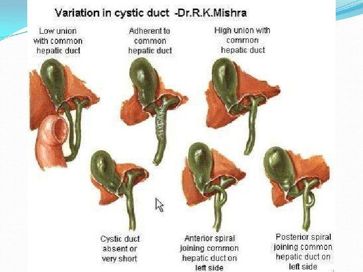

Congenital anomalies of GB

spleen
 Size 1 x 3 x 5")
spleen *size & weight ( odd No. ) Size 1 x 3 x 5 inches , weight about 7 ounces (200 g). Related to 9 -11 ribs * blood supply : -The splenic artery ( which is br. Of caeliac tranck ) it"s divides at the hilum into branches). * venous drainage 90% the portal v 10 % bypasses the cords and sinuses by direct arteriovenous connections. . Parts --. The white pulp has an immune function, the red pulp filters abnormal red cells from the circulation. Phagocytosis of blood-borne particles occurs in both areas.

Shape of spleen

Hilum of spleen Its longitudinal slit on the visceral surface Contents: 1 -terminal br. Of splenic art. 2 -tributeries of splenic v 3 -autonomic n &fat 4 -Its give attachment of 2 lig. --Lienorenal lig. which contain tail of pancreas --Gasterosplenic lig. (omentum) Peritoneal covering Spleen is completely covered by peritoneum except pancreatic impression

Hilum of spleen

Ligaments of spleen 1 -lieno-renal Lig. 2 -gasterosplenic lig. 3 -pherinicocolic lig. its support spleen

Stability of spleen 1 -ligements 2 -intra abd. Pressure 3 -positiion of surrounding organs Blood supply --splenic a(tortuous along upper pancreas). caeliac trunk Venous drainage enic vein behind pancreas unite sup. Mesenteric V portal v lymphatic's red pulp has no lymphatics Capsule & trabiculae pancreatico splenic LN Nerve supply Sympathetic caeliac plexus
- Slides: 37