Lips and alveolar processes gums Maxillary process forms

 - Maxillary process forms upper jaw, while")


 A glossa: failure of development")


: associated with polyhydramnios due")


 around the vertical")
 of the")


- Slides: 24
• Lips and alveolar processes (gums) - Maxillary process forms upper jaw, while mandibular process gives lower jaw. - Each primitive gum is divided by a horse-shoe groove (alveololabial groove –vestibule) into 2 parts; a- An outer part which gives rise to the lip and cheek. b- An inner part which gives the gum.
Development of tongue
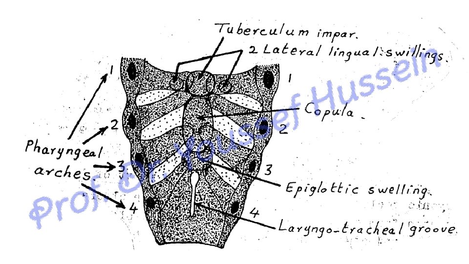
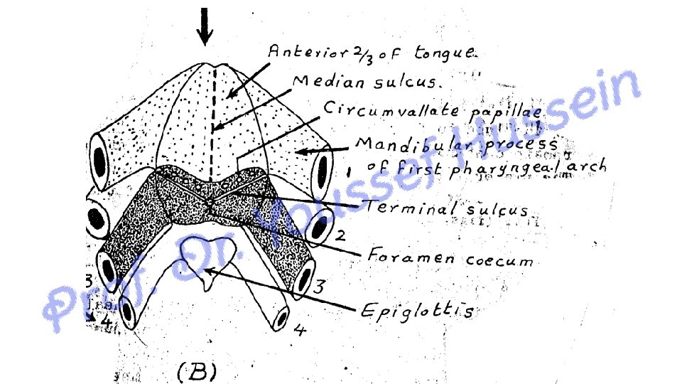
• Development of the mucous membrane of the tongue: • A- The anterior 2/3 of the tongue: • At 4 th week 3 endodermal swellings appears from 1 st pharyngeal arches • 1 - A median swelling called the tuberculum impare • 2 - Two lateral lingual swellings: The 2 lateral swellings proliferate and grow medially. • In the midline, they fused together at median sulcus and completely covered tuberculum impare forming mucous membrane of anterior 2/3 of the tongue. So the anterior 2/3 is supplied by Lingual nerve.
• Development of the mucous membrane of the tongue: • B- The posterior 1/3 of the tongue: • It develops as a large midline mass (copula of His) derived from endoderm of 2 nd, 3 rd and 4 th pharyngeal arches. • The part of the 3 rd arch proliferates and migrates forward overlying the 2 nd arch and forms the posterior 1/3 of the tongue that fused with 2/3 by terminal sulcus. So the posterior 1/3 is supplied by the glosspharyngeal nerve. • C- Root of the tongue; • From the part of the copula derived from the 4 th pharyngeal arch, So it is supplied by the vagus nerve (internal laryngeal nerve).
• Development of the muscles of the tongue: • The muscles of the tongue are derived from the occipital myotomes ( They migrate around the side of pharynx to reach their position under mucosa of tongue) except the palatoglossus muscle that develop from the mesoderm in situ. • So the muscles are supplied by the hypoglossal nerve except palatoglossal muscle supplied by pharyngeal nerve plexus • Separation of the tongue • At first the tongue is adherent to the floor of the mouth, then a horse-shoe (Cshaped) groove called alveolo-lingual groove separates the anterior 2/3 from the floor of the mouth except in the midline where the tongue is connected to the floor by the frenulum of the toungue.
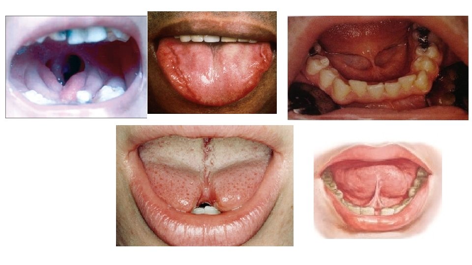
• ** Congenital anomalies of the tongue (I) A glossa: failure of development of the tongue, (II) Macroglossia: large sized tongue which protrudes from the mouth (as in mongolism and cretinism). (III) Microglossia: small sized tongue. (IV) Bifid tongue: due to failure of fusion of the 2 lingual swellings. (V) Short frenulum: due to incomplete separation of the anterior 2/3 of the tongue from the floor of the mouth (common). (VI) Long frenulum: due to excess separation of the tongue from the floor of the mouth. It causes the tongue to fall back and close the pharynx (suffocation, dangerous). (VII)Tongue-tie (Ankyloglossia): the tongue is adherent to the floor of the mouth due to failure of appearance of the alveolo-lingual groove.
Development of salivary glands
- Parotid ---- Submandibular ------ Sublingual glands - They arise at the 5 th-6 th weeks as ectodermal in origin - It begins as a solid cord ---- becomes grooved ----- duct - The distal end of the duct elongates and branched to form the acini - The proximal part form the duct of the glands
Development of esophagus
Development of esophagus - An endodermal diverticulum called tracheoesophageal groove. - The lips of the groove approximated and fused with each other forming tracheoesophageal tube. - It is divided by tracheoesophageal septum into laryngotracheal tube (ventral) and pharynx and esophagus (dorsal).
** Congenital abnormalities of the esophagus - Esophageal atresia (obstruction): associated with polyhydramnios due to failure of swallowing of the amniotic fluid - Esophageal stenosis (narrow): due to a- Posterior displacement of tracheoesophageal septum. b- Mechanical factors push pposterior wall of the tube forward.
• Tracheoesophageal fistula: 1 4 - An abnormal opening between esophagus and trachea caused by failure of complete closure of the tracheoesophageal septum. • Types of the fistula: 1. Proximal part of esophagus ends as a blind sac and distal part continues with the trachea. 2. Proximal part of esophagus continues with trachea and distal part ends as blind sac. 3. Proximal and distal parts of continue with trachea by single tube (fistula). 4. Proximal and distal parts of esophagus continue with trachea separately by double tubes. 2 3
Development of Stomach
Development of stomach
• STOMCH - It appears as a fusiform dilatation having 2 surfaces (right and left) and 2 borders (anterior and posterior). - It is connected to anterior abdominal wall by ventral mesogastrium and posterior abdominal wall by dorsal mesogastrium ** Changes during Development: 1 - The posterior border grows faster than the anterior border and becomes convex forming the greater curvature. 2 - The upper part of greater curvature bulges upward to form the fundus. 3 - The anterior border grows slowly and becomes concave to form the lesser curvature.
** Rotation of the stomach: - The stomach rotates 90° (clockwise) around the vertical axis. - Growth of the liver pushes the cranial end of the stomach to the left of the median plane and the caudal end moves to the right of the midline. ** Results of rotation: 1 - The greater curvature is directed to the left and the lesser curvature to the right. 2 - The left surface become anterior and right surfaces become posterior. 3 - A small recess of the peritoneal cavity is formed behind the stomach forming the lesser sac.
Congenital pyloric stenosis: due to excessive hypertrophy (thickening of the circular muscles) of the pylorus - It leads to pyloric obstruction and projecting vomiting in the new born
Hour-glass stomach: due to abnormal constriction of the middle of the body of the stomach
Congenital hiatus hernia, part of the stomach above the diaphragm due to short esophagus.
Th ank Qu you est ion s