Lipid bilayer Membrane proteins Multipass membrane Proteins Bacteriorhodopsin

Lipid bilayer, Membrane proteins, Multipass membrane Proteins, Bacteriorhodopsin
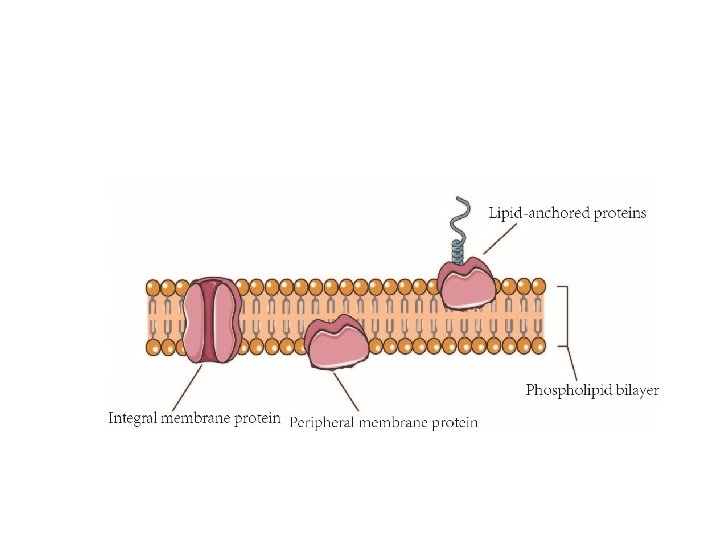

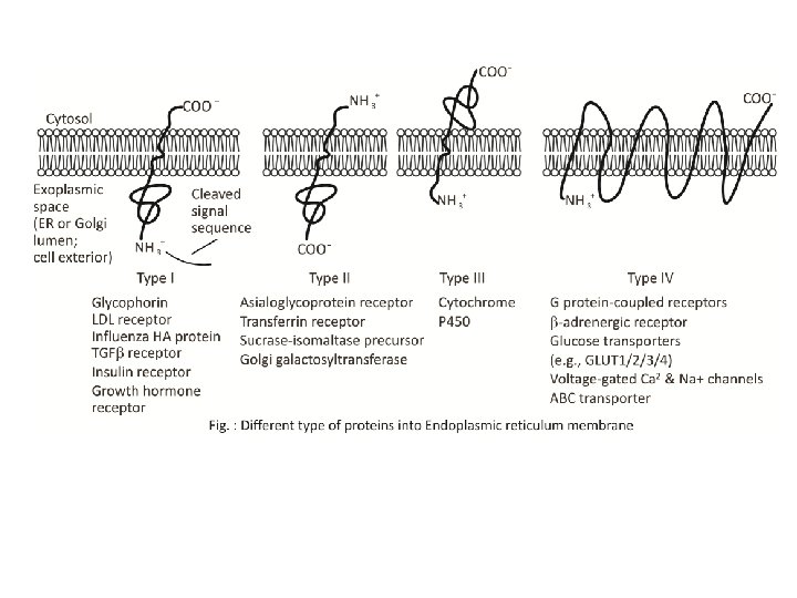
According to their relationship with the bilayer, integral membrane protein can be classified two primary types: Integral polytopic proteins and Integral monotopic proteins. Integral polytopic proteins • They are also known as “transmembrane proteins” which can span across the membrane at least once. • These integral membrane proteins may have different transmembrane topology which refers to orientations (locations of N- and C-termini) of membrane-spanning segments with respect to the inner or outer sides of the biological membrane occupied by the protein. • The first three types in the Fig. 2 are common forms in integral membrane proteins, such as, transmembrane α-helix protein, transmembrane α-helical protein and transmembrane β-sheet protein.

Integral monotopic proteins • They are one type of integral membrane proteins that are attached to only one side of the membrane and do not span the whole way across. • There are 4 types of interaction between Integral monotopic membrane protein and cell membranes: by an amphipathicα-helix parallel, by a hydrophobic loop, by a covalently bound membrane lipid and electrostatic or ionic interaction with membrane lipids (No. 4, 5, 6, 7 of Fig. 2). • Integral membrane proteins can be separated from the biological membranes only using detergents, nonpolar solvents, or sometimes denaturing agents. • Peripheral proteins dissociate following treatment with a polar reagent, such as a solution with an elevated p. H or high salt concentrations.
.")
The membrane is represented in yellow. 1. A single transmembrane α-helix (bitopic membrane protein). 2. A polytopic transmembrane α-helical protein. 3. A polytopic transmembrane β-sheet protein. 4. Interaction by an amphipathic αhelix parallel to the membrane plane (in-plane membrane helix). 5. Interaction by a hydrophobic loop. 6. Interaction by a covalently bound membrane lipid. 7. Ionic or electrostatic interactions with membrane lipids.

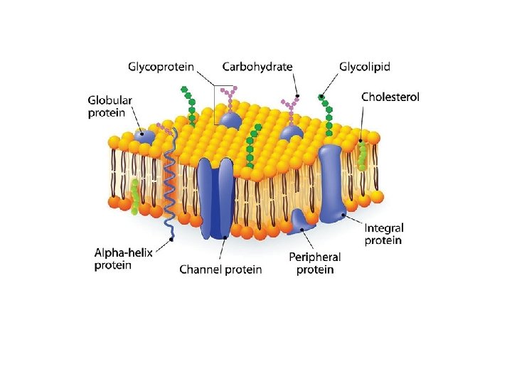
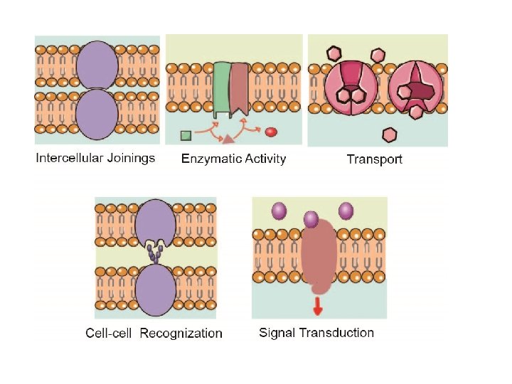
Function of Membrane Proteins Membrane proteins play crucial roles in all organisms, where they serve as, such as membrane receptors, ion channels, GPCR (G protein–coupled receptors) and various kinds of transport proteins. Membrane receptors that embedded in the cell membranes, which can transmit signals between the cell’s internal and external environments. Membrane transport protein (or transporter) is a kind of membrane protein that involved in the movement of ions, small molecules, or macromolecules across a biological membrane. Ion channels are one of most important type of membrane transport proteins. The functions of ion channel include establishing a resting membrane potential, shaping action potentials and other electrical signals by gating the flow of ions across the cell membrane, controlling the flow of ions across secretory and epithelial cells, and regulating cell volume. Some enzymes are also membranes proteins, for example oxidoreductase, transferase or hydrolase. Cell adhesion molecules that located on the cell surface involved in binding with other cells or with the extracellular matrix (ECM), allow cells to identify each other and interact. For example, proteins including Ig (immunoglobulin) superfamily involved in immune response.

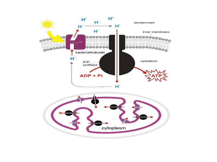
Bacteriorhodopsin • Bacteriorhodopsin is a proton pump found in Archaea, it takes light energy and coverts it into chemical energy, ATP, that can be used by the cell for cellular functions. • Bacteriorhodopsin forms chains, which contain retinal molecule (One of several yellow or red carotenoid pigments formed from rhodopsin by the action of light; retinene ) within, it is the retinal molecule that absorbs a photon from light, it then changes the confirmation of the nearby bacteriorhodopsin protein, allowing it to act as a proton pump. • While chlorophyll based ATP generation depends on a protein gradient, like bacteriorhodopsin, but with striking differences, suggesting that phototrophy evolved in bacteria and archaea independently of each other.

Bacteriorhodopsin is a protein used by Archaea, the most notable one being Halobacteria. It acts as a proton pump; ie, it captures light energy and uses it to move protons across the membrane out of the cell. The resulting proton gradient is subsequently converted into chemical energy.

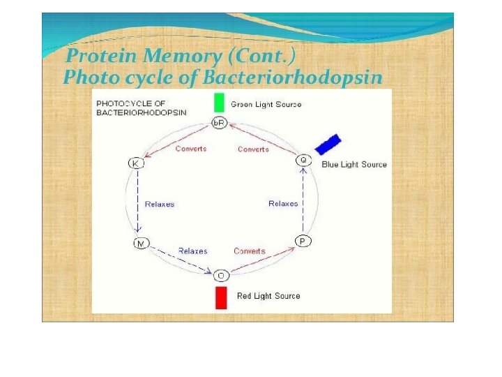
• Bacteriorhodopsin is an integral membrane protein usually found in twodimensional crystalline patches known as “purple membrane”, which can occupy up to nearly 50% of the surface area of the archaeal cell. • The bacteriorhodopsin forms repeating elements that are arranged in chains. • Each chain has seven transmembrane alpha helices and contains one molecule of retinal buried deep within, the typical structure for retinylidene proteins. • It is the retinal molecule that changes its conformation when absorbing a photon, resulting in a conformational change of the surrounding protein and the proton pumping action. • When a photon is absorbed, it causes a change in the conformation of the molecule. In bacteriorhodopsin, this is a change from a straight form to a bent form. This change in shape powers the pumping of protons. • This releases a proton from a “holding site” into the extracellular side (EC) of the membrane making the inside 10, 000 -fold more alkaline than the outside. • These protons are then allowed to flow back inwards through another protein, ATP synthase, building much of the ATP that powers the cell. • Reprotonation of the retinal molecule restores its original isomerized form. • This results in a second proton being released to the EC side. • The releases the proton from its “holding site, ” where a new cycle may begin. • The bacteriorhodopsin molecule is purple and is most efficient at absorbing green light (wavelength 500 -650 nm, with the absorption maximum at 568 nm).

• Bacteriorhodopsin belongs to a family of bacterial proteins related to vertebrate rhodopsins, the pigments that sense light in the retina. • Many molecules have homology to bacteriorhodopsin, including the lightdriven chloride pump halorhodopsin, and some directly light-activated channels like channel rhodopsin. All other phototrophic systems in bacteria, algae, and plants use chlorophylls or bacteriochlorophylls rather than bacteriorhodopsin. • These also produce a proton gradient, but in a quite different and more indirect way involving an electron transfer chain consisting of several other proteins. • Furthermore, chlorophylls are aided in capturing light energy by other pigments known as “antennas”; these are not present in bacteriorhodopsinbased systems. • Last, chlorophyll-based phototrophy is coupled to carbon fixation (the incorporation of carbon dioxide into larger organic molecules) and for that reason is photosynthesis, which is not true for bacteriorhodopsin-based system. • Thus, it is likely that phototrophy independently evolved at least twice, once in bacteria and once in archaea.
- Slides: 16