Light SCN direct via the retinohypothalamic RHT pathway
 pathway -indirect via geniculohypothalamic (GHT) pathway")


 and")


 injected in the adrenal gland")

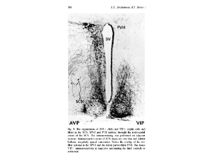
 A-C: in PVN oxytocin (red) D-E: in SCN vasopressin (red) F: in")
 pathway European")
 • Brain Res. 1972 Jul 13; 42(1): 201 -6. – Loss")
- Slides: 20
Light →SCN: -direct via the retinohypothalamic (RHT) pathway -indirect via geniculohypothalamic (GHT) pathway
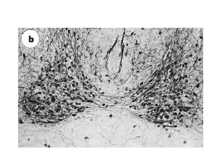
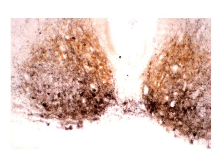
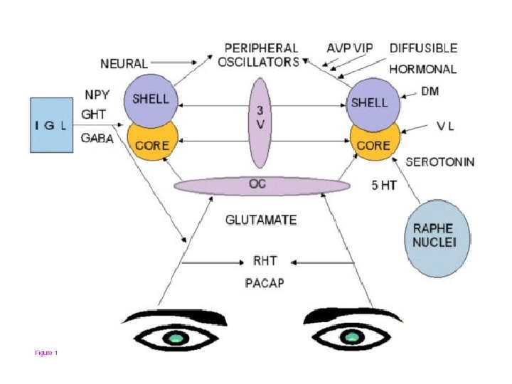
SCN: some morphological features • Parvocellular, paired structure, ~16 -20, 000 neurons in rodent, miniscule in man • Phenotypes: multiple potential transmitters – most express GABA (1993 proposal: SCN output is inhibitory) – Peptides: • vasoactive intestinal peptide (VIP) in cells in ventrolateral part; receives retinal input; forms part of the output projection • vasopressin (VP) in cells in dorsomedial part; forms part of the output projection • somatostatin (SS) in cells whose axons remain intrinsic to SCN
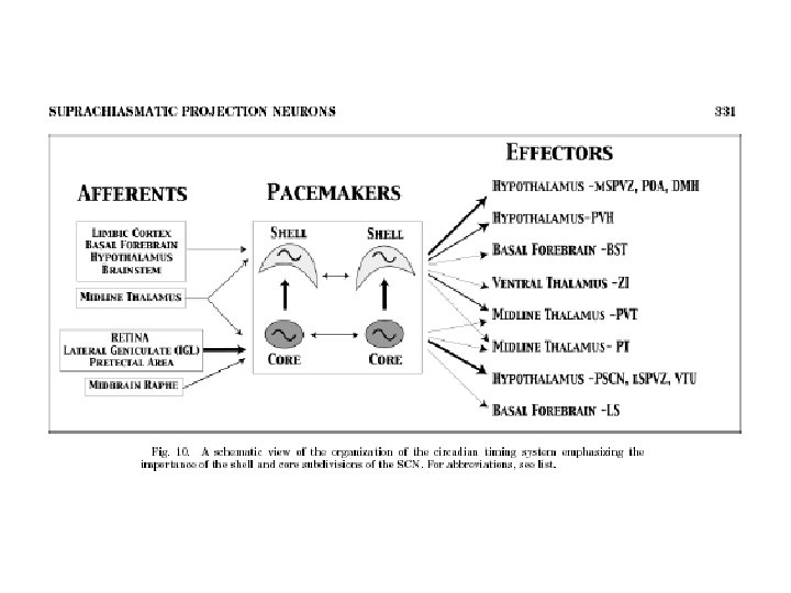
Lets consider the neural connections of SCN A schematic outline from Ibata et al Frontiers in Neuroendocrinology 20: 241 -268, 1999
Notes: 1 -Based on immunocytochemical grounds, SCN can be subdivided into dorsomedial (shell) and ventrolateral (core) segments 2 -Retinal input is to the VIPergic neurons in the ventrolateral SCN 3 -output pathways arise from both VIPergic and vasopressinergic neurons in SCN 4 -most projections are local, to hypothalamic sites (exceptions: LGB, TPV)
SCN: techniques to define inputoutput pathways • Using retrograde and/or anterograde transport of suitable markers e. g. wheat germ agglutin (WGA)
SCN: techniques to define inputoutput pathways • Using retrograde and anterograde transport of suitable markers e. g. WGA • Using viral retrograde transneuronal traceing (pseudorabies)
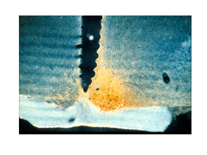
Retrograde transneuronal labeling with pseudorabies virus (PRV) injected in the adrenal gland
SCN: techniques to define inputoutput pathways • Using retrograde and/or anterograde transport of suitable markers e. g. WGA • Using viral retrograde transneuronal tracing (PRV) • Using double label immunocytochemistry to define phenotype (peptides)
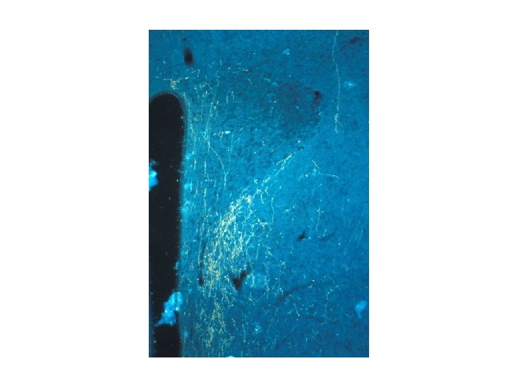
PRV (green) A-C: in PVN oxytocin (red) D-E: in SCN vasopressin (red) F: in SCN VIP (red)
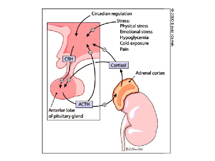
Anatomical and functional demonstration of a multisynaptic suprachiasmatic nucleus adrenal (cortex) pathway European Journal of Neuroscience 11: 15351544, 1999 RM Buijs, J Wortel, JJ van Heerikhuize, MGP Feenstra, GJ Ter Horst, HJ Romijn, A Kalsbeek
Suprachiasmatic nucleus (SCN) • Brain Res. 1972 Jul 13; 42(1): 201 -6. – Loss of a circadian adrenal corticosterone rhythm following suprachiasmatic lesions in the rat. Moore RY, Eichler VB.