Light microscopy Lysmikroskopi Optical microscopy MENA 3100 OBK

Light microscopy Lysmikroskopi Optical microscopy MENA 3100, OBK, 31. 01. 18

Grups from 9 th. February Forskningsparken
 Yi Hu")
Tomorrow (1 th. Febr. ) Yi Hu

Yes 1. 2 Instrumentation Yes 1. 3 Specimen Preparation 1. 4. 1 Bright-Field and Dark-Field 1. 4. 2 Phase-Contrast 1. 4. 3 Polarized-Light 1. 4. 4 Normarski 1. 4. 5 Fluorescence 1. 5 Confocal n. Optical Principles Mo re inf os oo 1. 1 Definetly Ma yb ew et hin kd iffe re ntl y ab o ut thi s. Yes Cursory: from Latin cursorius ‘of a runner’ rapidly and often superficially performed or produced Cursori Definetly We have it … Cursori (or not)

Only draw the ones that are ”important”

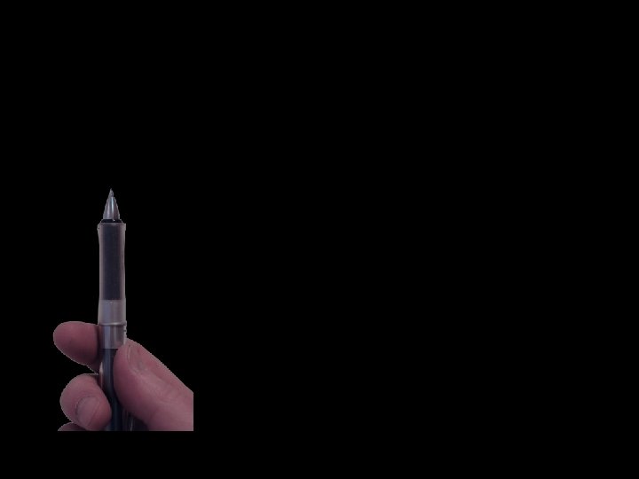
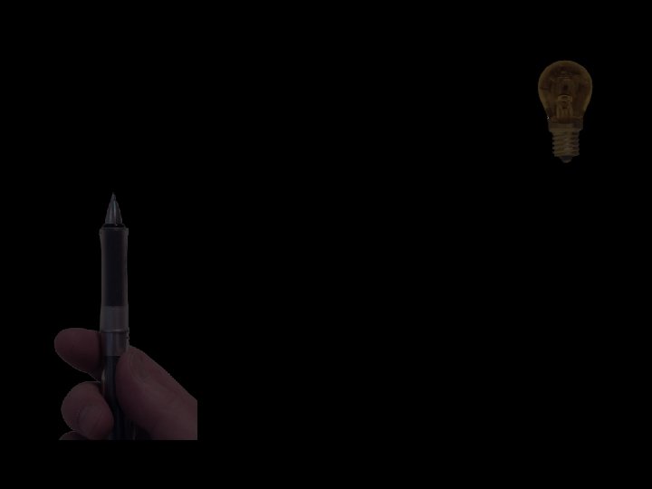
One lense Thin lense Projection

Through the centre is not deflected Parallell to the optical axis goes through back focal plane (Through focal plane before lense becomes parallel optical axis. ) f f Optical axis Projected onto a screen Distance one to two focal lengths

Focal plane
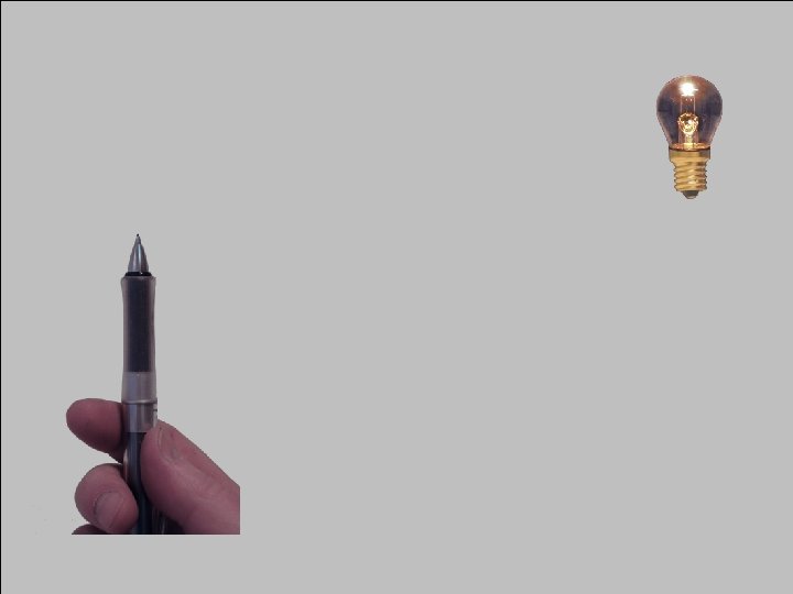
 f f M = s’ s f M= v− f")
s s’ (= v) f f M = s’ s f M= v− f f OBS! The image of the pencil has of cause no width in the direction parallell the optical axis! Optical axis
 s’ f 2 f > s > f Magnified, real image")
s (= v) s’ f 2 f > s > f Magnified, real image f f
 s’ f s > 2 f Demagnified, real image f f")
s (= v) s’ f s > 2 f Demagnified, real image f f
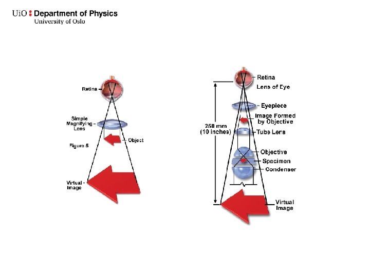
 s’ f s<f Magnefied, virtual image f Magnifying glass (forstørrelsesglass, lupe)")
s (= v) s’ f s<f Magnefied, virtual image f Magnifying glass (forstørrelsesglass, lupe) f
 s’ f f f")
s (= v) s’ f f f

Real image on retina Compound microscope Det sammensatte mikroskop Eyepiece Okular Real image (Primary image) OBS! Errors in figure Objective Object Usually corrected to «infinite» tubus Virtuell image

All rays spread in the same direction are collected here Image plane Diffraction plane Fourier plane All rays originated from the same point are collected here

Fourier transformation Read the text from Johan Taftø
 We want to see more details Resolution (Oppløsningsevne)")
Optical microscope Light microscope Magnification (Forstørrelse) We want to see more details Resolution (Oppløsningsevne) Contrast
 Virtual image Virtual: : very close to being")
Simple microscope Magnifying glass (forstørrelsesglass, lupe) Virtual image Virtual: : very close to being something without actually being it : existing or occurring on computers or on the Internet
")
Magnifying glass Antony van Leeuwenhoek (1632− 1723)
")
The compound microscope Det sammensatte mikroskop ca. 1595 Hans Janssen Robert Hooke (1635− 1703) Micrographia, 1664

Stereo mikroskope

Compound microscope for transmitted light Microscope for transmitted light

Mikroskope for reflected light
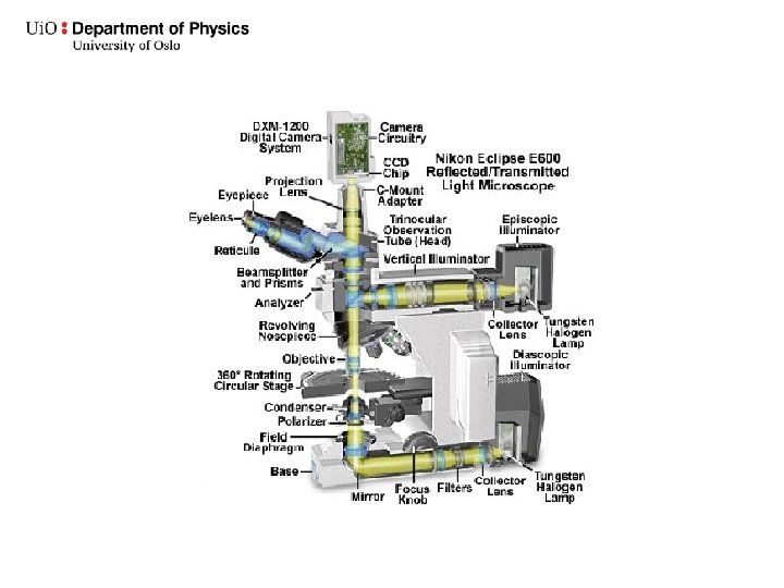

www. microscopy-analysis. com/files/ jwiley_microscopy/2006_Sept_Hammond. pdf

Illuminating system Sample Condensor lens Aperturblender Aperture diaphragm Feltblender Field diaphragm Filament

Belysningssystemet Prøve Kondensorlinse Aperturblender August Köhler 1866─1948 Köhler-belysning Jevn belysning av prøven Feltblenderen Bestemmer det belyste området Feltblender Filament Aperturblenderen Bestemmer vinkelen på lyskjeglen som treffer prøven

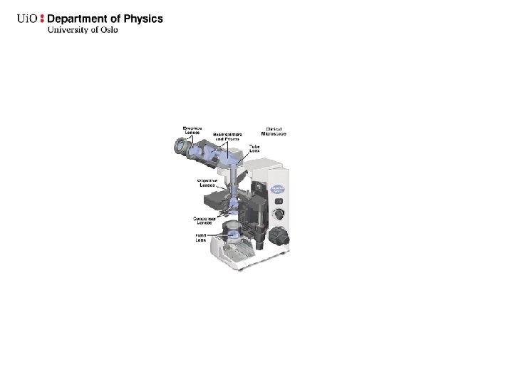
Avbildningssystemet Eyepiece Okular ”Tube lens” Objektiv Prøve

Primært bilde

Refraction Refraksjon ─ lysbrytning ϕ 1 2 Snell’s law: Sin ϕ 1 n 2 Sin ϕ 2 = n 1 = v 1 v 2

Chromatic aberation

Spherical aberasjon + astigmatisme, koma, fortegning. . .

Corrections
 Apochromate:")
Corrections Objective Acromate: Chromatic for red and blue, spherical for green Semi-acromate (fluorite) Apochromate: Chromatic for red, green, blue and dark blue, spherical for green and blue
")
More are needed than just the objective Kondensorsystem Eyepice Okular (ocular)

1000 X

2000 X

10 000 X

100 000 X

50 mm

Magnification vs. resolution Forstørrelse vs. oppløsningsevne

p. 5 Effective magnification Resolution of the eye is around 0. 2 mm Resolution limit is about 0. 2 µm Meff ≈ 0. 2 mm 0. 2× 10− 3 mm = 1000
 The limiting factor: Diffraction R = 0, 61 l/NA Rayleigh criterion")
Resolution (Oppløsning) The limiting factor: Diffraction R = 0, 61 l/NA Rayleigh criterion

Eric Betzig Stefan W. Hell William E. Moerner The Royal Swedish Academy of Sciences has decided to award Erik Betzig, Stefan W. Hell and W. E. Moerner the Nobel Prize in Chemistry 2014 for the development of super-resolution fluorescence microscopy.

Fluorescence microscopy Confocal microscopy
 NA = nsina Resolution Oppløsning R = 0, 61 l/NA")
Numerical aperture (NA) NA = nsina Resolution Oppløsning R = 0, 61 l/NA

Objectives for oil immersion

Optical anisotropy Calcite

Optically active crystals in the microscope

Can observe stress/strain

Optically active crystals in the microscope
- Slides: 61