
Lewy Bodies and Dementia John E Duda M
Lewy Bodies and Dementia John E. Duda, M. D. Director, Parkinson’s Disease Research, Education and Clinical Center Philadelphia VA Medical Center Assistant Professor of Neurology University of Pennsylvania School of Medicine Philadelphia, Pennsylvania
Objectives • Understand the relationship between dementia with Lewy bodies and Parkinson’s disease with dementia • Learn recent changes in the diagnosis and management of PDD and DLB • Understand the emerging role of neuroimaging in the diagnosis of PDD and DLB
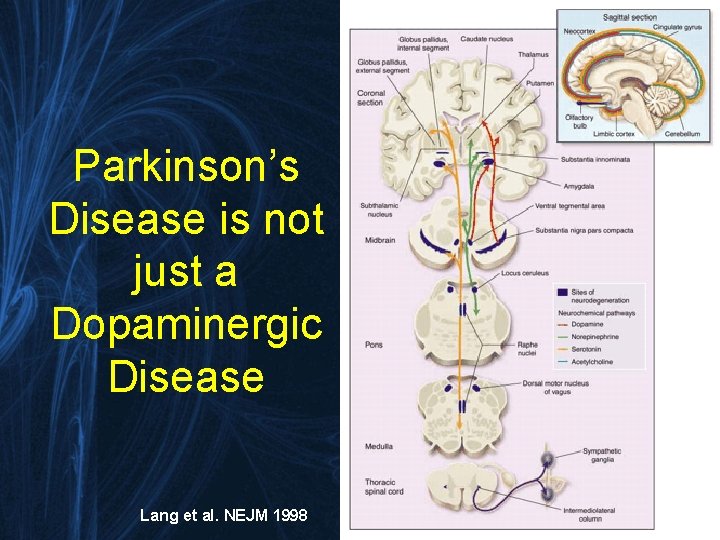
Parkinson’s Disease is not just a Dopaminergic Disease Lang et al. NEJM 1998
Diagnostic Criteria for PDD • Impairment in ≥ 2 core cognitive domains – Impaired attention, executive functions, visuospatial functions, and free recall memory which usually improves with cueing • Presence of at least one behavioral symptom (apathy, depressed or anxious mood, hallucinations, delusions, excessive daytime sleepiness) supports diagnosis • End result: - More sensitive (shift from focus on memory) - Bring in line with existing criteria for DLB Emre et al. Movement Disorders 2007; 22: 1689 -1707.
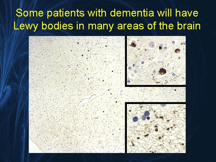
Some patients with dementia will have Lewy bodies in many areas of the brain
What do we call it? • Nomenclature – Diffuse Lewy body disease – Cortical Lewy body disease – Senile dementia of Lewy body type – Lewy body variant of Alzheimer’s disease – Parkinson’s disease dementia – Dementia with Lewy bodies • Proposed by the First International Workshop of the Consortium on Dementia with Lewy bodies – Neurology (1996) 47: 1113
Dementia with Lewy Bodies Consensus Criteria for the Clinical Diagnosis • Progressive cognitive decline of sufficient magnitude to interfere with normal social or occupational function • Core features (2 probable DLB; 1 possible DLB) – Fluctuating cognition – Recurrent visual hallucinations – Parkinsonism • Features supportive of the diagnosis are – – – Repeated falls Syncope Transient loss of consciousness Neuroleptic sensitivity Systematized delusions Hallucinations in other modalities • Diagnosis is less likely in the presence of – cerebrovascular disease – Any physical illness or brain disorder sufficient to account for the clinical picture Neurology (1996) 47: 1113
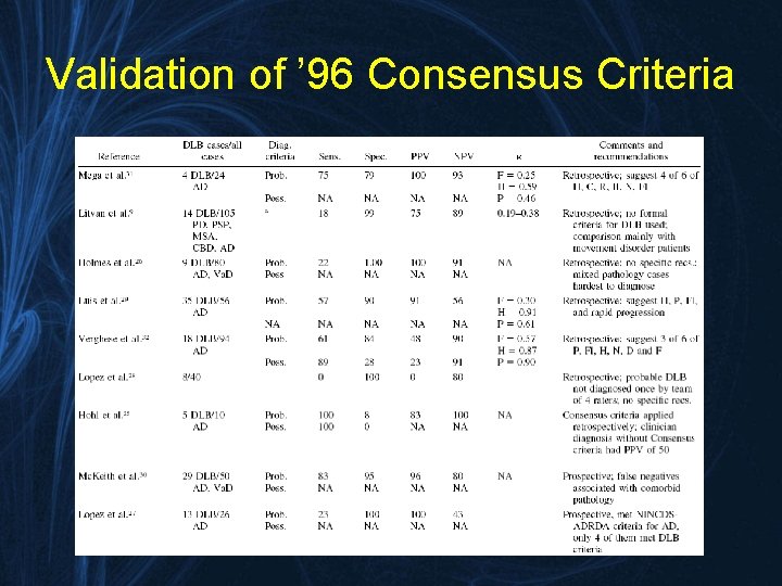
Validation of ’ 96 Consensus Criteria
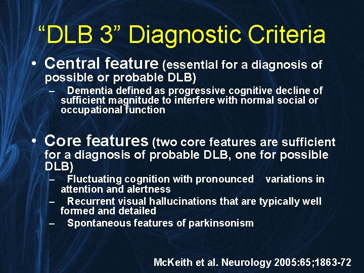
“DLB 3” Diagnostic Criteria • Central feature (essential for a diagnosis of possible or probable DLB) – Dementia defined as progressive cognitive decline of sufficient magnitude to interfere with normal social or occupational function • Core features (two core features are sufficient for a diagnosis of probable DLB, one for possible DLB) – Fluctuating cognition with pronounced variations in attention and alertness – Recurrent visual hallucinations that are typically well formed and detailed – Spontaneous features of parkinsonism Mc. Keith et al. Neurology 2005: 65; 1863 -72
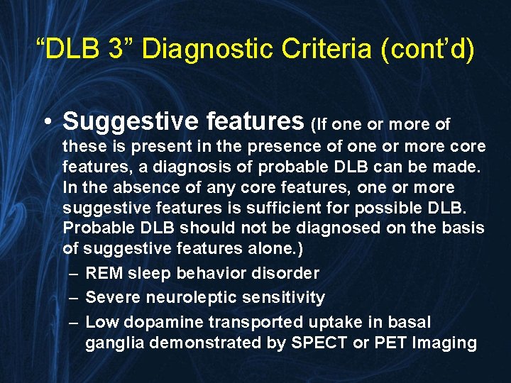
“DLB 3” Diagnostic Criteria (cont’d) • Suggestive features (If one or more of these is present in the presence of one or more core features, a diagnosis of probable DLB can be made. In the absence of any core features, one or more suggestive features is sufficient for possible DLB. Probable DLB should not be diagnosed on the basis of suggestive features alone. ) – REM sleep behavior disorder – Severe neuroleptic sensitivity – Low dopamine transported uptake in basal ganglia demonstrated by SPECT or PET Imaging
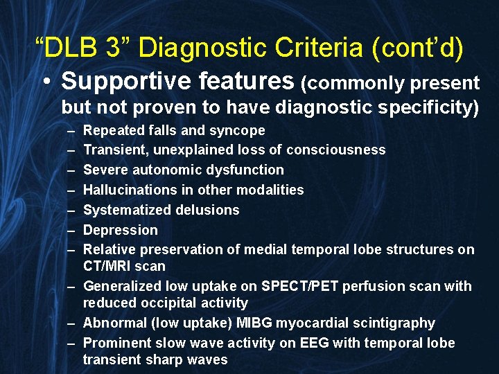
“DLB 3” Diagnostic Criteria (cont’d) • Supportive features (commonly present but not proven to have diagnostic specificity) – – – – Repeated falls and syncope Transient, unexplained loss of consciousness Severe autonomic dysfunction Hallucinations in other modalities Systematized delusions Depression Relative preservation of medial temporal lobe structures on CT/MRI scan – Generalized low uptake on SPECT/PET perfusion scan with reduced occipital activity – Abnormal (low uptake) MIBG myocardial scintigraphy – Prominent slow wave activity on EEG with temporal lobe transient sharp waves
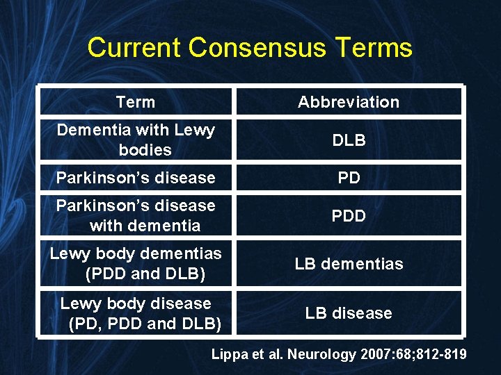
Current Consensus Term Abbreviation Dementia with Lewy bodies DLB Parkinson’s disease PD Parkinson’s disease with dementia PDD Lewy body dementias (PDD and DLB) LB dementias Lewy body disease (PD, PDD and DLB) LB disease Lippa et al. Neurology 2007: 68; 812 -819
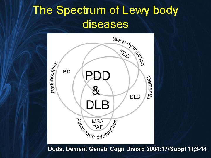
The Spectrum of Lewy body diseases Duda. Dement Geriatr Cogn Disord 2004: 17(Suppl 1); 3 -14
Prevalence of LBD • DLB causes 10 -15% of irreversible dementia • 25% of AD cases have parkinsonism and most of these cases have LBs at autopsy • PD affects 1 in 100 persons over the age of 60, with a yearly incidence of dementia increasing with age to 13. 7%/yr over the age of 70. • The combined sum of patients with parkinsonism and dementia may approach 2 million
Evaluation of LBD • History and physical examination • Psychometric testing – verbal and non-verbal memory, attention, concentration, abstraction, visuospatial abilities and construction • Neuroimaging
Screening Instruments
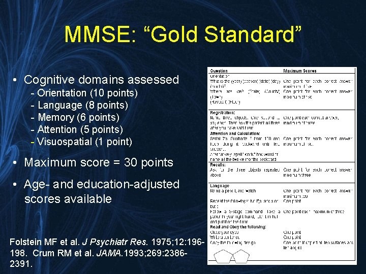
MMSE: “Gold Standard” • Cognitive domains assessed - Orientation (10 points) - Language (8 points) - Memory (6 points) - Attention (5 points) - Visuospatial (1 point) • Maximum score = 30 points • Age- and education-adjusted scores available Folstein MF et al. J Psychiatr Res. 1975; 12: 196198. Crum RM et al. JAMA. 1993; 269: 23862391.
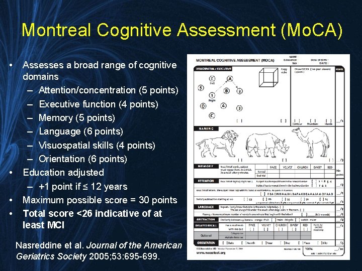
Montreal Cognitive Assessment (Mo. CA) • • Assesses a broad range of cognitive domains – Attention/concentration (5 points) – Executive function (4 points) – Memory (5 points) – Language (6 points) – Visuospatial skills (4 points) – Orientation (6 points) Education adjusted – +1 point if ≤ 12 years Maximum possible score = 30 points Total score <26 indicative of at least MCI Nasreddine et al. Journal of the American Geriatrics Society 2005; 53: 695 -699.
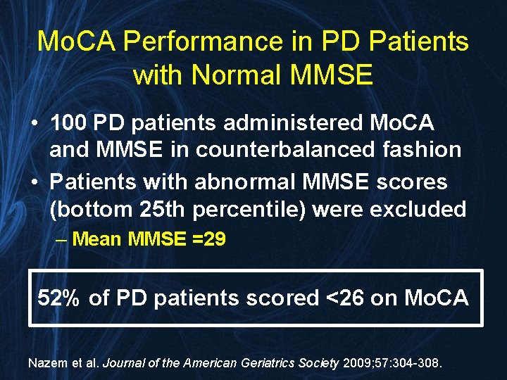
Mo. CA Performance in PD Patients with Normal MMSE • 100 PD patients administered Mo. CA and MMSE in counterbalanced fashion • Patients with abnormal MMSE scores (bottom 25 th percentile) were excluded – Mean MMSE =29 52% of PD patients scored <26 on Mo. CA Nazem et al. Journal of the American Geriatrics Society 2009; 57: 304 -308.
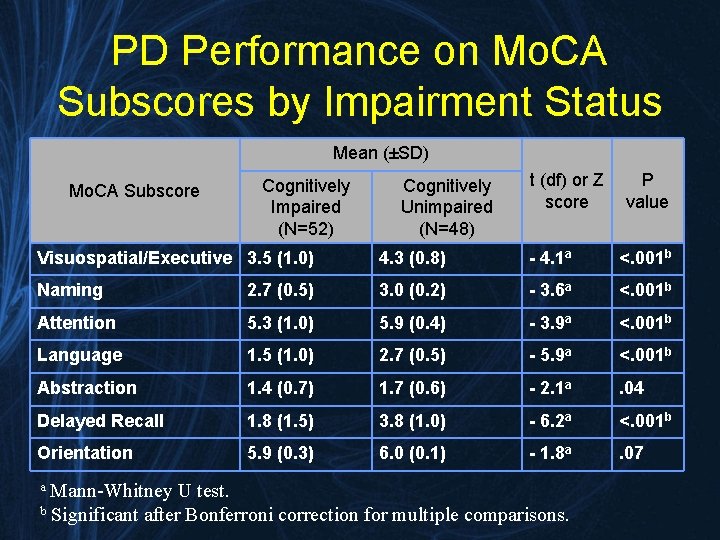
PD Performance on Mo. CA Subscores by Impairment Status Mean (±SD) Mo. CA Subscore Cognitively Impaired (N=52) Cognitively Unimpaired (N=48) t (df) or Z score P value Visuospatial/Executive 3. 5 (1. 0) 4. 3 (0. 8) - 4. 1 a <. 001 b Naming 2. 7 (0. 5) 3. 0 (0. 2) - 3. 6 a <. 001 b Attention 5. 3 (1. 0) 5. 9 (0. 4) - 3. 9 a <. 001 b Language 1. 5 (1. 0) 2. 7 (0. 5) - 5. 9 a <. 001 b Abstraction 1. 4 (0. 7) 1. 7 (0. 6) - 2. 1 a . 04 Delayed Recall 1. 8 (1. 5) 3. 8 (1. 0) - 6. 2 a <. 001 b Orientation 5. 9 (0. 3) 6. 0 (0. 1) - 1. 8 a . 07 Mann-Whitney U test. b Significant after Bonferroni correction for multiple comparisons. a
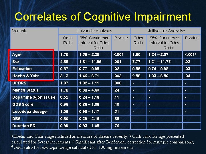
Correlates of Cognitive Impairment Variable Univariate Analyses Odds Ratio Multivariate Analysisa 95% Confidence Interval for Odds Ratio P value Odds Ratio 95% Confidence Interval for Odds Ratio P value Ageb 1. 75 1. 36 – 2. 25 <. 001 1. 60 1. 24 – 2. 07 <. 001 c Sex 4. 65 1. 81 – 11. 95 . 001 3. 77 1. 21 – 11. 73 . 02 Education 0. 87 0. 77 – 0. 98 . 02 0. 85 0. 74 – 0. 98 . 03 Hoehn & Yahr 3. 13 1. 46 – 6. 71 . 003 2. 58 1. 03 – 6. 50 . 04 UPDRS 1. 07 1. 02 – 1. 11 . 006 - - - Marital Status 1. 78 0. 68 – 4. 63 . 24 - - - Dopamine agonist use 0. 52 0. 24 – 1. 16 . 11 - - - GDS Score 0. 96 0. 86 – 1. 06 . 40 - - - Levodopa dosagee 1. 06 0. 95 – 1. 17 . 31 - - - DBS 0. 80 0. 29 – 2. 16 . 65 - - - Duration PD 0. 99 0. 93 – 1. 05 . 76 - - - a Hoehn and Yahr stage included as measure of disease severity; b Odds ratio for age presented calculated for 5 -year increments; c Significant after Bonferroni correction for multiple comparisons; d Odds ratio for levodopa dosage calculated for 100 -mg increments.
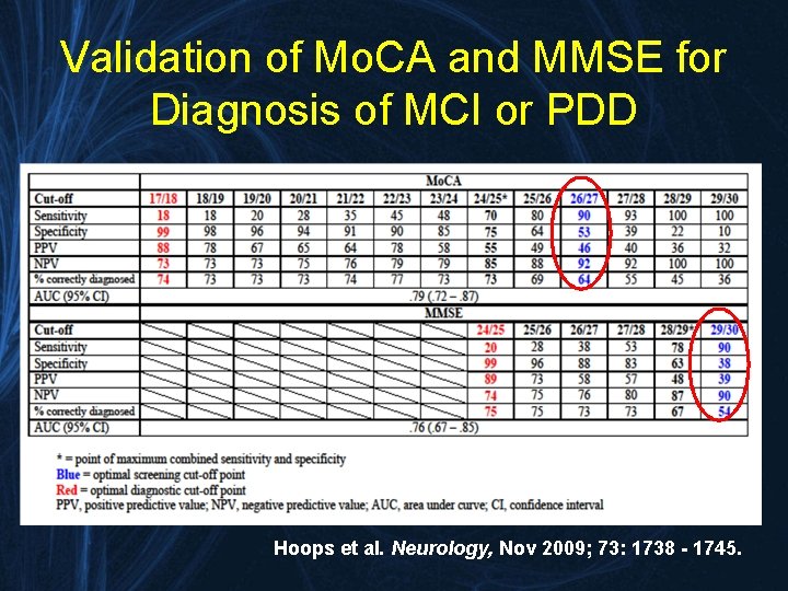
Validation of Mo. CA and MMSE for Diagnosis of MCI or PDD Hoops et al. Neurology, Nov 2009; 73: 1738 - 1745.
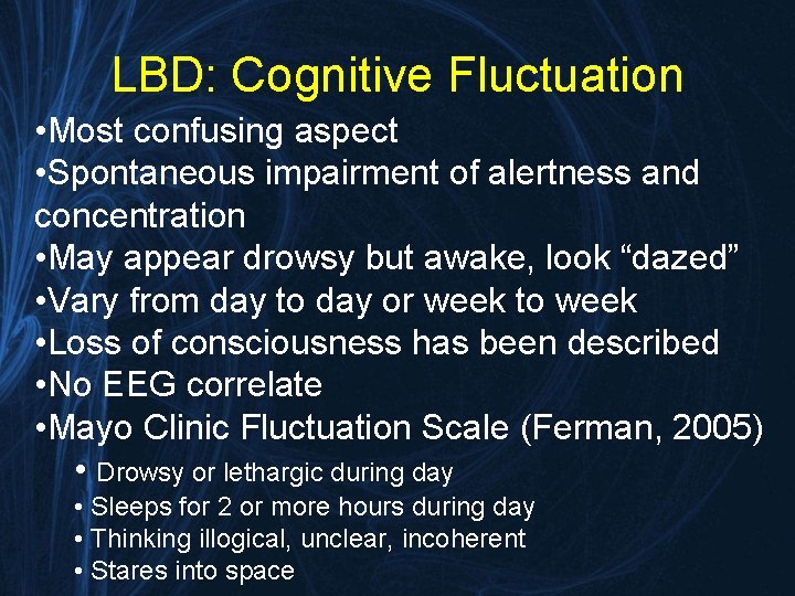
LBD: Cognitive Fluctuation • Most confusing aspect • Spontaneous impairment of alertness and concentration • May appear drowsy but awake, look “dazed” • Vary from day to day or week to week • Loss of consciousness has been described • No EEG correlate • Mayo Clinic Fluctuation Scale (Ferman, 2005) • Drowsy or lethargic during day • Sleeps for 2 or more hours during day • Thinking illogical, unclear, incoherent • Stares into space
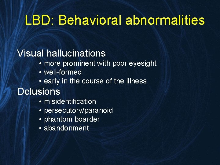
LBD: Behavioral abnormalities Visual hallucinations • more prominent with poor eyesight • well-formed • early in the course of the illness Delusions • misidentification • persecutory/paranoid • phantom boarder • abandonment
LBD: Behavioral abnormalities • Depression • Anxiety • Irritability • Apathy/Amotivational states • Aggression/violent behavior • Nocturnal confusion/insomnia
REM Behavior Disorder • Act out violent (attacking, chasing) dreams during rapid eye movement (REM) sleep • Shouting, kicking, punching • When they wake up, they remember the dream • Often hurt themselves or bed partners • Usually responds to low-dose clonazepam (0. 5 -1 mg qhs)

So what do we do with these patients?
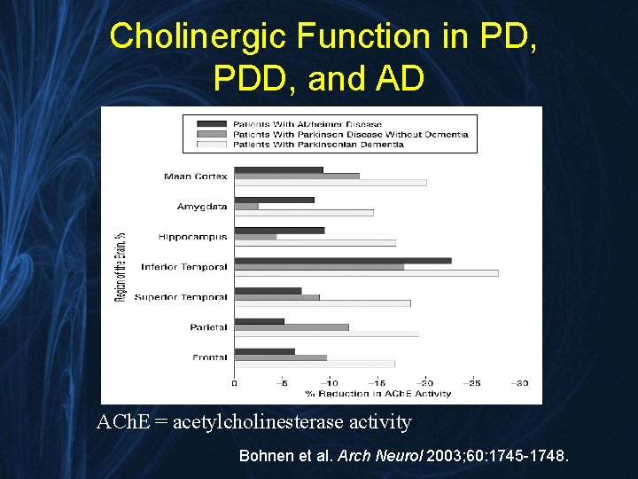
Cholinergic Function in PD, PDD, and AD ACh. E = acetylcholinesterase activity Bohnen et al. Arch Neurol 2003; 60: 1745 -1748.
Cognitive disorder • Central Acting Cholinesterase Inhibitors • • • modest improvement in cognition diminished hallucinations improvement in behavior may improve gait improvement in attention Rivastigmine only FDA-approved treatment for PDD • Role of Memantine is unclear
Movement Disorder • Becomes a balance between alleviating motor symptoms and worsening neuropsychiatric symptoms • Management often optimal with carbidopa/levodopa monotherapy • Dopamine agonists, COMT inhibitors, anti-cholinergics and MAO-B inhibitors often can worsen psychosis, hallucinations and delirium
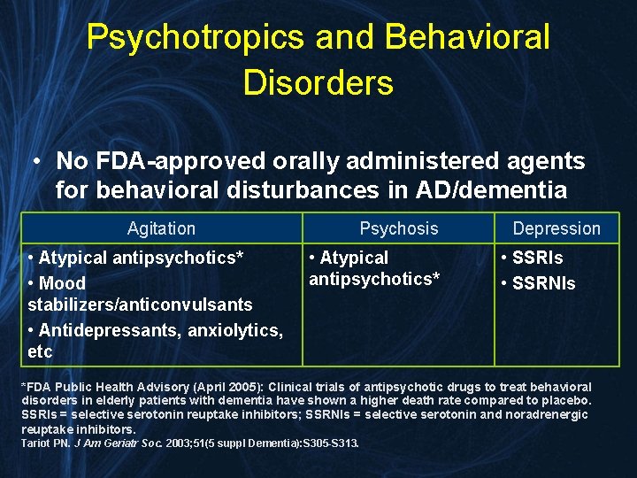
Psychotropics and Behavioral Disorders • No FDA-approved orally administered agents for behavioral disturbances in AD/dementia Agitation • Atypical antipsychotics* • Mood stabilizers/anticonvulsants • Antidepressants, anxiolytics, etc Psychosis • Atypical antipsychotics* Depression • SSRIs • SSRNIs *FDA Public Health Advisory (April 2005): Clinical trials of antipsychotic drugs to treat behavioral disorders in elderly patients with dementia have shown a higher death rate compared to placebo. SSRIs = selective serotonin reuptake inhibitors; SSRNIs = selective serotonin and noradrenergic reuptake inhibitors. Tariot PN. J Am Geriatr Soc. 2003; 51(5 suppl Dementia): S 305 -S 313.
Safety of Antipsychotics • FDA Public Health Advisory, April 2005: – Clinical trials of atypical antipsychotic drugs to treat behavioral disorders in dementia patients have shown a 1. 6 -1. 7 -fold higher death rate compared to placebo – Specific causes of death were primarily due to: • Heart-related events (eg, heart failure, sudden death) • Infections (mostly pneumonia) • Wang PS, et al. N Engl J Med. December 2005 – Conventional antipsychotics were associated with higher risk of death than atypical antipsychotics in elderly patients. – Risk is highest: • Soon after therapy is initiated • At high doses Schneider LS, et al. JAMA. 2005; 294: 1934 -1943; Wang PS, et al. N Engl J Med. 2005; 353: 2335 -2341; FDA Public Health Advisory. Available at: http: //www. fda. gov/cder/drug/advisory/antipsychotics. htm. Accessed October 2007.
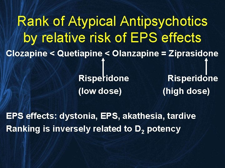
Rank of Atypical Antipsychotics by relative risk of EPS effects Clozapine < Quetiapine < Olanzapine = Ziprasidone Risperidone (low dose) (high dose) EPS effects: dystonia, EPS, akathesia, tardive Ranking is inversely related to D 2 potency
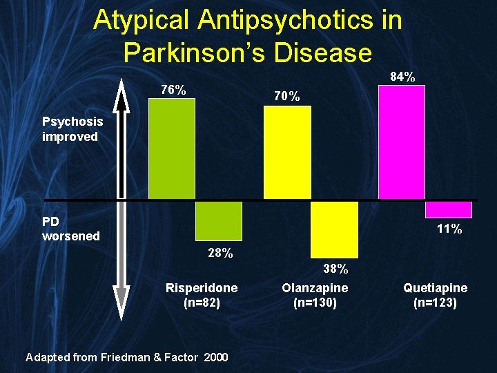
Atypical Antipsychotics in Parkinson’s Disease 84% 76% 70% Psychosis improved PD worsened 11% 28% 38% Risperidone (n=82) Adapted from Friedman & Factor 2000 Olanzapine (n=130) Quetiapine (n=123)
Management of Psychosis • Quetiapine and clozapine have become first line agents • LBD patients will often respond to doses MUCH lower than commonly used so ‘start low and go slow’ • Strenuously avoid typical neuroleptics to prevent neuroleptic sensitivity reactions (Aarsland et al. J Clin Psychiatry 2005: 66: 63 -7)
How imaging is beginning to play a role in the diagnosis and management of patients with PDD/DLB
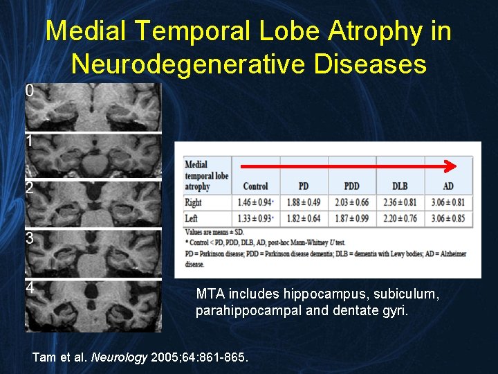
Medial Temporal Lobe Atrophy in Neurodegenerative Diseases MTA includes hippocampus, subiculum, parahippocampal and dentate gyri. Tam et al. Neurology 2005; 64: 861 -865.
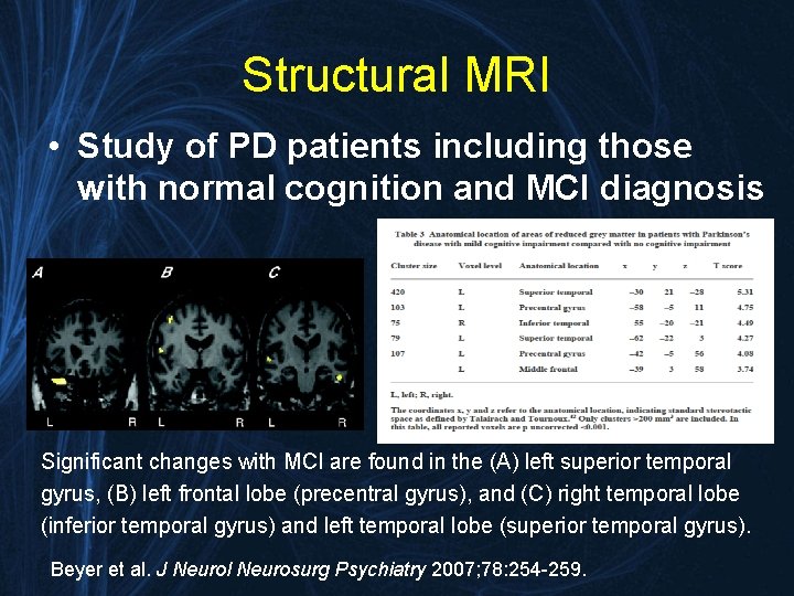
Structural MRI • Study of PD patients including those with normal cognition and MCI diagnosis Significant changes with MCI are found in the (A) left superior temporal gyrus, (B) left frontal lobe (precentral gyrus), and (C) right temporal lobe (inferior temporal gyrus) and left temporal lobe (superior temporal gyrus). Beyer et al. J Neurol Neurosurg Psychiatry 2007; 78: 254 -259.
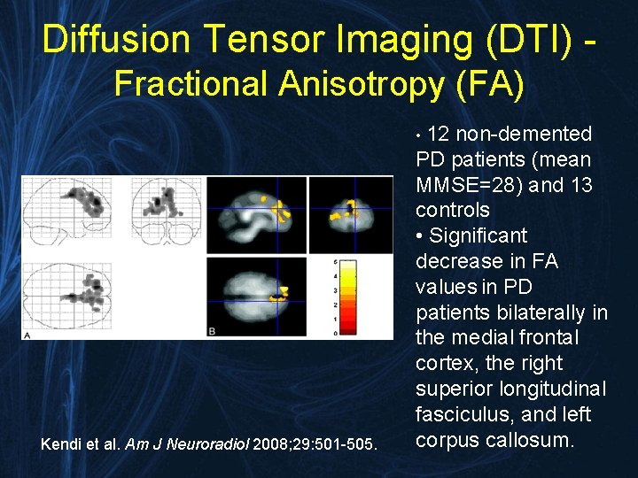
Diffusion Tensor Imaging (DTI) Fractional Anisotropy (FA) • 12 Kendi et al. Am J Neuroradiol 2008; 29: 501 -505. non-demented PD patients (mean MMSE=28) and 13 controls • Significant decrease in FA values in PD patients bilaterally in the medial frontal cortex, the right superior longitudinal fasciculus, and left corpus callosum.
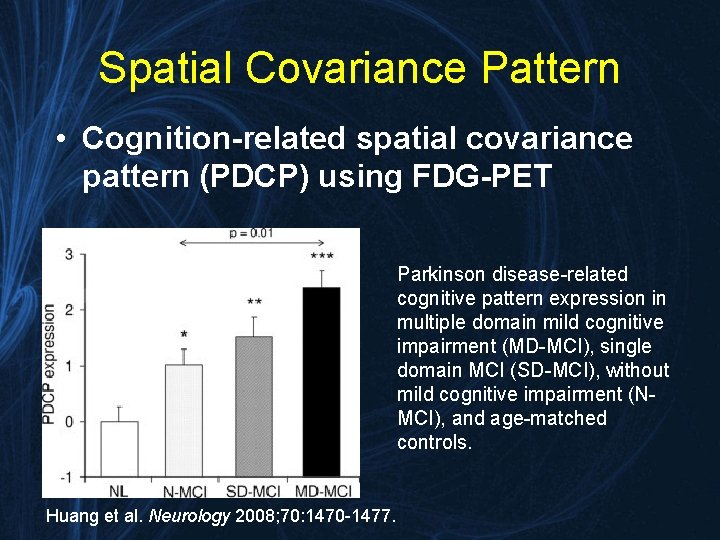
Spatial Covariance Pattern • Cognition-related spatial covariance pattern (PDCP) using FDG-PET Parkinson disease-related cognitive pattern expression in multiple domain mild cognitive impairment (MD-MCI), single domain MCI (SD-MCI), without mild cognitive impairment (NMCI), and age-matched controls. Huang et al. Neurology 2008; 70: 1470 -1477.
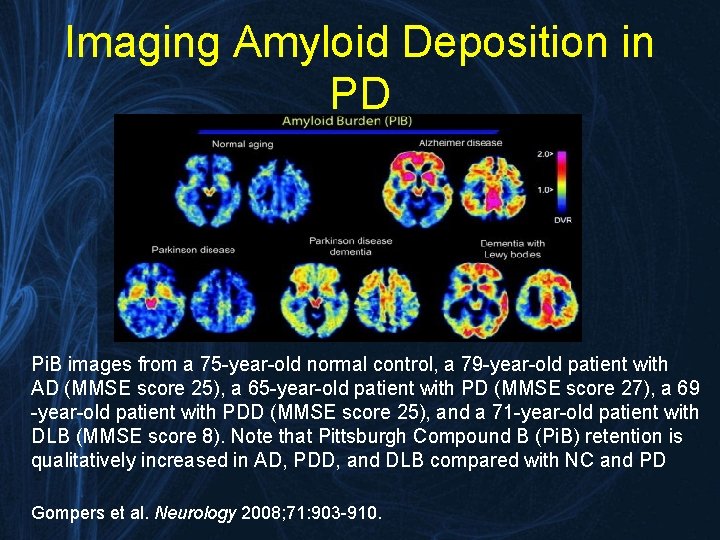
Imaging Amyloid Deposition in PD Pi. B images from a 75 -year-old normal control, a 79 -year-old patient with AD (MMSE score 25), a 65 -year-old patient with PD (MMSE score 27), a 69 -year-old patient with PDD (MMSE score 25), and a 71 -year-old patient with DLB (MMSE score 8). Note that Pittsburgh Compound B (Pi. B) retention is qualitatively increased in AD, PDD, and DLB compared with NC and PD Gompers et al. Neurology 2008; 71: 903 -910.
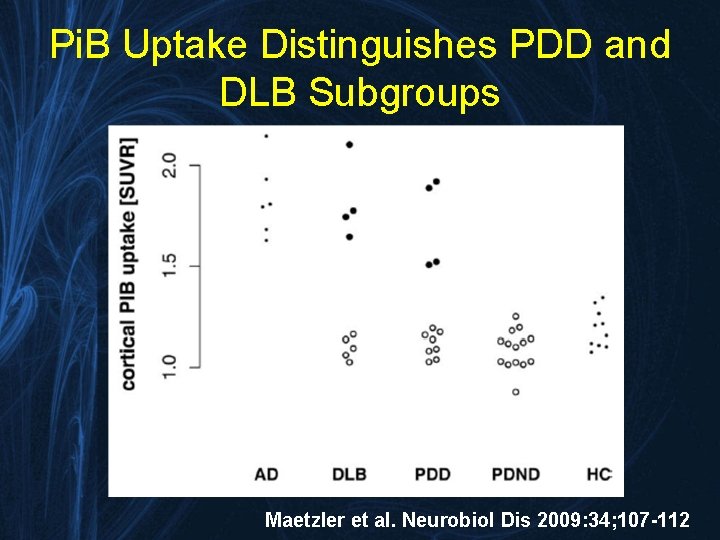
Pi. B Uptake Distinguishes PDD and DLB Subgroups Maetzler et al. Neurobiol Dis 2009: 34; 107 -112
Conclusions • PDD and DLB are similar disorders that may lie upon a spectrum of Lewy body diseases • Diagnostic screening of PD patients may be improved with the Mo. CA • Management requires a balance between alleviating motor and cognitive symptoms • Novel imaging techniques may soon play important roles in diagnosis and management
- Slides: 43