Leukopoiesis Dr Hytham Ahmed Abuagla Introduction The cellular

Leukopoiesis Dr. Hytham Ahmed Abuagla

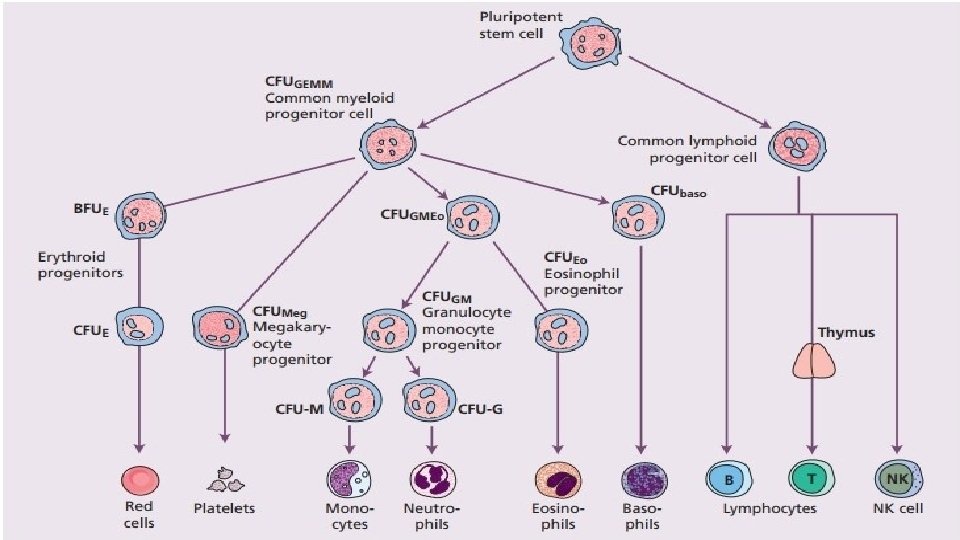
Introduction • The cellular elements of the blood are produced from a common, multipotential hematopoietic cell. This cell, the progenitor cell, undergoes mitotic division. • Subsequent maturation of progenitor cells produces the major categories of the cellular elements of the circulating blood: the erythrocytes, leukocytes, and thrombocytes. • On the basis of function, leukocytes can be divided into the granulocytic, monocytic, and lymphoid series.
 progenitor")
Granulocytic series • Production of Neutrophils, Eosinophils, Basophils • When the colony-forming-unit-granulocyte-erythrocyte-monocytemegakaryocyt (CFU-GEMM) progenitor cell differentiates into the colony-forming-unit-granulocyte-macrophage (CFU-GM) progenitor cell, the cell line becomes committed to developing into a myeloblast.

1. Myeloblast • Is the first identifiable cell in the granulocytic series. • It constitutes approximately 1% of the total nucleated bone marrow cells. • This stage lasts approximately 15 hours. • It has a diameter of 14 to 18μm. • The major part is occupied by a large oval nucleus composed of very fine nonaggregated chromatin and possesses 3 or more nucleoli. • The cytoplasm has basophilic character and is devoid of granules. • Mitochondria are present but have a rather small size.

2. Promyelocyte: • Constitutes approximately 3% of the nucleated bone marrow cells. • This stage lasts about 24 hours. • Promyelocytes are generally larger than myeloblasts, measuring approximately 12 to 20 microns. • The nucleus is similar in size to the myeloblast but the cytoplasm is more abundant at this stage. • The nucleoli will begin to close and become less prominent than in the myeloblast stage. • The chromatin strand texture in promyelocyte tends to become slightly more coarse and clumped than the chromatin pattern present in a myeloblast. • Promyelocyte cytoplasm will have a gritty basophilic color. • There will also be prominent non-specific primary granules. These granules will look like red/purple grains of sand.

3. Myelocyte: • It constitute about approximately 12% of the proliferative cells existing in this stage. • The stage from myelocyte to metamyelocyte lasts an average of 4. 3 days. • The cytoplasm of this cell begins to produce specific, secondary granules. • As the cell matures closer to a metamyelocyte, they fill the entire cytoplasm. • While the cytoplasm shifts to producing secondary granules it also looses the prominence of its primary granules. • Three types of myelocytes called eosinophilic, basophilic, and neutrophilic for the dyes the granules take are distinguished; these in turn give rise to leukocytes called, respectively, eosinophils, basophils, and neutrophils.

• At the same time the secondary granule production begins, the nucleus is shrinking and condensing. • The nucleoli close and disappear, the chromatin gets coarser/denser and more clumped. • Once the metamyelocyte stage has been reached, cells have undergone four or five cell divisions and the proliferative phase comes to an end. Neutrophilic Myelocyte Eosinophilic Myelocyte Basophilic Myelocyte

4. Metamyelocyte: • Myelocytes matures to a metamyelocyte with appearance of nuclear indentation. • Metamyelocyte can not divide. • The most pronounced change that takes place when a myelocyte transforms into a metamyelocyte is indentation of the nucleus. Neutrophilic Metamyelocyte Eosinophilic Metamyelocyte Basophilic Metamyelocyte

• 5. Band Granulocytes • The indentation of the nucleus characteristic of the metamyelocyte increases and the nucleus becomes ribbon shaped. • The nucleus of this cell has parallel borders for most of its length, like a band, and the cell is thus known as the band form. Band Neutrophil Band Eosinophil Band Basophil

• As the cell matures the nucleus becomes segmented. • The segments are connected by thin strands of nuclear material. • A constriction of more than half or two-third is accepted as segmentation by some. • Neutrophils usually have 2 -5 segments and eosinophils usually have 2 segments. • Segmentations of the basophil nucleus are obscured by the intensely granular cytoplasm. • The basophil nucleus ahs 2 -3 segments, appearing in the contain few granules

Monocyte-Macrophage series 1. Monoblast: • A typical monoblast is about 12 to 20 µm in diameter, has a nuclear to cytoplasm ratio of 4: 1 to 3: 1, and, like most myeloid blasts, has a round to oval nucleus with fine chromatin structure. • One to four nucleoli are usually visible. • The nucleus can be central or eccentric and it can show evidence of indentation or folding. • The cytoplasm is agranular, stains moderately to lightly basophilic, and often has an intensely stained periphery and a prominent perinuclear zone

2. Promonocyte • Three to four times larger than a mature RBC • Round Indented Nucleus with immature chromatin (not clumped; slightly more mature than monoblast) • Prominent nucleoli (less prominent than Monoblast) • Cytoplasm is scant but more than Myeloblasts, gray to pale blue and with rare or no granules • vacuoles maybe sometimes noted in the cytoplasm

Lymphopoiesis 1. Lymphoblast: • Lymphoblasts, the most immature cells of the lymphocytic series, measure 10 to 20 um in diameter and are usually round or oval, though sometimes may be irregular. • The nucleus is often round or oval but occasionally may be notched, cleft, folded or irregular in shape. It is usually centrally placed but may be eccentric. The chromatin is fine, but is less so than in myeloblasts. • One or more nucleoli may be present. The cytoplasm varies in amount, stains moderately basophilic and is usually agranular. It may, however, sometimes contain vacuoles. • The N: C ratio is 7: 1 to 4: 1

2. Prolymphocyte: • Prolymphocytes are generally the same size as lymphoblasts. • Usually centrally placed, a round, ovoid or slightly indented nucleus typically contains a single prominent nucleolus. The chromatin is somewhat condensed (coarser than in lymphoblasts but less dense than in mature lymphocytes). • The amount of cytoplasm, which stains homogeneously blue, is greater in the prolymphocytes than in lymphoblasts or mature lymphocytes. • The N: C ratio is 5: 1 to 3: 1
- Slides: 15