
Leukaemia Leukemia is uncontrolled monoclonal proliferation of hematopoietic
- Slides: 22
Leukaemia
Leukemia is uncontrolled monoclonal proliferation of hematopoietic precursor cell line, which replace the normal hematopoietic cells of the marrow.
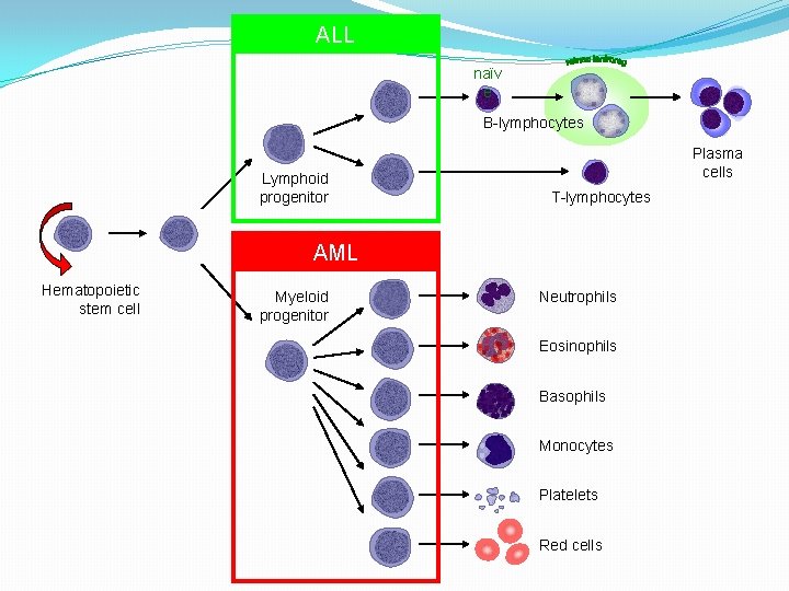
ALL naïv e B-lymphocytes Lymphoid progenitor Plasma cells T-lymphocytes AML Hematopoietic stem cell Myeloid progenitor Neutrophils Eosinophils Basophils Monocytes Platelets Red cells
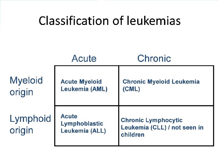
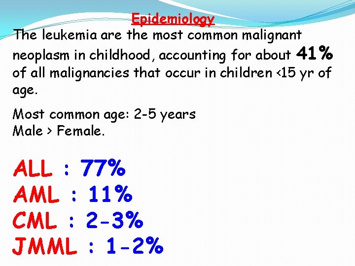
Epidemiology The leukemia are the most common malignant neoplasm in childhood, accounting for about 41% of all malignancies that occur in children <15 yr of age. Most common age: 2 -5 years Male > Female. ALL : 77% AML : 11% CML : 2 -3% JMML : 1 -2%
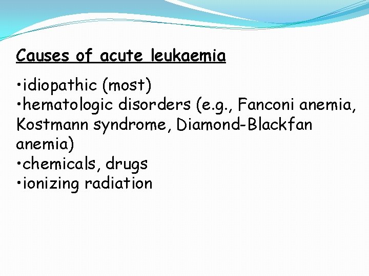
Causes of acute leukaemia • idiopathic (most) • hematologic disorders (e. g. , Fanconi anemia, Kostmann syndrome, Diamond-Blackfan anemia) • chemicals, drugs • ionizing radiation
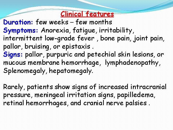
Clinical features Duration: few weeks – few months Symptoms: Anorexia, fatigue, irritability, intermittent low-grade fever , bone pain, joint pain, pallor, bruising, or epistaxis. Signs: pallor, purpuric and petechial skin lesions, or mucous membrane hemorrhage, lymphadenopathy, Splenomegaly, hepatomegaly. Rarely, patients show signs of increased intracranial pressure, meningeal irritation signs, papilledema, retinal hemorrhages, and cranial nerve palsies.
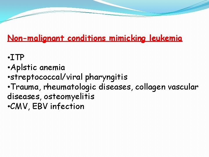
Non-malignant conditions mimicking leukemia • ITP • Aplstic anemia • streptococcal/viral pharyngitis • Trauma, rheumatologic diseases, collagen vascular diseases, osteomyelitis • CMV, EBV infection
Laboratory investigations CBP(complete Blood Picture) -Hb : mostly low <10 g/d. L (s. t. Normal) -Total WBCs: low, normal, or high (normal 4 -11 X 10^9/L). May be very high (hyperleukocytosis) > 100 X 10^9/L; causing increasing viscosity of blood, bleeding diathesis, ARDS, CVA. Leukopenia→ infection. -Platelets: usually thrombocytopenia ( plt<50 X 10^9/L cause mucocutaneous bleeding). -Blood film: normochromic normocytic anemia. Blasts in peripheral blood (%). -ESR: increased (not specific).
Bone marrow examination: necessary for diagnosis, useful for determining type of leukaemia ALL is defined by the presence of >25% blasts in bone marrow (% of nucleated marrow cells) AML is defined by the presence of >20% blasts Done in diagnosis, during therapy and during follow -up in certain spots; and when suspect BM relapse.
Classification of subtypes of leukemia depends on Morphology: FAB classification ALL : L 1, L 2, L 3 AML : M 0, M 1, M 2, M 3 v, M 4 Eo, M 5, M 6, M 7 Immunophenotype: flowcytometry; e. g ALL: Pre-B, B-cell , T -cell Cytogenetic: ALL : t(12, 21) AML : t(15, 17)in M 3 CML : t(9, 22) – Philadelphian chromosome
Biochemical investigations -S. uric acid : (NV: 120 -420 mmol/L). DNA (destructed by chemotherapy)→purine nucleotide→hypoxanthine→urate→ nephropathy -LFT: SGOT, SGPT, S. Alk Ph, TSB (direct & indirect) -RFT: S. creatinine, BUN -S. electrolyte: Na, Cl, K -S. minerals: Ca, Ph, Mg -Others: bl. Sugar, S. albumin
Imaging studies -Chest X-ray (AP & lateral): anterior mediastinal mass, pulmonary infiltrates, ARDS, pneumonia. X-ray of long bones and vertebrae: osteoporosis, pathologic fracture. - abdominal U/S: size and texture of liver, spleen, abdominal LN groups, kidneys, GB, UB, ascites, … -CT scan of brain (ICH), of lungs (pneumonia, pul. hemorrhage), of abdomen (liver & spleen) -MRI : rarely requested
Treatment Supportive measures -For anemia --- packed RBCs (10 m. L/Kg) -For infections--- broad-spectrum antibiotics(often 3 rd generation cephalosporin+ aminoglycoside) , antifungal, antiviral; systemic & local -For bleeding--- platelets concentrate, antifibrinolytic -Hydration --- twice maintenance i. e. , 3000 m. L/m 2/d (dilutional effect, increase GFR, corrects electrolyte imbalance, enhance excretion of toxic metabolites e. g. uric acid) -Nutrition
Specific measures(CHEMOTHERAPY) ALL Induction of remission: vincristine, prednisone, daunorubicin, L-asparginase CNS prophylaxis: intrathecal(methotrexate , cytosar, steroid), cranial radiation Consolidation of remission: cytosar, cyclophosphamide, methotrexate, etoposide Maintenance of remission: 6 -MP, oral MTX Total duration of Rx: for boys 3 years for girls 2. 5 years
Bone Marrow Transplantation Allogeneic : from matched related or unrelated donor S. E. : life-threatening infections, GVHD, secondary malignancies, …. Indication: very HR ALL, relapse, many cases of AML
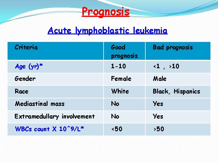
Prognosis Acute lymphoblastic leukemia Criteria Good prognosis Bad prognosis Age (yr)* 1 -10 <1 , >10 Gender Female Male Race White Black, Hispanics Mediastinal mass No Yes Extramedullary involvement No Yes WBCs count X 10^9/L* <50 >50
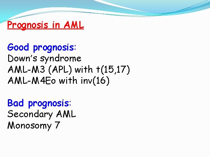
Prognosis in AML Good prognosis: Down’s syndrome AML-M 3 (APL) with t(15, 17) AML-M 4 Eo with inv(16) Bad prognosis: Secondary AML Monosomy 7
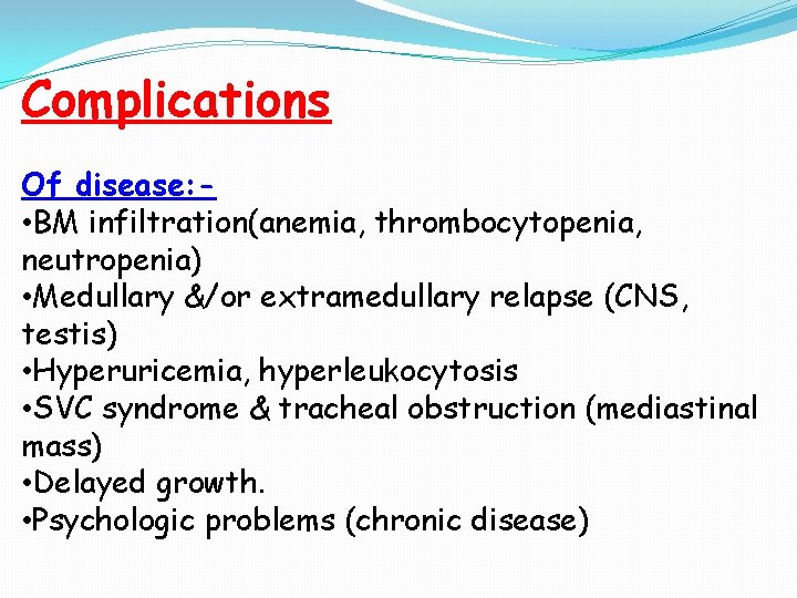
Complications Of disease: • BM infiltration(anemia, thrombocytopenia, neutropenia) • Medullary &/or extramedullary relapse (CNS, testis) • Hyperuricemia, hyperleukocytosis • SVC syndrome & tracheal obstruction (mediastinal mass) • Delayed growth. • Psychologic problems (chronic disease)
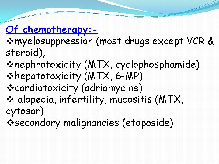
Of chemotherapy: vmyelosuppression (most drugs except VCR & steroid), vnephrotoxicity (MTX, cyclophosphamide) vhepatotoxicity (MTX, 6 -MP) vcardiotoxicity (adriamycine) v alopecia, infertility, mucositis (MTX, cytosar) vsecondary malignancies (etoposide)
Of radiotherapy: • neurocognetive defects • poor growth • psychatric disorders • poor school performance • Amnesia • secondary endocrine disorders • myelosuppression • secondary malignancies Of supportive measures: - transfusion-related infections (HBV, HCV, HIV, CMV), allergic reactions, fluid overload, S. E. of antibiotics
References LANZKOWSKY’S MANUAL OF PEDIATRIC HEMATOLOGY AND ONCOLOGY SIXTH EDITION (2016) Nelson TEXTBOOK of PEDIATRICS EDITION 20 (2016) EDITION 20