Lesson 10 Fluorescence Staining Fluorescent labeling Lecture overview

Lesson 10 Fluorescence Staining =Fluorescent labeling
 Fluorescence Staining (How? )")
Lecture overview 1. 2. 3. Fluorescence (What? / Why? ) Fluorescence Staining (How? ) Types & Examples

Why fluorescence? Contrast! Brilliant signals against dark background

Why Fluorescence staining ? widely used approaches for subcellular compartments and localizing proteins 1. Nonimmunological Fluorescent labeling 2. Immunological Fluorescent labeling (=Immunofluorescence)
 may be used to directly label specific subcellular")
Fluorescent stain � Fluorescent compounds (probe) may be used to directly label specific subcellular components and macromolecules.
 may be used to directly label specific subcellular")
Fluorescent stain � Fluorescent compounds (probe) may be used to directly label specific subcellular components and macromolecules. � Fluorescent compounds (probe) may be used to indirectly label specific subcellular components and macromolecules. ◦ Coupling fluorescent compounds to molecules that have specific affinity toward certain cellular components.

Specific interaction between molecules If you are specifically interested in B, then ……
! Use to identify B and determine its")
Tag with a label (e. g. fluorescence)! Use to identify B and determine its (B’s) location in cells and tissues
")
Procedure 1. 2. 3. 4. 5. Fixation (unless live cells are to be studied) Permeabilization Blocking Labeling Mounting
")
Procedure 1. 2. 3. 4. 5. Fixation (unless live cells are to be studied) Permeabilization To allow penetration of fluorescent compounds Blocking sites prone to nonspecific interactions Labeling Mounting

Permeabilization ◦ ◦ ◦ allow penetration of probes to gain access to the subcellular structures of interest Not required for the localization of a cell surface structure With detergent or organic solvents Triton X-100, NP-40 Methanol, acetone
, FITCWGA")
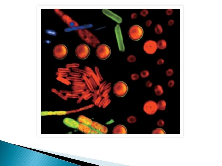
Labeling Plasma membrane ◦ NBD-PE, Fluorescent lipid analog (e. g. Di. I, …), FITCWGA Cytoskeleton ◦ phalloidin Nucleus ◦ DAPI, Hoechst, acridine orange, ethidium bromide Mitochondria ◦ Rhodamine 123, Mito Tracker

Di. I : Fluorescent lipid analog Phospholipid
 (e. g. RITC)")
Fluorescent dye (e. g. FITC) (e. g. RITC)

FITC- WGA fluorescein isothiocyanate Wheat Germ Agglutinin WGA is a plant lectin. � Different Lectins binds to specific sugars (glycoproteins, proteoglycans, glycoipids of the membrane). Fluorescently labeled lectins WGA (lectin) FITC (fluorescence) Sugar of the plasma membrane

DAPI � DNA phalloidin � F-actin phalloidin FITC F-actin
 F-actin")
DAPI 4’, 6 -diamino-2 -phenylindole � DNA phalloidin Rhodamine (RITC) F-actin

Acridine orange DNA & RNA
 Mito Tracker (tetramethylrhodamine) � Mitochondria � Membrane-potential-sensitive Labeling Live Cells!")
Hoechst � DNA (nucleus) Mito Tracker (tetramethylrhodamine) � Mitochondria � Membrane-potential-sensitive Labeling Live Cells!
 bovine pulmonary")
BODIPY FL C 5 -ceramide � Golgi apparatus Hoechst � DNA (nucleus) bovine pulmonary artery endothelial cell

Learning Resources 1. 2. Junqueira’s Basic Histology ; pp 5 & 12 Looking at the Structure of Cells in the Microscope http: //www. ncbi. nlm. nih. gov/books/NBK 28356/ ◦ “Specific Molecules Can Be Located in Cells by Fluorescence Microscopy”

LESSON 11 Detection Methods for specific proteins and genes Immunohistochemistry 2. In situ hybridization 3. Green fluorescent protein (GFP) 1.
 method of detecting the presence of specific proteins(/molecules) in cells (or tissues)")
Immunohistochemistry (IHC) method of detecting the presence of specific proteins(/molecules) in cells (or tissues) using modified (labeled) antibodies

Antibody ﺟﺴﻢ ﻣﻀﺎﺩ react specifically and bind to the antigen For IHC we must have an antibody against the protein to be detected (protein of interest).

• So these Antibodies will ‘stick’ to the antigens in our fixed cells. But how do we see them?
. – Fluorescent compounds")
• Of course we’re going to add a label (tag). – Fluorescent compounds – Enzyme (peroxidase, alkaline phosphatase……) – Electron-dense gold particles

Direct method Antibody against the protein of interest is tagged itself directly with a label

Indirect method � Primary Antibody specific for protein X. � Secondary Antibody tagged with a label , against the immunoglobulin class to which the primary antibody belongs. (e. g. rabbit anti-mouse antibodies) Amplification of Signals!
 • FITC-labeled Secondary Antibody • DAPI")
• Primary Antibody against Desmin (Intermediate filament) • FITC-labeled Secondary Antibody • DAPI

• Primary Antibody against lysozyme • Secondary Antibody labeled with peroxidase • Hematoxylin
 (e. g. peroxidase)")
(e. g. DAB) (e. g. peroxidase)

• Antibody against the amylase • Protein A coupled with gold particles. Protein A has high affinity toward antibody molecules.
 Method")
The Avidin–Biotin Complex (ABC) Method

Information flow • Can we detect specific sequence of DNA or RNA?
 method of detecting the presence of specific DNA or RNA")
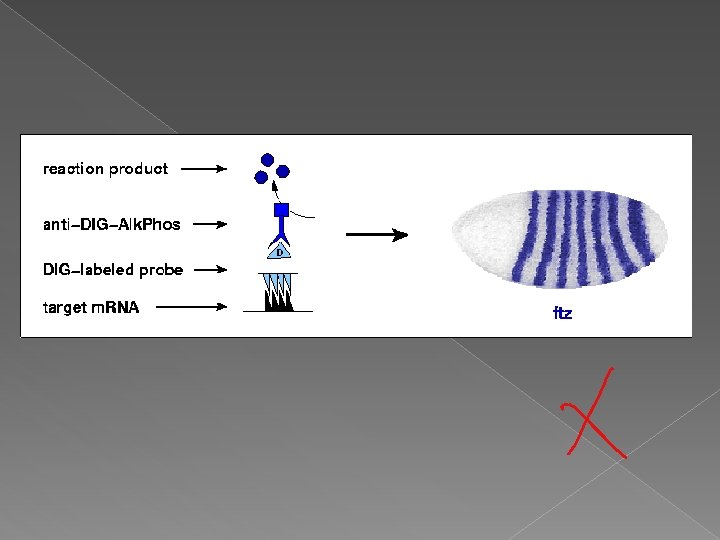
In situ hybridization (ISH) method of detecting the presence of specific DNA or RNA sequence in cells (or tissues) using labeled complementary DNA or RNA strand (probe)

1. Determining the localization of a gene in a specific chromosome

1. Determining the localization of a gene in a specific chromosome
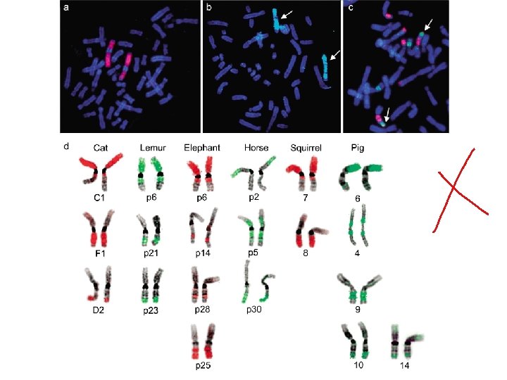

2. Identifying the cells containing specific m. RNAs

Whole mount in situ hybridization
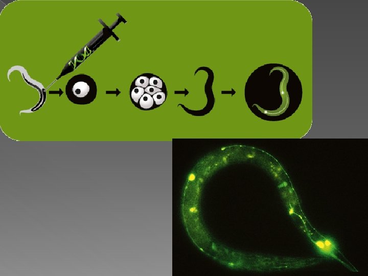
 ﺑﺮﻭﺗﻴﻨﺎﺕ ﻓﻠﻮﺭﻳﺔ ﺧﻀﺮﺍﺀ “A green guiding star for biosciences” •")
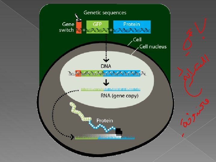
Green fluorescent protein (GFP) ﺑﺮﻭﺗﻴﻨﺎﺕ ﻓﻠﻮﺭﻳﺔ ﺧﻀﺮﺍﺀ “A green guiding star for biosciences” • to track protein of interest in cells (or tissues) • “Color your protein!”

Tumor surrounded by nourishing blood vessels

Living neruon GFP-mice

Learning Resources 1. Junqueira’s Basic Histology ; pp 12 - 15 2. Looking at the Structure of Cells in the Microscope http: //www. ncbi. nlm. nih. gov/books/NBK 28356/
- Slides: 48