Lesson 1 THE ANATOMICAL POSITION u In the

 - divides")


 1. A message originates")

 Occurs when the muscle fibers shorten. Eccentric: (Lengthening) Occurs")


 and")


 - generate energy slowly, are more")


")
 -")
 -")


- Slides: 73
Lesson #1 THE ANATOMICAL POSITION u In the same way that the maps of the world are universally oriented, anatomists and physiologists look at the human body from a standard starting point. u Anatomical position = the body in an upright, standing position, face and feet pointing forward, with the arms at the side, and the forearms fully supinated (palms forward).
Anatomical Planes The body moves in relation to 3 planes: Frontal (Coronal) - divides the body into anterior (front) and posterior (back) segments. Transverse – divides the body into superior and inferior segments. Sagittal – divides the body into medial and lateral segments.
Anatomical Planes
Anatomical Axes Describes how the rotation of the muscles and bones take place. Longitudinal (Polar) – refers to the north / south relationship. Horizontal (Bilateral) – refers to the east / west relationship. Anterior/Posterior – refers to the front of the body and the back of the body. Medial & Lateral – defined in relation to the sagittal plane. Closer to the midline of the body is medial, away from the midline is lateral. Proximal & Distal – Proximal is towards an attachment, Distal is away from the attachment. Superior/Inferior – Superior refers to one point being closer to the top, distal is lower.
Basic Movement Of Joints Flexion - decreasing an angle of a joint. Extension – increasing the angle of a joint. Adduction – adding to the midline Abduction – taking away from the midline. Internal Rotation – rotating towards the midline. External Rotation – rotating away from the midline. Circumduction – circular motion. Supination – lateral rotation of the forearm and hand. Pronation – medial rotation of the forearm and hand. Plantar Flexion – pointing the foot downward Dorsi Flexion – pointing the foot upward.
Basic Movement Of Joints
HELPFUL HINTS Axis of rotation is always perpendicular to the plane of movement. More involved movements are usually not in one specific plane but occur as a combination of motions from more than one plane.
Lesson #2 THE SKELETAL SYSTEM Axial vs. Appendicular Skeleton Axial bones include: 26 vertebral column 1 hyoid 22 skull 25 ribs and sternum = 74 bones Appendicualr bones include: 64 upper extremity 62 lower extremity 6 auditory = 132 bones = 206 TOTAL BONES in the HUMAN BODY
Meet GEORGE! Get to know him well!
And his Head!!
And His Hand
And finally, the foot and ankle!
Role of the Skeleton Protection 1. They protect vital organs and structures from trauma. The brain is packaged in a hard shell filled with cerebral spinal fluid, which helps absorb shock. Lungs and heart protected by the rib cage. Framework 1. Structural support for soft tissue, including muscles and viscera. Movement 1. Muscles attach to bones by tendons. Muscles contract and move bones to facilitate movement. Storehouse for Essential Nutrients 1. The bones store calcium and phosphorus for the body. Blood Cell Formation 1. Red blood cells and platelets are made in bones.
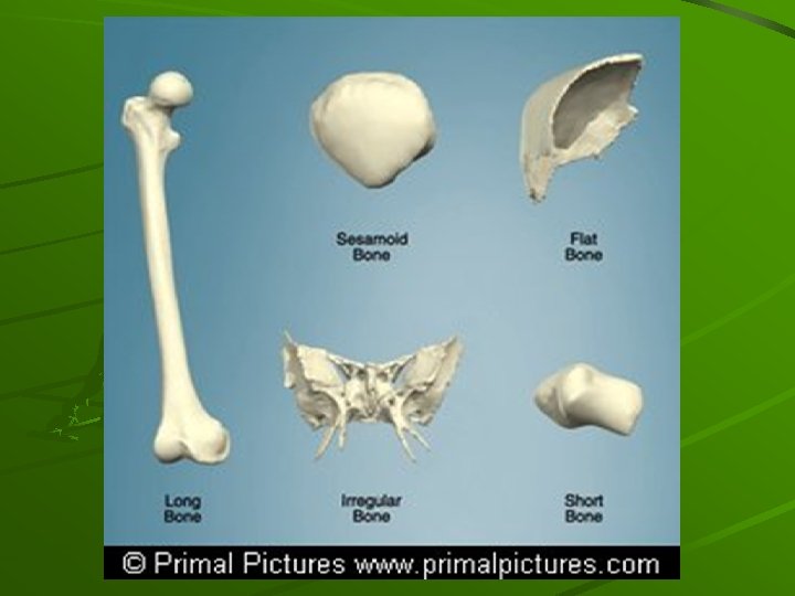
Classification of Bones 5 Types of bones classified according to their shape. 1. Long Bones: found in the arms and legs. Femur is a long bone. 2. Short Bones: most common in the wrists and ankles. Carpals, Tarsals 3. Flat Bones: flat and thin and often protect vital organs from injury. (Skull Bones) 4. Irregular Bones: odd looking bones. (Vertebrae) 5. Sesamoid Bones: unusual bones that are small and flat wrapped within tendons that move over bony surfaces. (patella)
ANATOMY OF A LONG BONE Composition: 1. Epiphysis: 2. 3. Diaphysis: The shaft of the bone. Articulating Cartilage: Located on both ends of long bones. It 4. Periosteum: 5. Medullary Cavity: 6. 7. Compact Bone: Dense part of the bone. Structural Integrity. Spongy Bone: Filled with marrow. Strengthens with specific Very ends of long bones, made up of compact bone. The part where the bone articulates with other bones. It is covered with cartilage. allows smooth movement within joints. Outer connective tissue that covers the entire length of the bone. Helps connect bone to bone and bone to muscle. Found inside the shaft of the bone and is filled with red and yellow bone marrow. Red marrow forms red blood cells. Yellow is fat. exercises. (Cancellous Bone)
Anatomy of a Long Bone
About Bone… Composed of 50% H 2 O and 50% organic and inorganic material. Elements include: phosphorus, zinc, calcium, magnesium, flourine, iron, and chlorine. Resists compression and tension. Bounded by joints – through ligaments. Muscles attach to bones by tendons, this produces movement.
Lesson #3 BONE FORMATION AND REMODELLING Osteocytes: Osteoblasts: Osteclasts: Bone cells Bone building cells. Bone destroying cells. Ossification of Bone is the growth of bones. Short bones have a single ossification center in the middle. Long bones of the arms and legs have 3, one at the center and one at each end. Growth continues until the epiphyseal plate has ossified and longitudinal growth is no longer possible. Bone Remodelling: During early growth, new deposits of bone replace old deposits so growth can continue.
Bone Fractures Simple / Greenstick : Occurs when there is no separation of the bone into parts, but a break or crack is detectable. Compound: Occurs when the bone breaks into separate pieces. Result of a major blow. Comminuted: Occurs when the broken ends of the bone have been shattered into many pieces. (Auto accidents) Transverse: Occurs when a powerful force causes a break across the bone width. Spiral: Occurs when a sharp, twisting force causes the bone to break diagonally across the shaft. Stress: Tiny cracks develop in the bone. Usually results from overuse injuries, shin splints.
Bone Fractures Cont.
FYI Osteoporosis: Most wide spread medical problem. A degenerative condition that involves low bone mass and deterioration. Leads to fractures, ect. (Silent Disease) Prevented by a balanced diet, exercise, healthy lifestyle, proper testing. Female Triad: Osteoporosis in young girls. Ephiphyseal Plates: Growth possible. Ephiphyseal Lines: Growth NOT possible.
Lesson #7 The Muscular System Muscle tissue – refers to a collection of cells that shorten during contraction, and in doing so, create tension that results in movement. Movement is generally achieved through tendons, tough bands of connective tissue that join muscle to bone. Over 650 muscles in the human body. 3 Types of Muscles: 1. Skeletal - Muscles that are attached to bone and are most prevalent in the human body. Make up 30%-40% of body weight. We have control over these muscles. Also called striated. (Voluntary muscles) 2. Cardiac - Found in the heart. Involuntary muscle, not controlled by the brain but directed by the autonomic nervous system. Also striated. 3. Smooth - Surround the body’s internal organs, including blood vessels, hair follicles, genitals, and digestive tracts. Involuntary, spindle shaped muscles arranged in dense sheets.
Properties of Muscle Cells 1. Irritability: 2. Contractibility: ability of a muscle to shorten and lengthen. 3. Elasticity: ability of a muscle to stretch and return to it’s normal position. 4. Extensibility: ability of a muscle to extend in length. 5. Conductivity: ability of a muscle to transmit nerve impulses. ability of a muscle to respond to a stimulus.
Muscles of the Human Body
ORIGINS, INSERTIONS, AND FUNCTIONS Origin: least moveable part, proximal attachment. Insertion: most moveable part, distal attachment. Function: what the muscle does when activated.
Lesson #8 MUSCLE ACTIONS Neuromuscular System - General term referring to the linkage between the muscular system and the nervous system. Messages are sent from the brain or spinal cord and a chemical reaction occurs causing an action in a muscle. (Neuromuscular Junction) - Nerves transmit impulses in waves to ensure smooth movements. A single nerve impulse and resulting contraction is a “twitch”. - One motor neuron (nerve), its axon (pathway) and muscle fiber it stimulates is the MOTOR UNIT.
How a Muscle Contracts 11 Basic Steps: (Sliding Filament Theory) 1. A message originates and is released from the CNS. 2. The message travels down the spinal cord and branches off at the specific area and travels to the PNS. 3. The message then travels from the axon branch to the axon terminal via the axon hillock. 4. The message is carried through the axon terminal via acetylcholine (Ach) to the sarcolemma of each muscle fiber involved. 5. Ach causes the sarcoplasmic reticulum to release calcium ions. 6. The calcium ions find their way to attachment sites on troponin, which are located on the actin.
How a Muscle Contracts cont. 7. The tropomyosin swivels, causing the binding sites for on the actin filament to be exposed. mysoin 8. The mysoin heads attach themselves to the binding sites on actin. 9. ATP is broken down by ATPase, causing the power stroke and the sliding of actin along the myosin filament. 10. Contraction of the filaments will continue until you decide to stop the activation. As long as calcium is present, the contraction will continue. 11. Stop. Calcium is removed from the binding sites by troponin and tropomysoin, which once again cover the binding sites. The muscle returns to a resting state.
Types of Contractions Concentric: (Shortening) Occurs when the muscle fibers shorten. Eccentric: (Lengthening) Occurs when the muscle fibers lengthen. Isometric: (Static) Occurs when the muscle fibers do not change in length. Isotonic exercise is when there is a controlled shortening and lengthening of the muscle. Isometric exercise is when the muscle stays a constant length throughout the exercise. Isokinetic exercise is when there is constant tension throughout the range of motion.
Lesson #9 NAMING MUSCLES Name given to a muscle often refers to the functional movement that the muscle permits. Adductor Muscles - squeeze limbs in towards the mid line of the body. (Adductor longus, adductor brevis, adductor magnus) Abductor Muscles - push out from the mid line of the body. (abductor pollicis longus, abductor pillicis brevis) Extensor Muscles - extend the limb and increase the angle between two limbs. Flexor Muscles - withdraw the limb decreasing the angle between two limbs.
NAMING MUSCLES 6 ways muscles are named: 1. Action of the muscle – flexion, extension. 2. Direction of the fibers – rectus abdominus, transverse. 3. Location of the muscle – tibialis anterior. 4. Number of Heads – Biceps brachii, triceps brachii. 5. Shape of the muscles – Deltoid, trapezius. 6. Point of Attachment – Sternum, clavicle.
How Muscles Attach to Bone Indirect Attachment – When attached indirectly, the epimysium extends past the muscle as a tendon and then attaches to the periosteum of bone. Direct Attachment. When - attached directly, the epimysium adheres to and fuses with the periosteum (outer membrane that covers bone)
FYI Muscle primarily responsible for movement is AGONIST. The muscle that counteracts is the ANTAGONIST. The point where muscle attaches to the more stationary of the bones of the axial skeleton is know as the ORIGIN. The other end, the point where the muscle attaches to the bone that is moved the most, is know as the INSERTION.
Lesson 13 JOINT MECHANICS INJURIES 3 TYPES OF JOINTS Classified according to their structure (what their made of) or function (what they do) 1. Fibrous Joints: bound tightly by connective tissue and allow NO movement. Bones of the skull, known as sutures. 2. Cartilaginous: the body of one bone connects to the body of another by means of cartilage, and slight movement is possible. The intervertebral discs of the spine are this type. Protection. 3. Synovial Joint: Allow the most movement. Bony surfaces are separated by a lubricating fluid (synovia) and by cartilage. Also joined by ligaments. Knee, shoulder, and ankle.
CHARACTERISTICS OF SYNOVIAL JOINTS Synovial Joints permit movement between two or more bones and can be distinguished by the following characteristics: 1. Articulating cartilage: is located on the ends of bones that come into contact with one another. This hyaline cartilage protects the ends of bone and allows for a smooth contact surface while acting as a shock absorber. 2. Joint Capsule: is a fibrous structure that consist of synovial membrane (allows certain nutrients to pass through) and a fibrous capsule (keeps synovial fluid from leaking) 3. Joint Cavity: located between the two bony surfaces. Filled with synovial fluid which acts as a lubricant for the joint. Reduces friction. 4. Bursae: Small, flattened fluid sacs found at friction points. 5. Intrinsic Ligaments: thick bands of fibrous connective tissue that reinforce the joint capsule. 6. Extrinsic Ligaments: are separate from the joint capsule and help to reinforce the joint by attaching the bones together.
TYPES OF SYNOVIAL JOINTS 1. Gliding Joints: This group connects flat or slightly curved bone surfaces. Joints in the foot between tarsals and carpals in the hand. 2. Hinge Joint: Have a convex portion of one bone fitting into a concave portion of another allowing movement in the plane. Joints between the bones of fingers, elbow and knee. 3. Pivot Joint: This joint allows rotation in one plane (uni-axial) a rounded point of one bone fits into a groove of another. 1 st two vertebrae of the neck. 4. Ellipsoid Joint: These joints allow movement in two planes. The wrist is an example. 5. Saddle Joint: Allow movement in two planes, but do not allow rotation. Thumb. 6. Ball and Socket Joint: The ball of the bone fits into the socket of another allowing movement around the axis. The hip and the shoulder.
TYPES OF SYNOVIAL JOINTS
Lesson 14 COMMON SPORT INJURY TERMS TISSUE PROPERTIES – Each tissue is unique in its design and role in the body. If exceeded beyond it’s capabilities it will sprain, strain or pull. 1. Ligaments: Attach bone to bone. Less rigid than bones made of tough fibrous tissue. Some stretch but not as much as tendons. Referred to as static stabilizers of joints while tendons are dynamic stabilizers. Can become stronger through proper conditioning programs. 2. Tendons: Attach muscle to bone. Composed of fibrous protein known as collagen. Have more stretch than ligaments. Vascularity – refers to the amount of supplied blood a tissue has. Ligaments and tendons do not get their nutrients from blood, they are Avascular.
COMMON SPORT INJURY TERMS Strains and Pulls 1. First Degree – are mild and considered least severe. Take a few days to heal. 2. Second Degree – are moderate and more severe. Require treatment from a specialist once diagnosed by a doctor. 3. Third Degree – Most severe. Require surgery and rehabilitation. Tendinitis - is an inflammation of a tendon caused by irritation due to prolonged or abnormal use. (Suffix “itis” means inflammation) Treatment involves PIER / RICE. Dislocation / Separation - occurs when a bone is displaced from its original location. Depending on the severity of the dislocation, tendons may also be damaged. General symptoms are: the joint looks awkward or deformed, the joint is painful when it is touched or moved, the joint is unusable. - A separation occurs when the bones held together by ligaments tear and separate from each other. Acromiocavicular joint – clavicle meets the sternum.
COMMON SPORT INJURY TERMS Cartilage Damage - Three main types: Hyaline cartilage is the most wide spread and found at the end of long bones, free moving joints, ribs, nose, trachea. Fibrocartilage is very tough and found between vertebrae in the spine. Elastic cartilage is in the ear and epiglottis. Shin Splints - refers to a painful condition occurring on the medial or lateral side of the tibia. Inflammation of the Tibialis Anterior. Stress Fracture.
PROPER TREATMENT Identify an injury with SHARP. S – Swelling, instantly or over time. H – Heat, or increased temp of the area. A – Altered tissue that will not function. R – Red area in colour. P – Painful to touch or move.
Lesson 15 SPECIFIC JOINT INJURIES The Shoulder Joint 1. Glenohumeral Joint, is a very unstable joint which allows the shoulder great movement. Synovial Ball and Socket joint is made up of two bones directly, scapula and humerous, and one indirectly, clavicle. Overhead actions, physical contact, and specific movements can all lead to injuries of the shoulder joint. Biceps Tendinitis: generally is an overuse injury and happens when adequate rest is not given to the biceps brachii muscle when it has been over worked. Symptoms include pain in the biceps making flexion hard.
SPECIFIC JOINT INJURIES 2. Shoulder Separation: is a tearing of the acromioclavicular ligament, which holds together the acromioclavicular joint. Results from direct falls on the shoulder. Surgery may be required. 3. Shoulder Dislocation: occurs when the humerous ‘pops out’ of the glenoid fossa causing a tear to the glenhumeral ligaments and joint capsule. Usually a result from a fall or contact. Numerous vital nerves “Brachial Plexus” and blood vessels in this area. Any attempt to put the shoulder back in is BAD. 3. Rotator Cuff Tears: Usually involve one or all four muscles involved. SITS. The Supraspinatus, infraspinitus, and teres minor share an insertion site (greater tubercle of humerous) so when a part of the tendon is torn, they are all affected. Injury makes it difficult to abduct and laterally/medially rotate the shoulder.
SPECIFIC JOINT INJURIES - The Knee Joint Made up of the articulation of the femur and tibia. This joint is held together by many ligaments. The Medial Collateral and Lateral Collateral ligaments provide the most stability for the knee. 1. Knee Ligament tears: most common involves a ‘blow’ to the lateral side of the knee. The severity of the blow determines the injury extent. A blow to the lateral side will result in an injury to the medial side. 2. Osgood-Schlatter Syndrome Result of a condition known as osteochondritis, a disease of the ossification centers of the bones of young children. Affects the epiphyseal plate of the tibial tuberosity. Growing pains for young children. More prevalent in males than females. 3. Patellofemoral Syndrome (PFS) Gradual onset of anterior knee pain or pain around the patella. Usually affects adolescents. Inflammation of the patella tendon.
SPECIFIC JOINT INJURIES - The Ankle Joint Is a modified hinge joint that comprises the distal ends of the tibia and fibula resting on the talus. Responsible for plantar flexion and dorsiflexion. Important ligaments include the anterior/posterior tibiofibular, anterior/posterior talofibular, and calcaneofibular ligament. On the medial side the Deltoid ligament is the strongest. 1. Inversion Sprains: are commonly called rolling the ankle sprains. The ankle is the most unstable during plantar flexion, landing, jumping, changing direction quickly all cause the ankle to invert past the normal range of motion. 2. Eversion Sprains: are very rare because of the strength of the deltoid ligament. This ligament attaches the medial malleolus to three bones of the foot. This ligament is so strong that instead of tearing completely, it tears off the tip of the malleolus. This is known as POTTS Fracture.
Lesson 19 ENERGY NUTRIENTS 3 Key Energy Nutrients - The food we eat is broken down into three key nutrients in the course of digestion: proteins, fats, and carbohydrates. - Bio-energetic Conversion is the process the body uses to break down nutrients for energy. Role of Carbohydrates - Carbs are the most abundant organic substance in nature, and essential for human survival. Come almost entirely from foods that originate from plants – green leafy vegetables, fruits, and grain based foods. - Humans assimilate carbs into glucose, which is stored in the liver as glycogen. The body breaks this down using enzymes to supply energy as needed. - The process in which the body breaks down food to use as energy is referred to as metabolism.
ATP – The Common Energy does come from the food we eat but it needs to be reconstituted into a free form that can be used for muscle contraction. Free energy is ATP ( Adenosine Triphosphate) In effect, ATP captures the chemical energy resulting from the breakdown of food and can be used to fuel the various cellular processes. At the molecular level, Adenosine triphosphate consist of 3 phosphates attached by a high energy bond to adenosine. Energy is released when a trailing phosphate is broken from the ATP molecule. This results in ADP plus energy. ATP ADP + Energy ATP energy supplies are used up very quickly. The problem is how to create new supplies for the body.
TWO ENERGY SYSTEMS There are two methods for resynthesizing ATP: Anaerobic (without oxygen) and Aerobic (with oxygen). Anaerobic System occurs relatively quickly in the muscle fibre and is used for short lived physical actions. (0 – 120 seconds) Aerobic System is a much more complicated process and takes place in the mitochondria. (120 seconds and above)
3 Metabolic Pathways Within these two systems there are 3 main metabolic pathways by which ATP energy reserves are restored: 1. ATP – PC (Anaerobic alactic) - this is the simplest of the systems. It yields enough ATP for about 10 -15 seconds of work and is performed without oxygen. - Also referred to as the phosphagen system, relies on the action of phosphocreatine (PC), a product normally stored in the muscle. - 100 m dash, Olympic Weightlifting. 2. Glycolysis (Anaerobic Lactic) - ATP energy produced will allow an athlete to perform intense exercise for up to 3 minutes. Also capable of producing ATP without Oxygen. - The main product of glycolysis is pyruvic acid, which is converted to lactic acid. Lactic acid builds up in the muscle and hampers the breakdown of glucose for energy, decreasing the muscles ability to contract.
3 Metabolic Pathways 3. Aerobic System - For any athlete to sustain intense activity longer than 3 minutes, a third energy system comes into play. - This energy system is what our bodies depend on for endurance type events. The aerobic pathway results in the complete breakdown of glucose. Cellular Respiration.
Lesson 20 MUSCLE FIBER TYPES Two types of muscle Fibres. 1. a. b. c. d. e. f. Slow Twitch Muscle Fibres: red or dark in colour generate and relax tension relatively slowly have the ability to maintain a lower level of tension for long durations. low levels of the enzyme myosin ATPase, which the body uses to provide instant energy for muscle contraction. low levels of glycolytic enzymes which permit the release of glycogen, and contain high oxidative enzymes. Most active during long distance activities.
MUSCLE FIBRE TYPES 2. Fast Twitch Muscle Fibres: a. more pale in colour b. generate and relax tension relatively quickly c. have the ability to generate large amounts of tension with low endurance levels. d. high levels of the enzyme myosin ATPase, which the body uses to provide instant energy for muscle contraction. e. high levels of glycolytic enzymes. f. Most active during fast, powerful muscle contractions for activites such as short sprints, weightlifting, and explosive jumping. .
The Importance of Myoglobin The difference in muscle fibre types are mainly due to the extent to which a particular muscle relies on O 2 in the production of energy. Slow twitch, red muscle fibres are high in myoglobin and ideal for long activities. Fast twitch fibres, low in myoglobin, are adapted for shorter bursts of energy.
Three Fibre Types 1. Type I (Slow Oxidative) - generate energy slowly, are more fatigue resistant, and primarily depend on aerobic processes. (Cellular Respiration) 2. Type IIA (Fast Oxidative Glycolytic) - Intermediate type muscle fibres that allow for high speed energy release as well as glycolytic capacity. (Glycolysis) 3. Type IIB (Fast Glycolytic) - These fibres store lots of glycogen and sufficiently high levels of enzymes necessary for quick contractions w/o requiring oxygen. (ATP-PC)
Lesson 21 NERVOUS SYSTEM The nervous system is the body’s way of gathering information, storing it, and responding to it. It’s main role is to assemble information about conditions external and internal to the body, to analyze the info, and to initiate responses that may be appropriate. The nervous system involves several interrelated systems. TWO MAIN COMPONENTS
CENTRAL NERVOUS SYSTEM Two Parts – Brain and Spinal Cord 1. The Brain - Main control center for movement, sleep, hunger, thirst, and virtually every type of activity. - The brain sends out commands to other parts of the body to perform. All human emotions are controlled by the brain. - 6 main parts
The Brain 1. The Cerebrum: Largest part of the brain, containing the 2. The Cerebellum: Second largest region. Lies behind and 3. The Brain Stem: Lying below the cerebrum and in front of nerve centers that control sensory and motor activities as well as intelligence. Has 4 lobes; frontal, temporal, parietal, and occipital lobes below the cerebrum. Main function is to coordinate muscle movement and control balance. the cerebellum. Links the cerebrum with the spinal cord. Houses the brain centers responsible for autonomic functions, postural control, muscle tone, and eye movement.
The Brain 4. The Diencephalon: Consists of the thalamus and 5. The Limbic System: Collection of structures that regulate 6. The Reticular Activating System: Network of neurons hypothalamus. Located between the cerebrum and brain stem. Relay center for most stimuli sent to the cerebral cortex and controls primitive awareness of pain, screening of signals, and focusing attention. Hypothalamus controls body temp, appetite, emotions. basic drives such as hunger, aggression, and emotional. that directs info to appropriate centers for interpretation. Its functioning is crucial for maintaining consciousness.
Spinal Cord – Vertebral Column Main pathway for information connecting the brain and peripheral nervous system. Spinal cord runs through the vertebral column, which protects it from damage. Starts at the base of the brain and travels to the second lumbar vertebrae. Spinal nerves carry sensory info to the CNS and motor commands away from the CNS.
PERIPHERAL NERVOUS SYSTEM Massive road network that carries info in and out of the CNS. 12 cranial nerves, 31 pairs of spinal nerves Efferent Nerves – motor nerves that carry info from the CNS to the body’s organs. Afferent Nerves – sensory nerves that carry info from sensory receptors to the CNS.
PERIPHERAL NERVOUS SYSTEM Autonomic and Somatic Autonomic - Involuntary contractions of the cardiac muscles (heart) and smooth muscles of our internal organs is regulated by the autonomic system. Has two branches: 1. Sympathetic System – causes localized bodily adjustments to occur (sweating) and it prepares the body for emergencies. This involves the release of adrenaline and increases heart rate, widens blood vessels, creates a ‘fight or flight’ response. 2. Parasympathetic System – returns the body to normal after it has been altered.
PERIPHERAL NERVOUS SYSTEM Autonomic and Somatic – Our awareness of the external environment, and the corresponding motor activity allowing us to cope with it, operates through this system. Contains both afferent and efferent nerve fibres. Somatic system handle the muscles extremities, which enables us to move. in our
SPINAL CORD INJURIES PARAPLEGIA AND QUADRIPLEGIA - There are 31 pairs of spinal nerves flowing from the spinal cord. Through these, the spinal cord sends messages to and from the brain and to parts of the body. - When the spinal cord receives a severe impact, damage to the spine can profoundly affect its ability to send impulses to body parts.
SPINAL CORD INJURIES When there is a serious injury, nerves above the injury keep working whereas nerves below may or may not work. Paraplegia – no use of the legs Quadriplegia – no use of arms and legs Rehab is extensive – Rick Hansen Facility
SPINAL CORD INJURIES FACTS: Estimated 900 Canadians sustain a spinal cord injury each year. 80% male. 50% or paraplegic, 50% quadriplegic. The following are common causes: 1. Car Accidents 35% 2. Falls 16. 5% 3. Medical 10. 8% 4. Sports 6. 7% 5. Vehicle Accidents 6. 2% 6. Driving 5. 3% 7. Industrial 5. 3% 8. Other 14. 2%
Lesson 24 BASICS OF TISSUE INJURIES Soft Tissue Injuries - Often called wounds, are very common in athletics. - A tissue can bleed, become inflamed, or produce extra fluid. - Sprains and strains are wounds that bleed internally. - Sprains are injuries to ligaments, a strain is an injury to a muscle or tendon. - 3 degree’s of each, 1 st, 2 nd, and 3 rd.
BASICS OF TISSUE INJURIES Stages of Soft Tissue Healing 1. Stage 1 (Acute Inflammatory) - When a body part is injured, cells in the area die from being ripped apart and separated from their food and O 2 supply. - During this stage an increased blood flow to the injured area brings cells and chemicals to begin the healing process. - Phagocytes eat up dead cells. - Leukocytes are white cells that fight infection. - Platelets carry blood clotting material. - Lasts for two days after the initial injury.
BASICS OF TISSUE INJURIES Stages of Soft Tissue Healing 2. Stage II (Repair) - Injured area is now filled with blood to begin repair. - Fibroblasts (fibre building cells) begin building fibre across the injury. - This forms are scar, can take 3 weeks to 6 months.
BASICS OF TISSUE INJURIES Stages of Soft Tissue Healing 3. Stage III (Remodelling) - Takes a year or more. The body builds tissue strength in the tendons, ligaments, and muscles.
FYI Healing Time – the greater the injury, the longer the healing time. Corticosteroids – Chemicals made in the body that help reduce inflammation. Can be synthetic. Skin Closures – A thin, tough piece of material that can bring the edges of a wound together. Deformity – A misalignment of a body part. Osteoporosis – A condition which bones are porous and fragile caused by a lack of calcium. Osteogenesis – Process of laying down new bone.
Lesson 25 BONE INJURIES Types of Injuries 1. Dislocation – Occurs when a significant force displaces the bone so the two ends of bone do not line up. 2. Fractures – The amount of energy required to cause a broken bone (fracture) IS CALLED THE FAILURE POINT.
BONE INJURIES Bone Fracture Healing 1. Stage I Acute – When a bone breaks, bleeding occurs. Osteoclasts eat the debri, osteoblasts begin to lay new bone immediately. 2. Stage II Repair – A bony splint, fibrous callus, forms in the bone. Continues for about three weeks. Aids in support. 3. Stage III Remodelling – Takes several years to complete. Callus is reabsorbed into the body.