Leprosy Update 2019 From the Basics to the

Leprosy Update 2019: From the Basics to the Latest AAFP Global Health Summit October 2019 Albuquerque. New Mexico Ronald Pust MD Professor Emeritus, Dept of Family & Community Medicine Office of Global & Border Health—College of Medicine University of Arizona--Tucson National Hansen’s Disease Program Arizona Clinic US Public Health Service Phoenix, Arizona

Activity Disclaimer It is the policy of the AAFP that all individuals in a position to control content disclose any relationships with commercial interests upon nomination/invitation of participation. Disclosure documents are reviewed for potential conflicts of interest (COI), and if identified, conflicts are resolved prior to confirmation of participation. Only those participants who had no conflict of interest or who agreed to an identified resolution process prior to their participation were involved in this CME activity. Ronald Pust has indicated that he has no relevant financial relationships to disclose. 2

Abstract • New leprosy treatment and prevention regimens issued by WHO in October 2018 are extensive and controversial. • In this session, we will delineate the basics of the recognition, diagnosis, and treatment of leprosy in the U. S-- --and abroad, where World Health Organization (WHO) recommendations are being implemented. … abstract is continued on next slide 3

…Abstract continued… • Using cases from the author's current clinical U. S. Public Health Service practice in Arizona-- ---we will discuss the clinician’s role in working with National Hansen's Disease Program (NHDP). • Working abroad, you will need to adapt to national leprosy programs which use the new WHO guidelines • We will provide the relevant primary resources and latest reviews. This presentation owes much to colleagues at Arizona Leprosy Clinic—next slide… 4

Leprosy in Clinical Practice Recognition, Regimens, Reactions, Rehabilitation and Resources …in the USA and Around the World Ronald Pust MD Tracy Carroll, PT, MPH Ronald Favors, Cert. Orthotist NHDP Arizona Clinic at… Wesley Community Health Center Phoenix, Arizona

Leprosy—Hansen’s Disease—is. . . but… … an infectious disease that, like HIV, can be a chronic disease/disability… but, unlike HIV, does not need lifetime drug therapy. Rates—not numbers of leprosy cases … a mycobacterial disease… Highest national rates are in Micronesia with a decreasing worldwide incidence… unlike tuberculosis, whose incidence is not decreasing… but leprosy’s clinical aspects are not at all like tuberculosis. … one of WHO TDR Program’s Six NTDs [Neglected Tropical Diseases. ]… but “Hansen’s Disease” historically was not a “tropical” disease. …easily diagnosed before disability by alert clinicians in LMICs… but often not diagnosed early by North American clinicians.

Numbers of new leprosy cases reported to WHO in 2017 * 2017 WHO map with case numbers not rates would go here. USA ~120 new cases/year ~6000 treated in past years New 2017 cases each in Brazil, India, Indonesia * In 2017, WHO began displaying leprosy burden by NUMBERS of cases in each nation [NOT rates]

Clinical features follow from 3 unique basic science facts about Mycobacterium leprae 1. COOL : M. leprae grows best at 82 F (28 C)—not 98. 6 (37 C). 2. LIKES NERVES, mainly the “cool nerves, ” i. e. , those closest to the skin surface—whether sensory, motor, or autonomic. 3. SLOW : M. leprae divides every 12 -21 days (compared to 12 -21 hours for many bacteria)
![The first [and least important !] update of this leprosy presentation is the… Current](http://slidetodoc.com/presentation_image/42abcc8889629ce18847d367f4c13b05/image-9.jpg "The first [and least important !] update of this leprosy presentation is the… Current")
The first [and least important !] update of this leprosy presentation is the… Current clinical status of “M. lepromatosis” per David Scollard, MD, Ph. D *, past Director of the USPHS National Hansen’s Disease Program in Baton Rouge, LA, August 2019: • This organism does have a unique molecular signature • Taxonomic issues of this mycobacterium are not decided Clinical experience with “M. lepromatosis” by 2019 has included all types of leprosy. With reference to classic M. leprae: • Similar range of clinical presentations and skin lesions • Identical appearance histologically—Infects nerves • Good response to standard HD drug regimens • Similar prognosis: not better or worse than the usual M. leprae Conclusion: M. lepromatosis is a variant of M. leprae, causes leprosy, and should be treated in the same manner. * personal communication, August 2019 9
![NHDP ALGORITHM FOR BIOPSY DIAGNOSIS, 2012 [current in 2019] Histological features of leprosy, and](http://slidetodoc.com/presentation_image/42abcc8889629ce18847d367f4c13b05/image-10.jpg "NHDP ALGORITHM FOR BIOPSY DIAGNOSIS, 2012 [current in 2019] Histological features of leprosy, and")
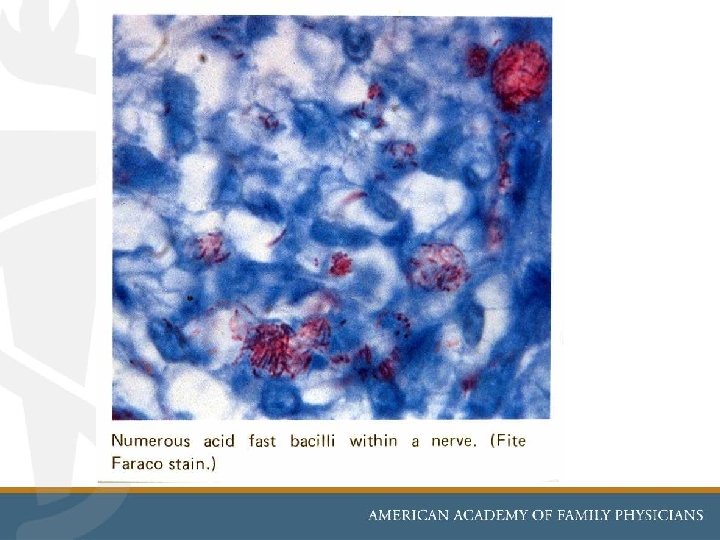
NHDP ALGORITHM FOR BIOPSY DIAGNOSIS, 2012 [current in 2019] Histological features of leprosy, and Acid-Fast Bacilli, seen in skin biopsy AFB in nerve No nerves/ No AFB in nerves Request PCR for M. leprae Positive Negative Request sequencing Diagnosis of Leprosy Identify M. lepromatosis No further studies necessary or other mycobacteria 10

Recognition—the first of five “Rs” LEPROSY IS A CHRONIC, INFECTIOUS HUMAN DISEASE WHICH PRIMARILY INVOLVES: • • SKIN PERIPHERAL NERVES (at their most superficial points) EYES (anterior chamber) NOSE Why these body areas? What do they have in common?
![SKIN 35 year old woman born in Hermosillo, Sonora, Mexico, with Borderline Tuberculoid [BT]](http://slidetodoc.com/presentation_image/42abcc8889629ce18847d367f4c13b05/image-12.jpg "SKIN 35 year old woman born in Hermosillo, Sonora, Mexico, with Borderline Tuberculoid [BT]")
SKIN 35 year old woman born in Hermosillo, Sonora, Mexico, with Borderline Tuberculoid [BT] leprosy. She had sought diagnosis for nearly two years in Tucson, before leprosy was diagnosed. The lesions are densely anesthetic. She also has facial nerve palsy and plantar ulcer. Except where otherwise credited, the patient photos are from the authors’ Arizona/NHDP clinic.

PERIPHERAL NERVES, especially where superficial Here: the recurrent auricular nerve, distal to its emergence from behind and under the S-C-Mastoid muscle. 40 yr old man diagnosed in Phoenix Arizona with borderline [BB] leprosy
![EYES [anterior chamber]: iritis in LL leprosy “reaction” [here] or damage to nerves V](http://slidetodoc.com/presentation_image/42abcc8889629ce18847d367f4c13b05/image-14.jpg "EYES [anterior chamber]: iritis in LL leprosy “reaction” [here] or damage to nerves V")
EYES [anterior chamber]: iritis in LL leprosy “reaction” [here] or damage to nerves V [sensory] or VII [motor] in BL, BB, or BT Photo from USPHS National Hansen’s Disease Program, Baton Rouge, Louisiana
 Photo R. Pust, Papua New Guinea, 1977")
MUCOUS MEMBRANES: mainly nasal (cooler temperature) Photo R. Pust, Papua New Guinea, 1977
![DIAGNOSTIC CRITERIA for Leprosy [HD] • ANESTHETIC SKIN LESIONS • “Leprosy until ‘ruled out’,](http://slidetodoc.com/presentation_image/42abcc8889629ce18847d367f4c13b05/image-16.jpg "DIAGNOSTIC CRITERIA for Leprosy [HD] • ANESTHETIC SKIN LESIONS • “Leprosy until ‘ruled out’,")
DIAGNOSTIC CRITERIA for Leprosy [HD] • ANESTHETIC SKIN LESIONS • “Leprosy until ‘ruled out’, ”especially if patient is from an endemic nation • ENLARGED PERIPHERAL NERVES • which usually have loss of motor and/or sensory function • ACID FAST BACILLI [AFB] in SKIN SMEARS • Mainly in multibacillary [ = LL, BB ] and/or SKIN BIOPSY may show granulomas in paucibacillary leprosy [ = BT, TT] AFB if within nerves is diagnostic of HD

TRANSMISSION & INCUBATION PERIOD • UNTREATED LEPROMATOUS PATIENTS – RESPIRATORY ROUTE? YES DIRECT SKIN CONTACT? Less likely • ARMADILLOS? Yes. What significance? • SOIL OR VEGETATION? Very unlikely • INCUBATION PERIOD: Averages 3 – 8 YEARS, but can be decades Epidemiology is hampered because no reliable skin test or serologic markers.

SUSCEPTIBILITY to M. Leprae • ONLY ~ 5% OF MOST POPULATIONS ARE SUSCEPTIBLE to becoming cases of clinical leprosy even if exposed • ATTACK RATE OF CASE SPOUSES IS 2 -10% [average 5%] • CHILDREN OR CONTACTS OF TREATED PATIENTS RARELY GET THE DISEASE • NUTRITIONAL FACTORS ? No evidence for this • GENETIC FACTORS ? Not proven • IMMUNE FACTORS ? —a unique immunological disease— See top line above re: 5% susceptibility
![“Immuno-Clinical” Spectrum of Leprosy [Ridley-Jopling]](http://slidetodoc.com/presentation_image/42abcc8889629ce18847d367f4c13b05/image-20.jpg "“Immuno-Clinical” Spectrum of Leprosy [Ridley-Jopling]")
“Immuno-Clinical” Spectrum of Leprosy [Ridley-Jopling]

Classifications within the Immuno-Clinical Spectrum of Leprosy Source: Pust R, Campos-Outcalt D, Postgraduate Med. , 1985 [based on several primary sources]
![WHO simplifies the clinical diagnosis of leprosy into paucibacillary [PB] or multibacillary [MB] types.](http://slidetodoc.com/presentation_image/42abcc8889629ce18847d367f4c13b05/image-22.jpg "WHO simplifies the clinical diagnosis of leprosy into paucibacillary [PB] or multibacillary [MB] types.")
WHO simplifies the clinical diagnosis of leprosy into paucibacillary [PB] or multibacillary [MB] types. For low resource nations, this chart uses number of lesions to Dx PB vs. MB, leading to 2 different treatment regimens. However—major and controversial UPDATE: In 2018 WHO proposes to treat both types with the same 3 drug regimen ! More on treatment options in later slides… Mc. Dougall AC, Yuasa Y. A New Atlas of Leprosy. Sasakawa Memorial Health Foundation; Tokyo; 2001.

Spectrum of Cases from TT to LL The next several clinical photos are from… • Browne S. Early Recognition of Leprosy [Only the 3 composite photo/slides introducing tuberculoid, borderline, and lepromatous types of leprosy] • Arizona Hansen’s Disease Clinic [all other clinical photos / brief patient histories]

Tuberculoid leprosy: High CMI, few AFB Circumscribed anesthetic macules or plaques Early loss of sensation (and perhaps of motor function—especially if Type I “reversal” immunologic reaction) Four photos via the late Dr. Stanley Browne [Uzuakoli Leprosy Hospital, Nigeria]

Tuberculoid Leprosy Filipino boy of 10, from Yuma, Arizona with giant hypopigmented, anesthetic macules; but no motor loss
![Motor nerve loss in leprosy Marshallese man, 52, who had tuberculoid [PB] leprosy](http://slidetodoc.com/presentation_image/42abcc8889629ce18847d367f4c13b05/image-26.jpg "Motor nerve loss in leprosy Marshallese man, 52, who had tuberculoid [PB] leprosy")
Motor nerve loss in leprosy Marshallese man, 52, who had tuberculoid [PB] leprosy many years ago. Type 1 leprosy reactions had caused classic “claw hand” and hypothenar atrophy due to ulnar nerve loss. Thumb shows median nerve damage. Left hand normal.

Tuberculoid: Woman, 78, Nogales, AZ: insensitive Charcot foot Charcot first described this in France in tertiary syphilis with loss of dorsal spinothalamic tract. This woman in 1999 was bearing all her Right leg’s weight and walking on her distal tibia.

Radiograph of this 78 yr old woman’s Charcot foot, Nogales, AZ

Borderline Leprosy: Moderate CMI, more diffuse and varied lesions, and a few AFB in skin smears Photos: Dr. Stanley Browne
![Borderline [BT]: Arizona M, 32: Frying pan burns to insensitive hands; motor nerves OK](http://slidetodoc.com/presentation_image/42abcc8889629ce18847d367f4c13b05/image-30.jpg "Borderline [BT]: Arizona M, 32: Frying pan burns to insensitive hands; motor nerves OK")
Borderline [BT]: Arizona M, 32: Frying pan burns to insensitive hands; motor nerves OK
![BT: [same man] Classic enlarging, insensitive macules of borderline leprosy with central healing](http://slidetodoc.com/presentation_image/42abcc8889629ce18847d367f4c13b05/image-31.jpg "BT: [same man] Classic enlarging, insensitive macules of borderline leprosy with central healing")
BT: [same man] Classic enlarging, insensitive macules of borderline leprosy with central healing
![19 yr old Tucson refugee from Somalia: Anesthetic, raised plaques of borderline lepromatous [BL]](http://slidetodoc.com/presentation_image/42abcc8889629ce18847d367f4c13b05/image-32.jpg "19 yr old Tucson refugee from Somalia: Anesthetic, raised plaques of borderline lepromatous [BL]")
19 yr old Tucson refugee from Somalia: Anesthetic, raised plaques of borderline lepromatous [BL] type of multibacillary [MB] leprosy. [MB~more than 5 lesions per WHO] He had no hand or foot damage, and only mild “stocking -glove” anesthesia.

Lepromatous Leprosy: Little to no CMI to M. leprae; Many AFB infiltrating lesions--and entire skin. Lesions may have no definitive borders. Sensation loss later, in “stocking/glove” pattern [like diabetes], may lead to limb damage Motor nerve loss rare. Clinical diagnosis may be more difficult than tuberculoid; but skin smears are always positive
![M, 40 s, ** Phoenix, Arizona Classic EENT features of lepromatous [LL] leprosy: •](http://slidetodoc.com/presentation_image/42abcc8889629ce18847d367f4c13b05/image-34.jpg "M, 40 s, ** Phoenix, Arizona Classic EENT features of lepromatous [LL] leprosy: •")
M, 40 s, ** Phoenix, Arizona Classic EENT features of lepromatous [LL] leprosy: • Saddle nose [due to collapsed nasal septum] • Madarosis [loss of eye brows] and eyelashes **Patient now ~ 66 years old, seen for HD limb problems.
![M, 40 s [same] Destruction/perforation of nasal septum. Differential of perforated nasal septum](http://slidetodoc.com/presentation_image/42abcc8889629ce18847d367f4c13b05/image-35.jpg "M, 40 s [same] Destruction/perforation of nasal septum. Differential of perforated nasal septum")
M, 40 s [same] Destruction/perforation of nasal septum. Differential of perforated nasal septum includes…. . ?
![M, 40 s, from Arizona and California. [born in Colima, MX] Severe, classic lepromatous](http://slidetodoc.com/presentation_image/42abcc8889629ce18847d367f4c13b05/image-36.jpg "M, 40 s, from Arizona and California. [born in Colima, MX] Severe, classic lepromatous")
M, 40 s, from Arizona and California. [born in Colima, MX] Severe, classic lepromatous leprosy facies and saddle nose. He also had disseminated coccidioidomycosis
![[same M, 40 s, from Arizona] Classic EENT features of advanced lepromatous [LL] leprosy](http://slidetodoc.com/presentation_image/42abcc8889629ce18847d367f4c13b05/image-37.jpg "[same M, 40 s, from Arizona] Classic EENT features of advanced lepromatous [LL] leprosy")
[same M, 40 s, from Arizona] Classic EENT features of advanced lepromatous [LL] leprosy …and cervical lymph adenitis due to coccidioidomycosis.

Borderline lepromatous leprosy in a 49 yr old Arizona cowboy in 1997 Sensory loss, plus R & L ulnar & R median motor nerve loss

This same Kingman, AZ cowboy at age 77, who had been on dapsone alone since diagnosis in 1964 at his Army discharge; he had served in Greenland Vietnam. He had been a cowboy in SE Texas, home to armadillos, before Army service. He attended our leprosy clinic in Phoenix once in 1995, but then was “lost to followup”. He was admitted to Tucson VA Hospital for treatment of vertebral osteomyelitis in July 2015 and is now back under our Arizona leprosy clinic care, where we gave him 3 drug, 2 year Rx.

His hands looked vaguely familiar…. …but much worse than in 1997.

Review of Leprosy Diagnosis • Clinical exam for enlarged/tender nerves • Clinical exam for skin anesthesia, best with graded- diameter [Semmes-Weinstein] filament set, if available • Slit-skin smears, from six sites, stained for AFB [also used to monitor treatment every 6 -12 months in multi-bacillary leprosy]. • Skin biopsy: Need a good AFB histopathologist • No useful blood or skin test [in contrast to TB] • Physical exam more useful than history or lab
![Population survey for Leprosy in [then] high prevalence population: Papua New Guinea, 1974 Leprosy](http://slidetodoc.com/presentation_image/42abcc8889629ce18847d367f4c13b05/image-42.jpg "Population survey for Leprosy in [then] high prevalence population: Papua New Guinea, 1974 Leprosy")
Population survey for Leprosy in [then] high prevalence population: Papua New Guinea, 1974 Leprosy is now decreasing world-wide, likely due to case-finding and improved, multi-drug treatment—and also due to BCG vaccine directed at tuberculosis—but BCG is more effective vs. leprosy than it is against TB…
 superficial nerves, during surveys or in")
Clinical exam protocol: to find enlarged (and/or tender) superficial nerves, during surveys or in evaluating persons suspected of having leprosy— --or Type 1 leprosy reaction [when they may be very tender].

Checking for anesthesia in a skin plaque [no instrument needed— though graded Semmes-Weinstein filament preferred] [PNG, 1988]
![Semmes-Weinstein filaments for quantitative sensory testing The 10 gram [5. 07] S-W filament, now](http://slidetodoc.com/presentation_image/42abcc8889629ce18847d367f4c13b05/image-45.jpg "Semmes-Weinstein filaments for quantitative sensory testing The 10 gram [5. 07] S-W filament, now")
Semmes-Weinstein filaments for quantitative sensory testing The 10 gram [5. 07] S-W filament, now in wide use to detect loss of protective sensation in diabetics’ feet, came from leprosy research using~20 filament sizes

Skilled user of Semmes-Weinstein filament set in sensory testing [Tracy Carroll PT, MPH at Arizona leprosy clinic] Filament set
![Slit-skin smear technique • Pressure [very firm] = hemostasis and “anesthesia” • With a](http://slidetodoc.com/presentation_image/42abcc8889629ce18847d367f4c13b05/image-47.jpg "Slit-skin smear technique • Pressure [very firm] = hemostasis and “anesthesia” • With a")
Slit-skin smear technique • Pressure [very firm] = hemostasis and “anesthesia” • With a No. 15 scalpel, make incisions 8 mm long x 4 mm deep at 6 standard sites: ear lobes, elbows, and knees bilaterally. • Apply the “interstitial fluid” to glass slide, then air dry. • AFB stain [similar to sputum for M. tbc] • Count AFB on log scale, from 1+ to 6 + Photo, Arizona Hansen’s Clinic, R. Pust

Slit Skin Smears: Log Scale 0 = NONE FOUND IN 100 OIF 1+ = 1 -10 PER 100 OIF 2+ = 1 -10 PER 10 OIF 3+ = 1 -10 PER OIF 4+ = 10 -100 PER OIF 5+ = 100 -1000 PER OIF 6+ = 1000+ PER OIF (OIF = OIL IMMERSION FIELD)
![Punch biopsy [4 mm] may be used to confirm or to classify leprosy where](http://slidetodoc.com/presentation_image/42abcc8889629ce18847d367f4c13b05/image-49.jpg "Punch biopsy [4 mm] may be used to confirm or to classify leprosy where")
Punch biopsy [4 mm] may be used to confirm or to classify leprosy where histologist is available, but biopsy [and especially PCR] are not mandatory tests. Thus they are rarely used in low-resource settings.

SKIN BIOPSIES IN LEPROSY • PUNCH BIOPSY: 4 -6 MM (IN FACE, 2 -3 MM) • MUST BE DEEP ENOUGH TO INCLUDE SUBCUTANEOUS TISSUE AND FAT • FIX PROMPTLY IN AT LEAST 10 VOLUMES OF 10% FORMALIN [10 -30 ml] • MAIL [at no charge for histology] to: NATIONAL HANSEN’S DISEASE PROGRAM 1770 Physicians Park Drive [attn: lab] Baton Rouge, LA 70816 1 -800 -642 -2477 Toll-free for all consults
![Regimens WHO LEPROSY REGIMENS PAUCIBACILLARY [PB]: Rx 2 drugs for 6 Months Dapsone 100](http://slidetodoc.com/presentation_image/42abcc8889629ce18847d367f4c13b05/image-51.jpg "Regimens WHO LEPROSY REGIMENS PAUCIBACILLARY [PB]: Rx 2 drugs for 6 Months Dapsone 100")
Regimens WHO LEPROSY REGIMENS PAUCIBACILLARY [PB]: Rx 2 drugs for 6 Months Dapsone 100 mg daily Rifampin 600 mg monthly 2018: WHO proposes the 3 MB drugs/12 months regimen for ALL new cases: MB and PB—very controversial UPDATE ! MULTIBACILLARY [MB]: Rx 3 drugs for 12 Months Dapsone 100 mg daily Rifampin 600 mg monthly [by DOT = directly-observed therapy] Clofazimine 300 mg monthly [by DOT] plus 50 mg daily USA LEPROSY REGIMENS: All medications daily PAUCIBACILLARY: 12 Months [“ 2 drugs for 1 year”] Dapsone 100 mg daily Rifampin 600 mg daily MULTIBACILLARY: 24 Months [“ 3 drugs for 2 years”] Dapsone 100 mg daily Rifampin 600 mg daily Clofazimine 50 mg daily

WHO REGIMEN for multibacillary leprosy: Monthly “blister” pack Dapsone 100 mg daily Rifampin 600 mg monthly--DOT Clofazimine 300 mg monthly—DOT plus 50 mg daily If 2018 WHO recommendations take hold, ALL patients would get this monthly packet for 12 months. Article in press [2019] by Diane Lockwood et. al. strongly opposes this one-size-fits-all approach. WHO standard is unlikely to be adopted by NHDP in USA
![Update: Single dose Rifampin [SDR] to prevent leprosy in close/ household contacts of all](http://slidetodoc.com/presentation_image/42abcc8889629ce18847d367f4c13b05/image-53.jpg "Update: Single dose Rifampin [SDR] to prevent leprosy in close/ household contacts of all")
Update: Single dose Rifampin [SDR] to prevent leprosy in close/ household contacts of all new cases… Recommended in the same WHO 2018 leprosy guideline, based largely on the COLEP study: Moet FJ, et al. Effectiveness of single dose rifampicin in preventing leprosy in close contacts of patients with newly diagnosed leprosy. BMJ 2008; 336; 761 -4. But is disputed by… Lockwood DNJ, et al. Single-dose rifampicin chemoprophylaxis protects those who need it least and is not a cost-effective intervention. PLo. S Neglected Tropical Diseases 2018; 12: e 0006403 53

MONITORING RESPONSE TO TREATMENT IN LEPROSY • EXAMINATION OF EYES, HANDS AND FEET for complications [due to insensitivity] • EXAMINATION OF SKIN for lesion regression • EXAMINATION for Type II “reactions”—see next slides • REPEAT SKIN SMEARS q. 6 -12 months in multibacillary [ LL, BB] patients • REPEAT BIOPSY in 6 -12 months in paucibacillary [BT, TT] patients [rarely needed if patient is adherent and improving on the drug regimen]
![Reactions Immunologic reactions in leprosy Type 1 = “upgrading” reaction [as CMI increases] •](http://slidetodoc.com/presentation_image/42abcc8889629ce18847d367f4c13b05/image-55.jpg "Reactions Immunologic reactions in leprosy Type 1 = “upgrading” reaction [as CMI increases] •")
Reactions Immunologic reactions in leprosy Type 1 = “upgrading” reaction [as CMI increases] • May occur in BT, BB, BL [ie, any borderline case] • Inflammation, edema of nerves and “old” skin lesions • If severe, it can lead over days to permanent loss of motor nerve function. Rx: Prednisone 60 -80 mg/day • The only true emergency in leprosy, esp. when there is weakness and swelling of motor nerves. Type 2 = ENL [Erythema Nodosum Leprosum] • May occur in LL & BL: unpredictable in onset, duration • Large subdermal nodules; often edema, fever, systemic malaise; sometimes iritis or orchitis • Rx: Thalidomide 400 mg/day; may start with steroids
![Type 1 reaction = “upgrading” [~ Coombs type 4 = Delayed-type hypersensitivity] Occurs as](http://slidetodoc.com/presentation_image/42abcc8889629ce18847d367f4c13b05/image-56.jpg "Type 1 reaction = “upgrading” [~ Coombs type 4 = Delayed-type hypersensitivity] Occurs as")
Type 1 reaction = “upgrading” [~ Coombs type 4 = Delayed-type hypersensitivity] Occurs as cell-mediated immunity increases, i. e. , “upgrades, ” shifting toward tuberculoid end of spectrum. 58 year old baker with Borderline [BB], first seen when in reaction: Flat lesions are now inflamed, raised. Many leprosy patients are first seen in reaction ! The only leprosy emergency, Type 1 reactions need prednisone 60 -80 mg/day
![SKIN…[duplicate of a prior slide in this presentation] Now, what else do you notice](http://slidetodoc.com/presentation_image/42abcc8889629ce18847d367f4c13b05/image-57.jpg "SKIN…[duplicate of a prior slide in this presentation] Now, what else do you notice")
SKIN…[duplicate of a prior slide in this presentation] Now, what else do you notice about her large, anesthetic skin plaques? She, too, has Type 1 reaction. She also has prior facial nerve palsy…

JB, 19 year old baseball outfielder on Milwaukee Brewers Phoenix farm team. Recruited from Dominican Republic, he bats and throws right-handed. Note hypopigmented , anesthetic macules on his forehead and right cheek, typical of BT leprosy, though not currently in reaction.

JB came to us after 3 months of management--by the Brewers’ hand surgeon--of acute right hand dysfunction and dorsal swelling… What do you see ? [including classic PB anhidrosis in his R hand] The sequelae of 3 months of his untreated Type 1 reaction: atrophic loss of R ulnar & median motor nerve function—and “iatrogenic” career loss. He returned to the Dominican Republic…
![Type 2 reaction = ENL [Erythema Nodosum Leprosum] Classic, and extensive ENL on extensor](http://slidetodoc.com/presentation_image/42abcc8889629ce18847d367f4c13b05/image-60.jpg "Type 2 reaction = ENL [Erythema Nodosum Leprosum] Classic, and extensive ENL on extensor")
Type 2 reaction = ENL [Erythema Nodosum Leprosum] Classic, and extensive ENL on extensor surfaces of arms [and legs]. Patient may be quite systemically ill with fever, malaise and other inflammatory manifestations of Type 2 reaction [eyes, testes, etc] Mc. Dougall AC, Yuasa Y. A New Atlas of Leprosy. Sasakawa Memorial Health Foundation; Tokyo; 2001.
![Type II Reaction [ ~ Coombs type 3 = immune complex] is called Erythema](http://slidetodoc.com/presentation_image/42abcc8889629ce18847d367f4c13b05/image-61.jpg "Type II Reaction [ ~ Coombs type 3 = immune complex] is called Erythema")
Type II Reaction [ ~ Coombs type 3 = immune complex] is called Erythema Nodosum Leprosum [ENL]. M/23 migrated from Guam, where his MB leprosy treatment was completed in 2017. Seen in Sept 2019 in Arizona, with new fevers, malaise, and many red [and brown] subdermal nodules in stages of evolution over past 3 months of undiagnosed ENL. His ENL responded rapidly to thalidomide, confirmed when seen in Arizona NHDP Clinic on 1 October, 2019.

Classic distribution of ENL: extensor surface of thighs & forearms [These ENL lesions have regressed after ~ 5 years, leaving some subdermal atrophy] M/33 from Arizona and Sonora. First diagnosed with MB leprosy and ENL by an inquisitive naturopath, after failure of allopathic Arizona MDs to diagnose.
![Treatment of choice for Type 2 [ENL] reactions is usually daily Thalidomide, in decreasing](http://slidetodoc.com/presentation_image/42abcc8889629ce18847d367f4c13b05/image-63.jpg "Treatment of choice for Type 2 [ENL] reactions is usually daily Thalidomide, in decreasing")
Treatment of choice for Type 2 [ENL] reactions is usually daily Thalidomide, in decreasing doses until ENL is no longer clinically apparent…several days to a few years. Thalidomide's history … and current dermatological applications Anthony J Perri III, and Sylvia Hsu MD Dermatology Online Journal 9 (3): 5 In 1956, thalidomide was introduced … as a sedative…. By 1958, it permeated the world market, and its use expanded beyond simply combating insomnia. Pregnant women frequently treated their nausea of pregnancy with thalidomide. [3] Soon thalidomide's teratogenic effects became apparent, and it was withdrawn from the market in 1961. Fortunately, thalidomide had never been approved in the United States [1, 3]… Thalidomide resurfaced in 1965 when Sheskin, a dermatologist from Israel, made a fortuitous discovery while treating his leprosy patients with thalidomide. Prescribing thalidomide to his leprosy patients for its sedative properties, Dr. Sheskin noticed that those with erythema nodosum leprosum had resolution or improved markedly within 2 days…

P. A. 33 yr old Hilton hotel worker came to Tucson in 2005 from Republic of The Marshall Islands [in Micronesia, which has highest leprosy rate in world. Woodall P, Scollard D, Ryan L. Hansen Disease among Micronesian and Marshallese persons living in the United States. Emerging Inf Dis 2011; 17: 1202 -8 Oct 2010, he stopped work due to fever, malaise, and 70 -80 red nodules, treated as Staph furuncles. None of these nodules had ever burst or drained. You and a medical student now meet P. A. on November 26, 2010

Many of the nodules are regressing. P. A. said he had a similar, milder episode in 2007 that resolved “on its own. ” [ENL may do that. ] Note his enlarged, nodular left ear. Eyebrows and nose appear intact. How to diagnose? How to treat? What else would you do—since you are also a public health professional?
![You send a Case Report to USPHS National Hansen’s Disease Program [NHDP] in Baton](http://slidetodoc.com/presentation_image/42abcc8889629ce18847d367f4c13b05/image-66.jpg "You send a Case Report to USPHS National Hansen’s Disease Program [NHDP] in Baton")
You send a Case Report to USPHS National Hansen’s Disease Program [NHDP] in Baton Rouge, LA. Then, since he has MB leprosy with 6+ skin AFB smears, you examine his household contacts. Would you treat his 11 contacts with a single dose of rifampin [WHO]?

Rehabilitation Ronald Favors, Pedorthotist at the Arizona Hansen’s Disease Clinic

Shoes made for the deformed and/or insensitive foot in leprosy—similar in diabetes Shoes shown are produced by R. Favors, Az NHDP Clinic.

At first glance, a classic plantar ulcer of an insensitive foot in leprosy—or diabetes…but this one is harder to treat because…

Pressure has destroyed the plantarflexion tendon, leading to dorsal subluxation of the great toe…

…requiring a surgical approach: arthroplasty to restore plantar flexion at the first metatarso-phalangeal joint [which was successful]

Resources & Referrals for Leprosy Arizona Hansen’s Disease Clinic at Wesley CHC in Phoenix, Arizona Ronald E. Pust MD rpust@email. arizona. edu cell (520) 668 -6441 Office (520) 626 -1992 www. globalhealth. arizona. edu in Tucson • National Hansen’s Disease Program (NHDP) https: //www. hrsa. gov/hansens-disease/index. html (Toll free 1 -877 -464 -4772 for consults) • American Leprosy Mission. http: //www. leprosy. org/ • The British Leprosy Relief Association (LEPRA) http: //www. lepra. org. uk (includes journal, Leprosy Review) • International Federation of Anti-Leprosy Associations. http: //www. ilep. org. uk/ • World Health Organization Action Program for the Elimination of Leprosy. http: //www. who. int/lep
![Summary of Updates [buried ? ] in this Presentation M. lepromatosis, clinically, is a](http://slidetodoc.com/presentation_image/42abcc8889629ce18847d367f4c13b05/image-73.jpg "Summary of Updates [buried ? ] in this Presentation M. lepromatosis, clinically, is a")
Summary of Updates [buried ? ] in this Presentation M. lepromatosis, clinically, is a variant of M. leprae, and should be treated the same as M. leprae. Scollard DM. Infection with Mycobacterium lepromatosis. Am J Trop Med Hyg 2016; 95: 500 -501. Armadillos: [an “Update” that is not so new] • May have leprosy • May transmit leprosy to humans, but rarely. [Can humans transmit leprosy to armadillos? This is the author’s favorite irrelevant question. ] • Are a great animal model for researching leprosy in vivo Truman RW, et al. Probable Zoonotic Leprosy in the Southern United States. NEJM 2011; 364: 1626. WHO: Global Leprosy • WHO and USA leprosy drug treatment regimens differ—and will likely differ even more as 2018 WHO protocols are adopted by many nations. WHO. Guidelines for the diagnosis, treatment and prevention of leprosy. New Delhi: WHO Regional Office for South East Asia; 2018 Lockwood DNJ, et al. Little evidence to support a major change in paucibacillary leprosy treatment in the 2018 WHO treatment guidelines. Leprosy Mailing List Dec. 23, 2018. • WHO in 2018 proposed that all household or “close” contacts of new cases of all types [MB—and PB!] of leprosy be given a single dose of rifampin [SDR] as a means of preventing leprosy and decreasing future population incidence. Moet FJ, et al. Effectiveness of single dose rifampicin in preventing leprosy in close contacts of patients with newly diagnosed leprosy. BMJ 2008; 336; 761 -4. Lockwood DNJ, et al. Single-dose rifampicin chemoprophylaxis protects those who need it least and is not a cost-effective intervention. PLo. S Neglected Tropical Diseases 2018; 12: e 0006403 73

REFERENCES Pust RE, Scollard D. Leprosy In: Control of Communicable Disease Manual: CCDM Clinical Manual APHA [in press 2019] Scollard D. Leprosy. In: Up. To. Date. Current at: www. uptodate. com Pust RE. Leprosy. In: Rakel RE. Saunders Manual of Medical Practice. WB Saunders; 2000. pp. 1162 -5. …and from prior slides in this presentation: WHO. Guidelines for the diagnosis, treatment and prevention of leprosy. New Delhi: WHO Regional Office for South East Asia; 2018. Lockwood DNJ, et al. Little evidence to support a major change in paucibacillary leprosy treatment in the 2018 WHO treatment guidelines. Leprosy Mailing List [Dec. 23, 2018. ] [not yet formally published] Moet FJ, et al. Effectiveness of single dose rifampicin in preventing leprosy in close contacts of patients with newly diagnosed leprosy. British Med J 2008; 336; 761 -4. Lockwood DNJ, et al. Single-dose rifampicin chemoprophylaxis protects those who need it least and is not a cost-effective intervention. PLo. S Neglected Tropical Diseases 2018; 12: e 0006403 Scollard DM. Infection with Mycobacterium lepromatosis. Am J Trop Med Hyg 2016; 95: 500 -501. Truman RW, et al. Probable Zoonotic Leprosy in the Southern United States. NEJM 2011; 364: 1626. Woodall P, Scollard D, Ryan L. Hansen’s Disease among Micronesian and Marshallese persons living in the United States. Emerging Infectious Diseases 2011; 17: 1202 -8 74

Review—The final “R”
![One final case: [from National Hansen’s Disease Program case archives]: What type of clinical](http://slidetodoc.com/presentation_image/42abcc8889629ce18847d367f4c13b05/image-76.jpg "One final case: [from National Hansen’s Disease Program case archives]: What type of clinical")
One final case: [from National Hansen’s Disease Program case archives]: What type of clinical leprosy does this man have? How would you confirm your diagnosis?
![Leprosy…and its stigma: Boot Hill Cemetery, Tombstone, Arizona [80 miles SE of Tucson] “Two](http://slidetodoc.com/presentation_image/42abcc8889629ce18847d367f4c13b05/image-77.jpg "Leprosy…and its stigma: Boot Hill Cemetery, Tombstone, Arizona [80 miles SE of Tucson] “Two")
Leprosy…and its stigma: Boot Hill Cemetery, Tombstone, Arizona [80 miles SE of Tucson] “Two Chinese died of leprosy” about 1882—the most prosperous year in Tombstone [ photograph by R. Pust, MD]

Have you ever met this man? Working with leprosy in USA or abroad can be a most satisfying aspect of global health. We thank you for viewing this leprosy presentation… R. E. Pust MD—for the USPHS National Hansen’s Disease Program & AAFP Photo from USPHS NHDP case archives
- Slides: 78