
LEISHMANIASIS INTRODUCTION Flagellated protozoa found in peripheral blood
LEISHMANIASIS
INTRODUCTION � Flagellated protozoa found in peripheral blood circulation � Belong to phylum Sarcomastigophora � Two genera of medical importance - Leishmania - Trypanosoma � They have oval to elongated body, a nucleus and a single flagellum arising from a kinetoplast.
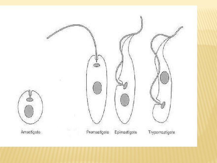
� The parasites occur in 4 morphological forms: 1. Amastigote 2. Promastigote 3. Epimastigote 4. Trypomastigote � The suffix mastigote indicates the presence of flagella. � It is derived from Greek word Mastix which means WHIP.
LEISHMANIA � Is an obligatory intracellular parasite � Affects the reticuloendothelium system of vertebrate host � Causes Leishmaniasis which encompasses a spectrum of various clinical syndromes � Mainly a zoonotic disease � Transmitted by the bite of female sandfly
CLASSIFICATION � According to the geographical area leishmaniasis can be classified in two 1) Old world leishmaniasis 2) New world leishmaniasis � Most common pathogenic species associated with old world leishmaniasis is L. donovani
� CLINICAL spectrum of the disease consist of - Visceral leishmaniasis - PKDL - Cutaneous leishmaniasis - Diffuse cutaneous leishmaniasis - Mucocutaneous leishmaniasis
LEISHMANIA DONOVANI � Named after two scientist who discovered the parasite in 1903 – Sir William Boog Leishman and Sir Donovan � It causes visceral leishmaniasis. � Also known as kala azar. � Charles Nicolle was able to cultivate the agent in vitre and was awarded a noble prize for that in 1928.
EPIDEMIOLOGY � Present in most developing countruies in tropical regions. � India constitutes the largest foci of infection along with the neighbouring countries like Nepal, Bangladesh, Sudan and Brazil. � In India, Bihar is the worst affected state followed by Jharkhand, West Bengal and Uttar pradesh.
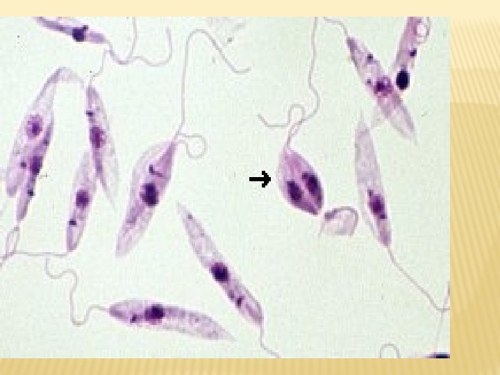
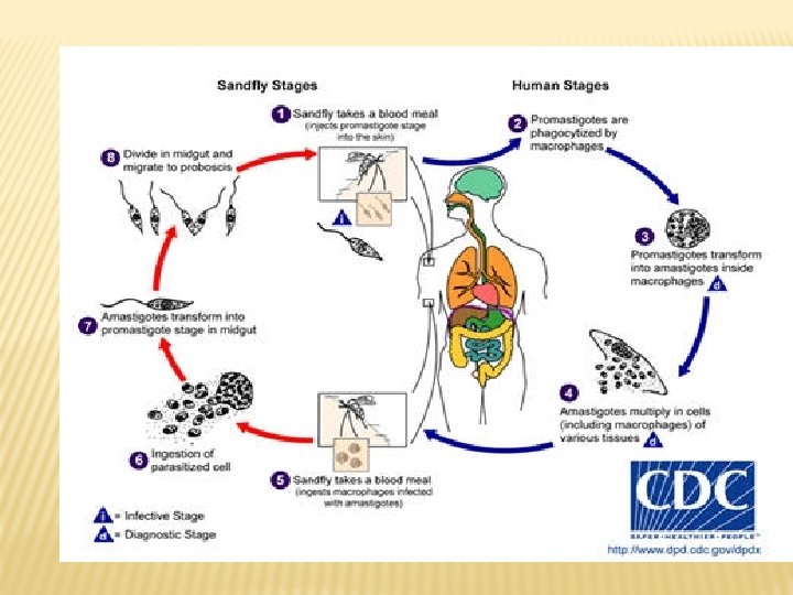
MORPHOLOGY � Occurs in two forms 1) Amastigote- obligate intracellular infective to sandfly found in RE cells round to oval, 3 -5 microns 2) Promastigote- extracellular form - infective to human - seen in sandfly & cultures - motile with single anterior flagella - pear shaped 8 -15 microns in length.
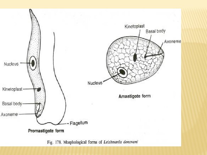
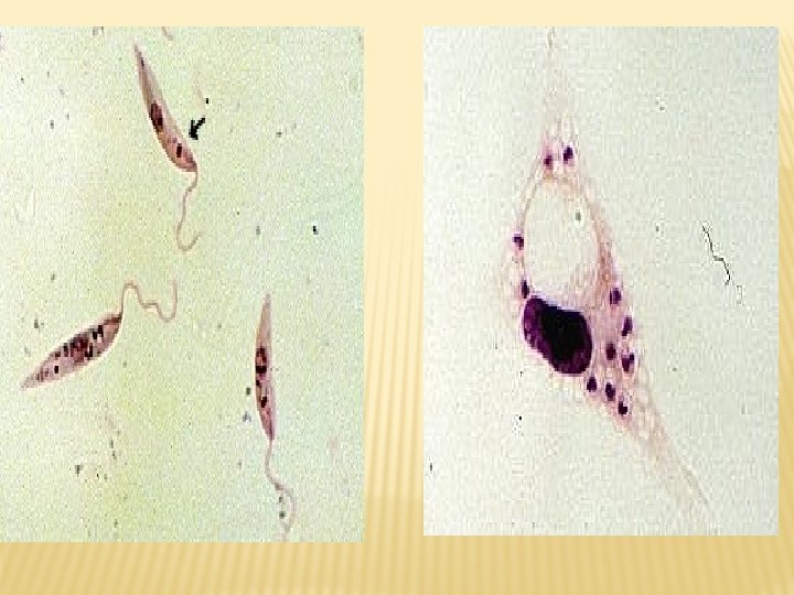
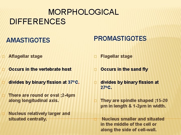
MORPHOLOGICAL DIFFERENCES AMASTIGOTES PROMASTIGOTES � Aflagellar stage � Flagellar stage � Occurs in the vertebrate host � Occurs in the sand fly � divides by binary fission at 37 o. C. � divides by binary fission at 27 o. C. � There are round or oval ; 2 -4µm alongitudinal axis. � They are spindle shaped ; 15 -20 µm in length & 1 -2µm in width. Nucleus relatively larger and situated centrally. � Nucleus smaller and situated in the middle of the cell or along the side of cell-wall. �
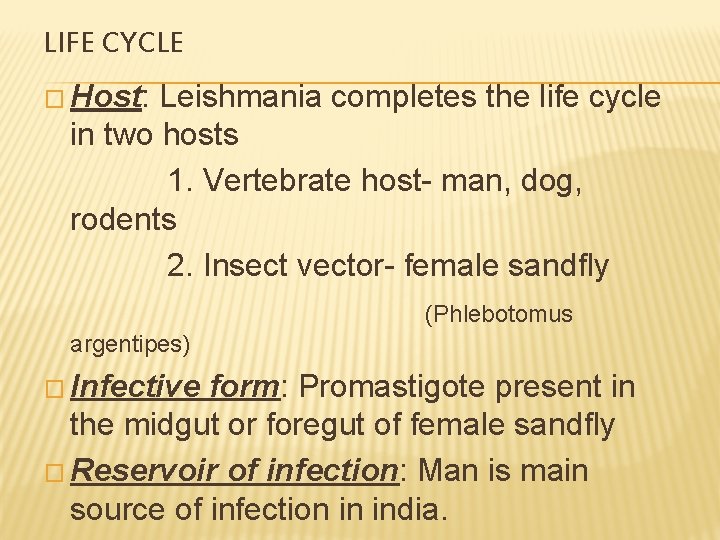
LIFE CYCLE � Host: Leishmania completes the life cycle in two hosts 1. Vertebrate host- man, dog, rodents 2. Insect vector- female sandfly (Phlebotomus argentipes) � Infective form: Promastigote present in the midgut or foregut of female sandfly � Reservoir of infection: Man is main source of infection in india.
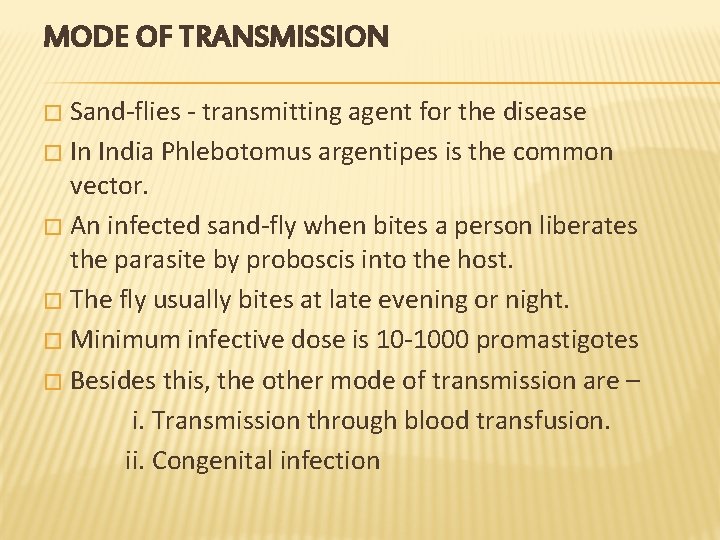
MODE OF TRANSMISSION Sand-flies - transmitting agent for the disease � In India Phlebotomus argentipes is the common vector. � An infected sand-fly when bites a person liberates the parasite by proboscis into the host. � The fly usually bites at late evening or night. � Minimum infective dose is 10 -1000 promastigotes � Besides this, the other mode of transmission are – i. Transmission through blood transfusion. ii. Congenital infection �
CLINICAL FEATURES � Incubation period is 3 -6 months. May extend upto 2 years in some � The disease is characterised by triad of - Fever - Hepatospleenomegaly - Pancytopenia � Other features include lymphadenopathy, hyperpigmentation of skin, pedal oedema and ascites, weight loss, leishmanioma
� Various hematological abnormalities are also present like anaemia, leucopenia, thrombocytopenia, hypergammaglobulinemia. � The disease usually gets complicated by the secondary infections like measles, pneumonia, tuberculosis, bacillary or amoebic dysentery and gastroenteritis.
LABORATORY DIAGNOSIS � MICROSCOPY: - - Specimen: spleenic aspiration, bone marrow, lymph node, liver biopsy, Peripheral blood smear, biopsy sample - Stains used: Leishman, Giemsa or Wright stains can be used - The amastigotes can be demonstrated in the macrophages - The cell consisting of amastigotes is called as Leishman Donovan or LD bodies.
� CULTURE: - - Specimen: spleenic aspiration, bone marrow aspiration - Medium used NNN medium (Novy, Mc. Neal, Nicolle), Schneider’s liquid medium. - Specimen inoculated and incubated at 22 C upto 4 weeks. - Cultures positive in 75% of cases.
� ANTIBODY DETECTION: - ELISA, IFA, IHA, Direct agglutination test, Immunochromatic tests are done for demonstration of antibodies in patients serum. � NON SPECIFIC TESTS: - These are done for detection of hypergammaglobulinaemia. Two tests: 1. Napier’s Aldehyde test 2. Chopra’s Antimony test � Molecular methods like PCR
� LEISHMANIN TEST or MONTEGRO TEST: - delayed hypersensitivity skin test. - Suspension of killed L. donovani promastigote injected intradermally - Positive test is indicated by induration of >= 5 mm in 72 hrs. - Positive in people with prior exposure and those with good CMI - It is negative in active visceral leishmaniasis and diffuse cutaneous leishmaniasis.
� ANIMAL INOCULATION � NON SPECIFIC TESTS - Complete blood count - Elevated liver enzymes - Reversal of albumin globulin ratio
TREATMENT � Specific Anti. Leishmanial Therapy: 1 Pentavalent antimony compounds - Sodium stibogluconate - Meglumine antimoniate 2 Amphotericin B 3 Paromomycin 4 Miltefosine � Supportive Therapy
PREVENTION � No effective vaccine available. � Both killed and lived vaccines are under trial. � Vector control measures. � Reservoir control measures. � Personal prophylaxis- use of insect repellants and bed nets.
- Slides: 26