Leicester Warwick Medical School Neoplasia 1 What is


 Hodgkins Disease BONE MARROW Acute and chronic leukaemia")

- Slides: 42
Leicester Warwick Medical School Neoplasia 1 What is a Tumour? Professor Rosemary A Walker raw 14@le. ac. uk Department of Pathology
WHAT IS A TUMOUR? a swelling inflammatory – abscess neoplasm - growth
NEOPLASM • Abnormal growth of cells which persists after initiating stimulus has been removed • Cell growth has escaped from normal regulatory mechanisms • Benign • Malignant
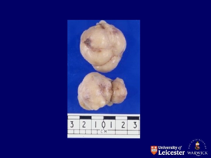
BENIGN NEOPLASM Cells grow as a compact mass and remain at their site of origin
MALIGNANT NEOPLASM Growth of cells is uncontrolled Cells can spread into surrounding tissue and spread to distant sites Cancer = a malignant growth
HOW DO TUMOURS DEVELOP? • There has to be a change to DNA • The change must cause an alteration in cell growth and behaviour • The change must be non-lethal and be passed onto daughter cells
HOW DO TUMOURS DEVELOP? • Alteration is to more than one gene • Genes concerned are oncogenes/tumour suppressor genes • Sequence of gene alterations from normal to benign to malignant • Intrinsic and extrinsic / inheritance and environment key factors
CLONALITY Alterations in genes regulating growth and behaviour occur in every cell – monoclonal population Evidence from studying G 6 PD In heterozygotes cells contain either G 6 PD A or G 6 PD B, but tumours in those people consist of cells that all have the same enzyme A B B A A NORMAL A A A B B OR B CANCER B
HOW DO NEOPLASTIC CELLS DIFFER FROM NORMAL CELLS? Alterations in growth control • proliferation • cell death • factors regulating growth and response Alterations in cellular interactions • cell-cell • cell-stroma
GROWTH CONTROL • Increased cell proliferation more cells enter cell cycle “speeded up” • Cells have changed life span • Alterations in cell death-decreased apoptosis • Modification of cell metabolism • Angiogenesis
GROWTH CONTROL • Increased or decreased growth factor receptors or altered receptors • Synthesis of growth factors – autocrine or paracrine effect • Excess/modified growth control proteins e. g. oncoproteins
Autocrine Increased DNA synthesis and proliferation Growth factor receptor Paracrine = Growth factor
CELLULAR INTERACTIONS Cell-cell interactions Cell-stromal interactions with basement membrane Important for cell and tissue differentiation, embryogenesis, growth regulation
Desmosomes Ordered Cytoskeleton Basement membrane Cell receptors Disorganised Cytoskeleton Loss of cell receptors
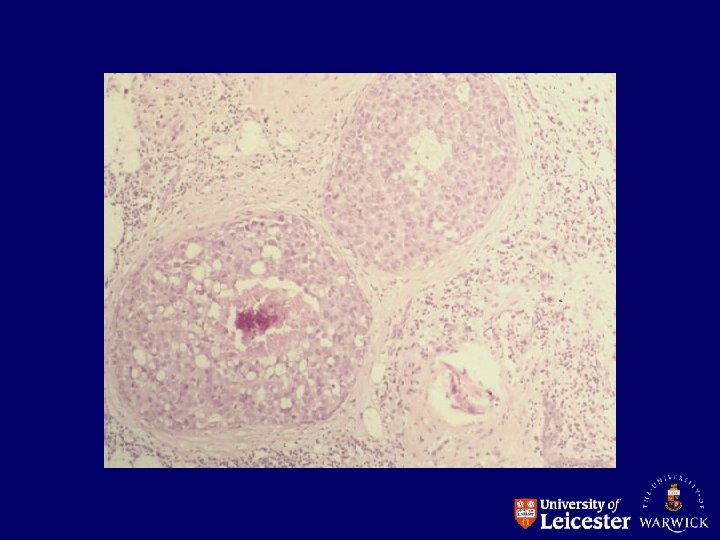
DIFFERENCES BETWEEN BENIGN AND MALIGNANT NEOPLASMS • • • Size Growth characteristics Vascularity/necrosis Function Invasion/metastasis
DIFFERENCES BETWEEN BENIGN AND MALIGNANT NEOPLASMS BENIGN MALIGNANT Nuclear variation in size and shape minimal to marked, often variable Diploid Range of ploidy Low mitotic count, normal mitosis Low to high mitotic count, abnormal mitosis Retention of specialisation Loss of specialisation
DIFFERENCES BETWEEN BENIGN AND MALIGNANT NEOPLASMS BENIGN MALIGNANT Structural differentiation retained shows wide range of changes Organised Not organised Functional differentiation usually Functional differentiation often lost
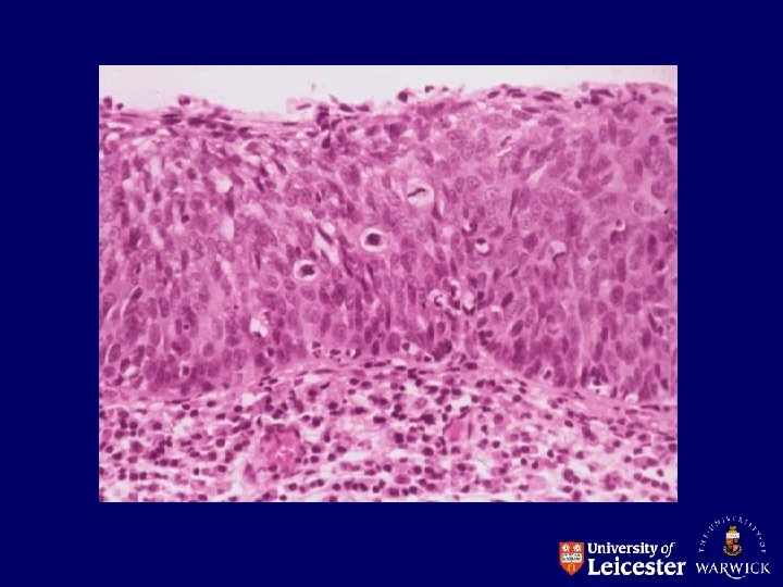
DYSPLASIA • • • Premalignant condition Increased cell growth Cellular atypia Altered differentiation Can range from mild to severe Sites -cervix -bladder -stomach
IN-SITU MALIGNANCY Epithelial neoplasm with features of malignancy • altered cell growth • cytological atypia • altered differentiation BUT-no invasion through basement membrane
POSSIBLE EVENTS Benign Dysplasia In-situ Benign Dysplasia In-situ Invasive Invasive
TYPES OF NEOPLASMS Benign Malignant Epithelial Connective tissue Lymphoid /haemopoietic Germ cell
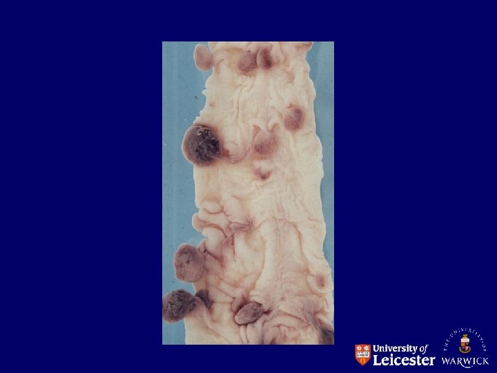
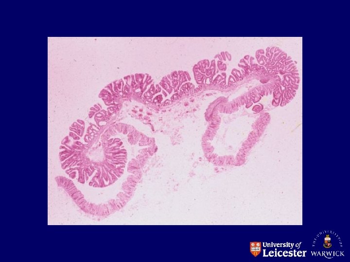
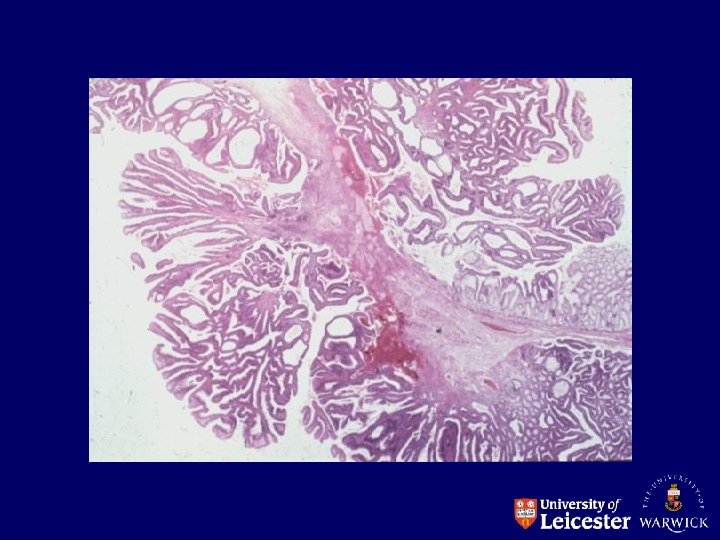
BENIGN EPITHELIAL NEOPLASMS Papilloma • squamous • transitional Adenoma • glandular
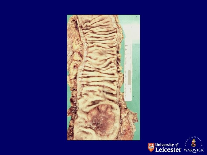
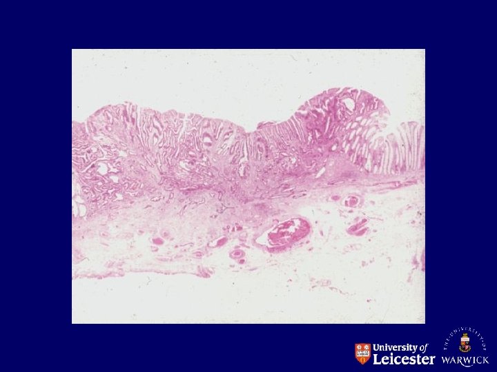
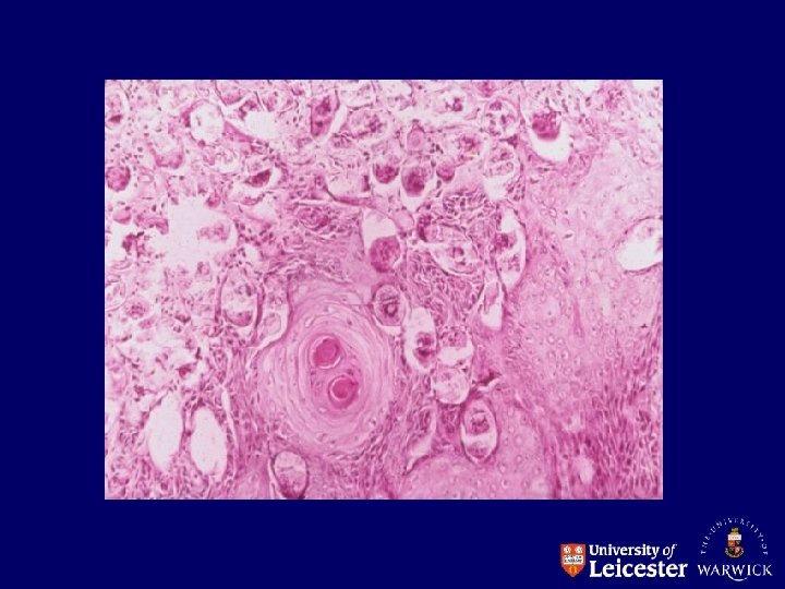
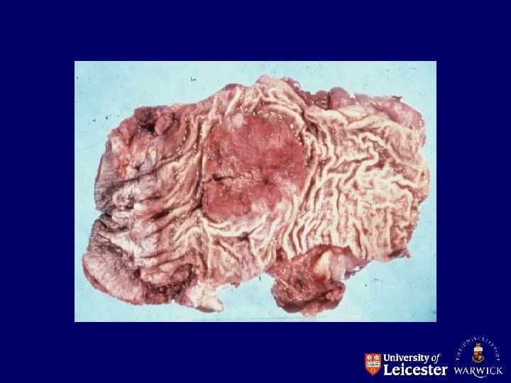
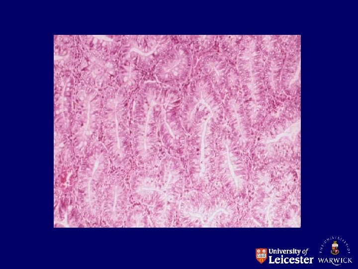
MALIGNANT EPITHELIAL NEOPLASMS Carcinomas Squamous: Transitional: Adeno: colon Basal cell: skin bladder stomach, skin
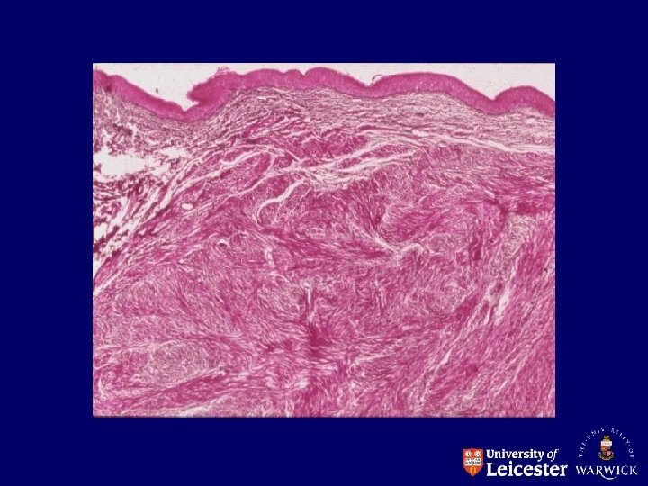
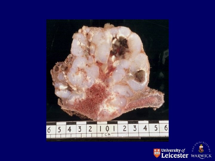
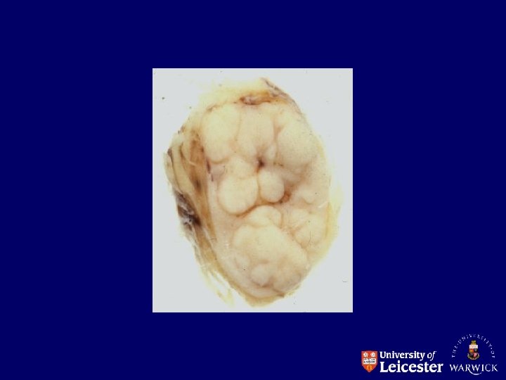
CONNECTIVE TISSUE NEOPLASMS Smooth muscle: Leiomyoma Fibrous tissue: Fibroma Bone: Osteoma Cartilage: Chondroma Fat: Lipoma Nerve: Neurofibroma Nerve sheath: Neurilemmoma Glial cells: Glioma
CONNECTIVE TISSUE NEOPLASMS Smooth muscle: Leiomyosarcoma Bone: Osteosarcoma Fibrous tissue: Fibrosarcoma Cartilage: Chondrosarcoma Fat: Liposarcoma Nerve: Neurofibrosarcoma Nerve sheath: Neurilemmosarcoma Glial cells: Malignant glioma
LYMPHOID Malignant lymphoma (B and T) Hodgkins Disease BONE MARROW Acute and chronic leukaemia
GERM CELL Testis Teratoma Seminoma Ovary Dermoid Cyst