
Lecturer Reham M M ALMosawi Medical Microbiology Bacteriology
Lecturer Reham M. M. AL-Mosawi Medical Microbiology (Bacteriology) Lec. Part 1 -b

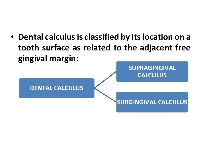
• Dental calculus is classified by its location on a tooth surface as related to the adjacent free gingival margin: SUPRAGINGIVAL CALCULUS DENTAL CALCULUS SUBGINGIVAL CALCULUS

Inorganic components are also found in dental • plaque; largely calcium and phosphorus which are primarily derived from saliva. The inorganic content of plaque is greatly • increased with the development of calculus. The process of calculus formation involves the • calcification of dental plaque. The practical consequences of calculus formation • are that the deposit is significantly more difficult to remove once calcified, and it leaves a rough surface on the root which is easily colonized by plaque. .

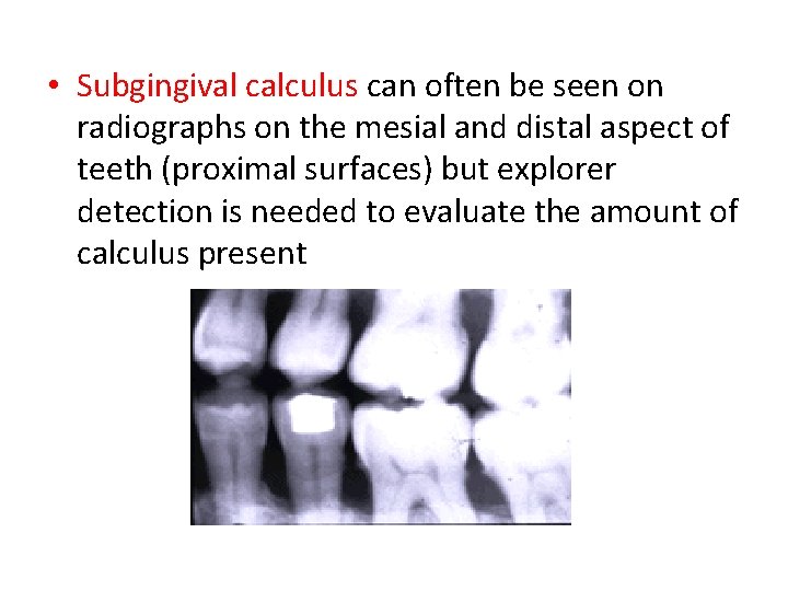
• Subgingival calculus can often be seen on radiographs on the mesial and distal aspect of teeth (proximal surfaces) but explorer detection is needed to evaluate the amount of calculus present
BACTERIAL CONTENT- • The percentage of gram positive and gram- negative filamentous organisms is greater within calculus than in the remainder of oral cavity. 7 • The microorganisms at the periphery are predominantly gram-negative rods and cocci. • Most of the organisms within the calculus is nonviable.
SUPRAGINGIVAL CALCULUS ü Predominance of gram-positive filaments. ü Next in frequency; gram-negative filaments and cocci. ü Gram-positive cocci seen in calculus about which suppuration had taken place.
SUBGINGIVAL CALCULUS • Superficial layers : gram- negative filaments most numerous • Deep and middle zones : gram-positive filaments predominant.
ULTRASTRUCTURE OF CALCULUS • LAYERS – calculus forms in layers that are more or less parallel with the tooth surface. 6 • The layers are separated by a line that appear to be a pellicle that was deposited over previously formed calculus, and as mineralization progressed, the pellicle became embedded. • The lines between the layers of calculus can be called incremental lines. The lines are evidence that calculus grows, or increases by apposition of new layers.
- Slides: 10