LECTURE INTEGUMENTARY SYSTEM Integumentary system The skin along

 is called integumentary")


 cells which are sometimes")

 • This layer is named papillary because it has papillae that")
 • Just beneath the epidermis the reticular fibers form fibrils which")


- Slides: 33
LECTURE: INTEGUMENTARY SYSTEM
Integumentary system The skin along with some of its specialized derivatives(appendages) is called integumentary system. The appendages include • Hairs • Sweat glands • Sebaceous glands • nails
SKIN • The skin also called integument or cutis forms the external covering of the body. • Consist of two layers of completely different types of tissue Classification of skin: Generally classified into two types, thick and thin skin, the term thick and thin is used for the thickness of epidermis only and do not refer to the thickness of the skin as a whole. 1. Thick skin This thick type covers palms and soles. 1. Thin skin is found on remainder of the body.
EPIDERMIS The epidermis is a continuously self-replacing stratified squamous, keratinized epithelium. It contain four types of cells 1. Keratinocytes 2. Melanocytes 3. Langerhans 4. Merkel cells
EPIDERMIS Keratinocytes: • They are the principle cells of the epidermis, epidermis is composed of many superimposed layers of these cells. • They undergo constant renewal throughout life through continuous mitotic division in the basal layer replacing those shed at the surface.
EPIDERMIS Melanocytes: • Melanocytes are stellate cells and have dendritic cells. • Melanocytes form a brown-black pigment celled melanin that protects the skin from harmful effects of the electromagnetic radiations of sun. • Melanocytes are produced by membrane bound bodies called melanosomes. • Melanosomes are produced by Golgi apparatus of the cell and they contain enzyme tyrosinase. • Tyrosinase act on tyrosine and convert it to melanin.
EPIDERMIS Langerhans: • They have dendritic processes like melanocytes. • No melanocytes or filaments are present in cytoplasm of these cells. Function: • These cells participate in the body’s immune responses performing the same function as macrophages. • Langerhans cells take up antigen and present it to the T -Lymphocytes in a form to which they can respond
MERKEL CELLS • These cells are found in or near the stratum basale of the epidermis in association with free nerve endings. • Their cytoplasm stains darkly because of the presence of numerous small granules. • They serve as mechanoreceptors.
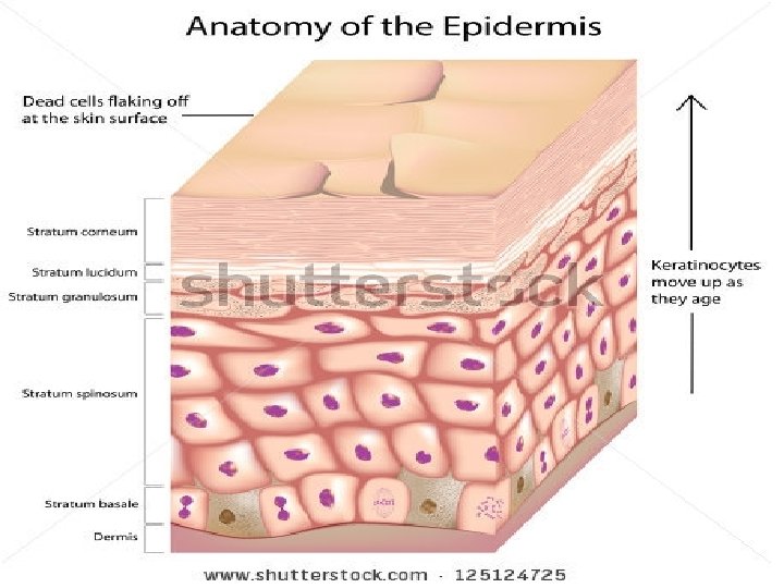
LAYERS OF EPIDERMIS • Under ordinary L/M the epidermis is seen to consist of the following five strata (layers) from deepest to superficial 1. Stratum basale 2. Stratum spinosum 3. Stratum granulosum 4. Stratum lucidum 5. Stratum corneum
STRATUM BASALE • Also called stratum germinatum consist of a layer of columnar or cuboidal keratinocytes resting on a basement membrane which lies on the dermis. • Each cell contain an oval nucleus. • The cytoplasm contain keratin intermediate filaments which are called tonofilamenst and bundles of these filaments are visible under L/M as fibrils also called tonofibrils. Function: • This layer is responsible for the production of new keratinocytes hence the alternative name stratum germinatum is used for this layer.
STRATUM SPINOSUM • It comprises of several layers of irregular polyhedral keratinocytes containing spherical nuclei. • Towards the surface the cells become flattened. • Their cytoplasm also contain bundles of keratin filaments. • The stratum basale and stratum spinosum are collectively known as stratum Malpighi.
STRATUM SPINOSUM……. FUNCTION: • Production and differentiation of new keratinocytes takes place here. • The stratum Malpighi lodges melanocytes, langehans and Merkel cells.
STRATUM GRANULOSUM • This stratum consist of 3 -5 layers of flattened cells. • The cytoplasm of these keratinocytes contains coarse granules called keratohyaline granules. • Keratin filaments are also found in cells of this layer.
STRATUM GRANULOSUM • The cells becomes dead before moving to next layer. • The cells of this layer also contain small granules called membrane coating granules or keratinosomes. • These granules discharge their contents into the intercellular spaces and act as a barrier foreign material and provide very important sealing effect in the skin.
STRATUM LUCIDUM • This layer is apparent only in the skin of palms and soles(thick skin). • It is usually absent in the other regions of the body. • It appears as a thin translucent zone composed of three to five layers of closely packed and extremely flattened cells. • The cells are not distinguishable as separate entities. • Their nuclei and organelles are absent and the cytoplasm consist primarily of densely packed filaments.
STRATUM CORNEUM • Consist of many layers of flattened keratinized(horny) cells which are sometimes called as corneocytes. • These are devoid of nuclei and organelles. • Cytoplasm has keratin in the form of bundles of intermediate filaments. • Keratinized cells become more and more flattened as they approach to surface. • These cells are constantly shed off from the surface of the skin.
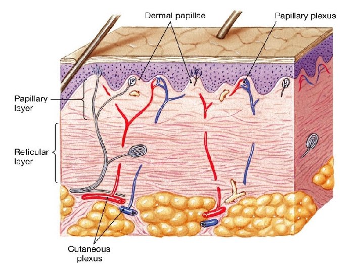
DERMIS • A sheet of connective tissue that supports the epidermis and binds it to the subcutaneous tissue is called dermis or corium. • Thickness of the dermis varies from o. 5 -3 mm. • The dermis hair follicles, sweat glands, sebaceous glands. • The dermis also contains abundant blood vessels, nerves and nerve endings • It is composed of two layers 1. Papillary layer(outer layer) 2. Reticular layer(deeper layer)
PAPILLARY LAYER(OUTER LAYER) • This layer is named papillary because it has papillae that protrude into the epidermis. • This layer is thinner and is composed of loose connective tissue. • It contains fibroblasts, mast cells, macrophages, and an extensive network of fine collagenous, elastic and reticular fibers.
PAPILLARY LAYER(OUTER LAYER) • Just beneath the epidermis the reticular fibers form fibrils which insert into the basal lamina of the epidermis and extend perpendicularly into dermis. • These fibrils are called anchoring fibrils, they bind the epidermis to dermis. • Those papillae that have capillary plexus are called vascular papillae while those containing special nerve endings are termed as nervous papillae.
RETICULAR LAYER • This layer is thicker and consist of dense , irregularly arranged connective tissue. • Mainly contain of collagenous fibers and some elastic fibers • Scattered smooth muscle fibers are also found.
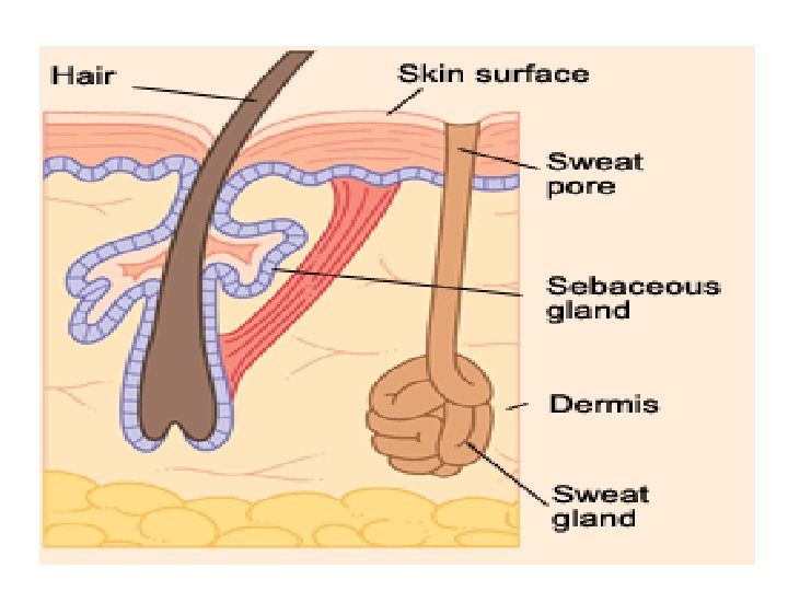
THE HAIR • Hairs are elastic keratinized threads derived from epidermal epithelium. • Found over entire skin except on the palms, soles and dorsal surfaces of distal phalanges. • Each hair consist of two portions Shaft: The shaft projects above the skin. Root: • The root is embed in the skin. • Surrounding the hair root is a tubular hair follicle. • At this deeper end the hair follicle is expanded into a hair bulb.
Glands of the skin There are types of glands in the skin 1. Sebaceous glands 2. Sweat glands 1: Sebaceous glands: • Sebaceous Glands - also known as "Oil Glands" • Most are associated with hair follicles. They secret either into the neck of a hair follicle, or directly onto the surface of the skin via a pore. Where : • Sebaceous glands are located over most of the surface of the skin/body but not in the palms of the hands or the soles of the feet. Secretion(s): • Sebaceous glands secret an oily substance called sebum.
1: Sebaceous glands: function: Sebum helps to protect skin and hair by: • preventing hairs from becoming too dry and brittle • preventing the skin from becoming too dry, hence also helping to keep the skin soft.
SWEAT GLANDS "SUDORIFEROUS GLANDS " • Sweat glands, also known as sudoriferous glands, are distributed over the entire body surface but are particularly abundant on the palms of hands, soles of feet, and on the forehead. • Its primary function is body temperature regulation. Sweat is primarily composed of water (99%), various salts. • It contains minute amounts of fatty materials, urea, and other wastes. • The presence of sodium chloride gives sweat a salty taste. • Sweat glands are coiled tubular glands leading directly to the most superficial layer of the epidermis but extending into the inner layer of the skin (dermis layer).
SWEAT GLANDS "SUDORIFEROUS GLANDS " ECCRINE SWEAT GLANDS • Eccrine sweat glands are distributed all over the body, but the density varies greatly according to body regions; the highest density is found on soles, palms, and scalp. • Eccrine sweat glands are smaller sweat glands that do not extend into the dermis. • They are coiled tubular glands that discharge their secretions directly onto the surface of the skin. • Eccrine glands have three primary functions: I. Thermoregulation II. excretion III. protection.
SWEAT GLANDS "SUDORIFEROUS GLANDS " APOCRINE SWEAT GLANDS • Apocrine sweat glands are limited to axilla (armpits) and perianal areas in humans and are larger than eccrine sweat glands. • These sweat glands are coiled tubular glands that produce a viscous, cloudy and potentially odorous secretion. • Apocrine sweat glands discharge in the canals of hair follicles. • They begin secreting at puberty.
NAILS • They are horny plates covering the dorsal surface of the terminal phalanges of the fingers and the toes. • The nail plate consist of fused, highly keratinized cells. • This plate has three following parts BODY: • The uncovered and visible part which rests over the nail bed. • The body of the nail is translucent. • The crescent shaped whitish area on the proximal part of the nail body is known as Lunuda. It is a reflection of the partially keratinized cells in this region of the nail. • The fold of skin present around the proximal and lateral borders of the nail is known as nail fold. • The furrow between the nail fold and nail bed is called nail groove.
NAILS BODY……. . • The epidermis of the nail bed consist only oh the stratum basale and strataum spinosum. • The epidrmis of the nail bed is very thick proximally and here it is known as matrix of the nail. • The matrix exhibits a high rate cell division and is responsible for the growth of the nails(the growth rate of the hand nails is about 0. 5 mm/week while foot nails grow at a much slower rate). Free edge: Part projecting beyond the skin distally Root: The proximal part of nail that lies beneath the skin fold.
Blood vessels/nerves of the skin • The larger arteries running in the subcutaneous tissue give rise to branches which form two arterial plexuses • One of these plexuses is located between subcutaneous tissue(hypodermis) and dermis while the other lies at the junction of the reticular and papillary layers of the dermis. • Thin branches leave these plexuses and vascularize the dermal papillae. • The veins are disposed in three plexuses, two of these plexuses lie in the same position as the arterial plexuses while third lies in the middle of the dermis. Nerves • The skin is abundantly supplied by sensory nerves. • Discussed in detail in the nervous tissues.
COLOR OF THE SKIN • The final color of the skin is determined by four pigments which are present at various levels and places in the skin. • These pigments are 1. Melanin(brownish black): present nearly in all layers of the epidermis. 2. Carotene(yellowish pigment): Present in the stratum corneum and the adipose cells of the dermis and superficial fascia. 3. Hemoglobin (purple) 4. Oxyhemoglobin (red) Contained in the blood circulating in vascular plexuses of the skin.